Cardiac rhythm problems during space flight

Last updated

Heart rhythm disturbances have been seen among astronauts. Most of these have been related to cardiovascular disease, but it is not clear whether this was due to pre-existing conditions or effects of space flight. It is hoped that advanced screening for coronary disease has greatly mitigated this risk. Other heart rhythm problems, such as atrial fibrillation, can develop over time, necessitating periodic screening of crewmembers’ heart rhythms. Beyond these terrestrial heart risks, some concern exists that prolonged exposure to microgravity may lead to heart rhythm disturbances. Although this has not been observed to date, further surveillance is warranted.

Contents

The incidence and clinical significance of cardiac arrhythmias during long-term exposure to microgravity experienced on the International Space Station (ISS) or during a prolonged (that is, up to 3 years) sojourn to Mars or on the Moon are a concern for the National Aeronautics and Space Administration (NASA). At present, there are only anecdotal reports of cardiac arrhythmias in space, including one documented episode of non-sustained ventricular tachycardia. However, the potential catastrophic nature of a sudden cardiac death in the remote, but highly public, environment of space flight has led to continued concern since the early days of the space program over the possibility that space flight might be arrhythmogenic. Indeed, there are known and well-defined changes in the cardiovascular system with space flight:

  1. plasma volume is reduced;
  2. left ventricular mass in decreased;
  3. the autonomic nervous system adapts to the microgravity environment.

Combined, these physiologic adaptations suggest that changes in cardiac structure and neurohumoral environment during space flight could alter electrical conduction, although the evidence supporting this contention consists mostly of minor changes in QT interval in a small number of astronauts after long-duration space flight. Concurrent with efforts by Flight Medicine to improve screening techniques, as NASA enters the era of exploration class missions, it will be critical to determine with the highest degree of certainty whether space flight by itself alters cardiac structure and function sufficiently to increase the risk for arrhythmias. This undertaking must be done in a highly systematic way.

Introduction

At present, there is little evidence suggesting that cardiovascular adaptation to microgravity or space flight increases susceptibility to life-threatening arrhythmias in astronauts. From a clinical perspective, according to the “biological model” of sudden cardiac death, [1] both the substrate and the trigger for arrhythmias should be considered to determine whether long-term space flight could lead to an increased risk of sudden death. In this model, structural abnormalities interact with functional alterations, such as exercise, electrolyte disturbances, or neurohumoral modulation, to create an environment in which arrhythmias can be initiated and/or sustained. In patients with coronary artery disease, the substrate is clear: a myocardial infarction (MI) and/or scar leading to focal areas of slowed conduction, a necessary condition for re-entry. For patients with apparently normal ventricular function, the potential substrate is less certain. In fact, reentry often is not the mechanism of arrhythmia development in these clinical cases: the arrhythmias may be caused by delayed after-depolarizations, and the triggered activity may be mediated via catecholamines. [2] The published report of non-sustained ventricular tachycardia during prolonged space flight [3] supports this hypothesis, in that initiation of tachycardia by a late diastolic premature ventricular contraction (PVC) is more consistent with triggered activity than it is with re-entry.

While there are no definitive data showing that long-duration space flight is associated with cardiac arrhythmias, there are observational data that have been documented over many years that are suggestive of cardiac electrical changes during long flights. For example, during Skylab, all 9 American crewmembers exhibited some form of rhythm disturbance. Most of these rhythm disturbances consisted of single PVCs and were clinically insignificant. However, one crewmember experienced a 5-beat run of ventricular tachycardia during a lower-body negative pressure protocol, and another had periods of “wandering supraventricular pacemaker” during rest and following exercise. More recently, it has been shown that the corrected QT interval (QTc), a marker of ventricular repolarization, was prolonged slightly in a small number of astronauts after long-duration space flight. In-flight Holter monitoring was not performed during these space flights. Thus, it is not known whether this prolongation was associated with any known arrhythmias. In-flight Holter monitoring was undertaken in the early Space Shuttle era.

Virtually no changes in arrhythmias were documented in flights of 4 to 16 days during either intravehicular or extravehicular operations compared to preflight measurements. [4] [5] Indeed, in these studies, the frequency of arrhythmias may actually have been reduced in flight, though the day-to-day variability of these arrhythmias, which is known to be quite wide, was not quantified. However, aboard the Mir space station, PVCs were detected that were not present before flight [6] and a 14-beat run of ventricular tachycardia was documented. [3]

More recently, several conditions that may predispose crewmembers to arrhythmias have been identified. D’Aunno et al. [7] found that after long-duration missions QTc intervals are slightly prolonged in crewmembers who did not have prolonged QTc intervals after their short-duration Space Shuttle flights, and several investigators have found decreases in left ventricular mass following space flight. [8] [9]

All of these findings raise the concern that cardiac rhythm disturbances may become an issue during the long in-flight tours of duty planned for ISS and interplanetary missions. The degree to which space flight and its many variables can be considered arrhythmogenic is not clear, but the possibility that serious cardiac rhythm disturbances might occur during space flight is a concern to NASA.

Spaceflight evidence

There have been no systematic studies of the arrhythmogenic potential of long-duration space flight, and only two studies of short-duration space flight. There have been, however, a number of published reports detailing in-flight arrhythmias. Table 1 includes a summary of some of these reports.

Table 1. Summary of anecdotal reports of cardiac arrhythmias during U.S. human space flight programs.
ProgramLaunchFlightEVARe-entry or landingPost-flight
MercuryPVCs, PACsSinus Dysrhythmia, 1 PVC, 1 PAC, One fusion beat
GeminiRare PACs
ApolloLunar surface: atrial bigeminal rhythm (extreme fatigue), PVCs, PACs
SkylabPVCs, AV block, ectopic beats, AV junctional rhythm, ST segment and Twave alterations during max stress, ventricular couplet, 3-beat V-tachVentricular Tachycardia
Space ShuttlePVCs, PACsPVCs, PACs
Table adapted from Charles, JB, Frey, MA, Fritsch-Yelle JM, Fortner GW. Chapter 3: Cardiovascular and Cardiorespiratory Function in Space Biology and Medicine. Nicogossian AE, Mohler SR, Gazenko OG, Grigoriev AI, eds. AIIA, Reston VA. 1996. p. 73.

Leguay and Seigneuric also compiled some of the reports from the pre-Shuttle era of crewed space flight. [10] Several of these reports are briefly described below.

One crewmember during Apollo 15 experienced a 22-beat nodal bigeminal rhythm, which was followed by premature atrial beats. [5] This crewmember reported extreme fatigue during the incident, but only when questioned about it by crew surgeons; thus, it was not severe enough to impact the mission. Twenty-one months later the crew member suffered from coronary artery disease and a cardiac infarction without suggestive ECG changes. [10]

In the Skylab missions, several instances of ventricular PVCs, supraventricular PVCs, and nodal arrhythmia were recorded. The arrhythmias occurred during effort tests, extravehicular activities (EVAs), lower body negative pressure sessions, and throughout the entire mission. These included two consecutive PVCs in one astronaut during exercise and an episode of atrioventricular dissociation preceded by sinus bradycardia in two astronauts. [10]

In addition, an isolated incident of a non-sustained 14-beat ventricular tachycardia (Figure 1), with a maximum heart rate of 215 beats per minute, was recorded using in-flight Holter monitoring aboard the Mir. [3] Although not part of a systematic scientific study, this case provides additional evidence of arrhythmias during long-duration space flight. [11]

Figure 1. Record of a non-sustained tachycardia from a Mir crewmember. Cardiac Figure 1.jpg
Figure 1. Record of a non-sustained tachycardia from a Mir crewmember.

Systematic studies of cardiac rhythm disturbances have been performed during short-duration space flight. [4] [5] These studies were conducted in response to medical reports of arrhythmias occurring in 9 to 14 Space Shuttle EVA astronauts between 1983 and 1985. Rossum et al. [5] used 24-hour Holter recordings acquired during and after high altitude chamber activity, 30 days before launch, during and after each extravehicular activity performed, and on return to Earth. The investigators observed no change in the number of premature ventricular contractions of premature atrial contractions per hour during flight compared to preflight or postflight (Figure 2). Likewise, arrhythmias were not observed by Fritsch-Yelle et al. in 12 astronauts studied before, during and after 6 Space Shuttle missions. [4] Given the fact that these data disagreed with previous reports, the investigators suggested that further study was required.

Figure 2. Number of premature ventricular and atrial contractions seen before, during and after space flight. Cardiac Figure 2.jpg
Figure 2. Number of premature ventricular and atrial contractions seen before, during and after space flight.

It is unknown whether long-duration exposure to microgravity itself may precipitate cardiac arrhythmias. Based on observations and clinical judgement, medical operations personnel have suggested that some of these incidents have been related to pre-existing, undiagnosed coronary artery disease. Additional pre-selection crew screening tests, including calcium scoring, have been added to reduce such occurrences in the future.

Contributing factors

Left ventricular mass

Figure 3. Left ventricular mass before and after space flight. Lines represent individual crew members and circles with error bars represent the mean. Cardiac Figure 3.jpg
Figure 3. Left ventricular mass before and after space flight. Lines represent individual crew members and circles with error bars represent the mean.

Recent evidence suggests that the development of apoptosis, or “programmed cell death” in response to pathological, physiologic, and/or genetic signals, may be a key developmental factor in causing cardiac arrhythmias. [12] [13] For example, apoptosis associated with atrophy and fibrofatty replacement of right ventricular tissue has been identified as the likely mechanism forarrhythmia development in arrhythmogenic right ventricular dysplasia, a condition that may lead to sudden death in otherwise healthy young individuals. [14] [15]

Two publications have reported decreases in left ventricular mass after short-duration space flight. In one of these publications, [9] cardiac MRI was used and showed a reduction in left ventricular mass on landing day; however, extended recovery data were not obtained (Figure 3). In the other publication, echocardiography was used and showed a similar decrease in mass on landing day with full recovery 3 days after landing. [8]

Figure 4. Left ventricular mass before and after short-duration space flight [based on(8), n=38]. * = P <= 0.05. Cardiac Figure 4.jpg
Figure 4. Left ventricular mass before and after short-duration space flight [based on(8), n=38]. * = P ≤ 0.05.

Unpublished data (also measured with ultrasound) show decreases in left ventricular mass after 6-month missions aboard the ISS. These decreases are double those observed after short flights and do not fully recover by the third day after landing (Figure 5).

Figure 5. Left ventricular mass before and after long-duration space flight. Cardiac Figure 5.jpg
Figure 5. Left ventricular mass before and after long-duration space flight.

There is some disagreement over the mechanism of the decrease in mass, especially after short-duration missions. While there is evidence to support the idea that tissue dehydration contributes to the loss in mass after short-duration space flights, [9] there are data from bed rest studies showing that the decrease in mass can be prevented with exercise and/or nutritional countermeasures. [16] However, there is agreement that the greater loss of mass with long-duration flight is most likely due to atrophy.

QT prolongation

The QT interval is a measure of the combined duration of ventricular depolarization (QRS) and repolarization (T-wave). The QRS complex is usually of fixed duration in healthy individuals and does not change during long-duration space flight. Thus, changes in QT duration represent alterations in ventricular repolarization. The QT interval of the surface ECG is a spatial and temporal summation of all cardiac cellular action potentials. Not all cells within the heart share identical action potentials; therefore, a certain degree of variability, or inhomogeneity, in their repolarization time exists. The degree of inhomogeneity during repolarization directly correlates with the overall morphology of the QT waveform (primarily the T-wave) and in most cases with the QT interval duration. A clear association between the magnitude of inhomogeneity of repolarization and the risk for the development of ventricular arrhythmias has been established. [17] [18] [19]

The QT interval is often corrected for heart rate and is shown as QTc. Some conditions that can prolong the QTc interval are ischemic heart disease, autonomic dysfunction, bradycardia, electrolyte abnormalities, cardiac remodeling, and dehydration medications that interfere with the cardiac potassium ion channels. [20] [21] [22] [23] Which of these factors are seen in long-duration astronauts?

The environment created by the combination of factors listed above might cause or exacerbate the prolongation of the QT interval.

Prolongation of QTc interval does not itself guarantee an increase in ventricular arrhythmias. For example, sleep, hypothyroidism, and use of the anti-arrhythmic drug amiodarone all prolong QTc without increasing the incidence of ventricular arrhythmias. It is possible that space flight presents a similar situation. However, at this time, that determination cannot be made due to lack of data. Therefore, the data must be collected.

Ground-based evidence

In general, subjects in bed rest studies do not exhibit increases in ventricular ectopy, although numerous studies have shown decreases in left ventricular mass and/or volume. [9] [16] [28] [29] During bed rest, left ventricular mass has been shown to decrease by eight percent after 6 weeks, which was thought to be related to decreased physiological loading. [9]

Ground-based animal studies also have been used to determine the effects of microgravity on the cardiovascular system. Tachycardia has been observed in standing rats, after hindlimb-unloading for 28 days. [30] A trend in decreased cardiac mass has also been documented in studies of hindlimb-suspended rats. [31] However, hemodynamics in humans differ from hemodynamics in quadrupeds; thus, the rat is not the most appropriate model in which to examine the effects of microgravity on cardiovascular adaptations. [32]

Computer-based simulation information

A systems analysis using the computer model of human physiology developed at the University of Mississippi Medical Center also predicts a loss in left ventricular mass following short-duration space flight. According to the model predictions, the reductions in left ventricular mass observed after short-duration exposure to microgravity may be the result of a contraction of the myocardial interstitial fluid space secondary to a loss in the plasma volume (see Figure 6). [8] [33]

Figure 6. Model predictions of myocardial interstitial fluid spaces preflight, on landing day, and after landing day. Cardiac Figure 6.jpg
Figure 6. Model predictions of myocardial interstitial fluid spaces preflight, on landing day, and after landing day.

The finding of QTc prolongation in astronauts has been of concern from the clinical operations perspective. Such prolongation has been documented on several occasions but it is not clear if these findings have any clinical significance or portend risk. [7] [34]

Risk in context of exploration mission operational scenarios

Cardiac rhythm disturbances could jeopardize mission objectives and, at the most extreme, the life of crewmembers. The worst-case scenario would be a life-threatening arrhythmia during a Mars exploration mission, where return to Earth would take months. Under these conditions, other crewmembers would need to treat the affected crewmember with the limited supplies available on the spacecraft.

Gaps

The data are compelling enough that this risk cannot be retired until a systematic evaluation of cardiac structure and function is made on the ISS. This is considered a high priority activity.

Conclusions

Very little research has systematically evaluated the prevalence (or potential risk) of cardiac arrhythmias during space flight. There are several observational reports of non life-threatening but potentially concerning arrhythmias. At least two potential risk factors for arrhythmias have been reported either during or immediately after space flight: cardiac atrophy and a prolonged QTc interval. The potential severity of the mission impact of a serious arrhythmia requires that a systematic evaluation be conducted of the risk of arrhythmia due to space flight.

See also

Acronyms and abbreviations

Acronym/AbbreviationDescription
AVAtrionetricular
ECG Electrocardiogram
LV Left Ventricle
LVMLeft Ventricular Mass
MI Myocardial Infarction
MRI Magnetic Resonance Imaging
NASANational Aeronautics and Space Administration
P-WaveAtrial Depolarization
PAC Premature Atrial Contraction
PVC Premature Ventricular Contraction
QRSVentricular Depolarization
QTMeasure of time between ventricular depolarization and repolarization
QTcCorrected QT Interval
R+0Landing Day
R+3Three days post-landing (Recovery)
T-WaveVentricular Repolarization
V-tach Ventricular Tachycardia

Related Research Articles

Electrocardiography Method to record the electrical activity of the heart through passive electrodes placed over the skin.

Electrocardiography is the process of producing an electrocardiogram. It is a graph of voltage versus time of the electrical activity of the heart using electrodes placed on the skin. These electrodes detect the small electrical changes that are a consequence of cardiac muscle depolarization followed by repolarization during each cardiac cycle (heartbeat). Changes in the normal ECG pattern occur in numerous cardiac abnormalities, including cardiac rhythm disturbances, inadequate coronary artery blood flow, and electrolyte disturbances.

Tachycardia Heart rate that exceeds the normal resting rate

Tachycardia, also called tachyarrhythmia, is a heart rate that exceeds the normal resting rate. In general, a resting heart rate over 100 beats per minute is accepted as tachycardia in adults. Heart rates above the resting rate may be normal or abnormal.

Long QT syndrome Medical condition

Long QT syndrome (LQTS) is a condition in which repolarization of the heart after a heartbeat is affected. It results in an increased risk of an irregular heartbeat which can result in fainting, drowning, seizures, or sudden death. These episodes can be triggered by exercise or stress. Some rare forms of LQTS are associated with other symptoms and signs including deafness and periods of muscle weakness.

Micro-g environment

The term micro-g environment is more or less synonymous with the terms weightlessness and zero-g, but with an emphasis on the fact that g-forces are never exactly zero—just very small. The symbol for microgravity, μg, was used on the insignias of Space Shuttle flights STS-87 and STS-107, because these flights were devoted to microgravity research in low Earth orbit.

Antiarrhythmic agent

Antiarrhythmic agents, also known as cardiac dysrhythmia medications, are a group of pharmaceuticals that are used to suppress abnormal rhythms of the heart, such as atrial fibrillation, atrial flutter, ventricular tachycardia, and ventricular fibrillation.

Short QT syndrome Medical condition

Short QT syndrome (SQT) is a very rare genetic disease of the electrical system of the heart, and is associated with an increased risk of abnormal heart rhythms and sudden cardiac death. The syndrome gets its name from a characteristic feature seen on an electrocardiogram (ECG) – a shortening of the QT interval. It is caused by mutations in genes encoding ion channels that shorten the cardiac action potential, and appears to be inherited in an autosomal dominant pattern. The condition is diagnosed using a 12-lead ECG. Short QT syndrome can be treated using an implantable cardioverter-defibrillator or medications including quinidine. Short QT syndrome was first described in 2000, and the first genetic mutation associated with the condition was identified in 2004.

Torsades de pointes Type of abnormal heart rhythm

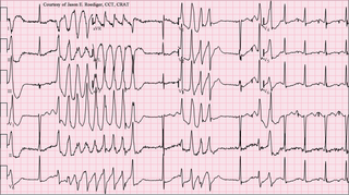
Torsades de pointes, torsade de pointes or torsades des pointes (TdP) is a specific type of abnormal heart rhythm that can lead to sudden cardiac death. It is a polymorphic ventricular tachycardia that exhibits distinct characteristics on the electrocardiogram (ECG). It was described by French physician François Dessertenne in 1966. Prolongation of the QT interval can increase a person's risk of developing this abnormal heart rhythm, occurring in between 1% and 10% of patients who receive QT-prolonging antiarrhythmic drugs.

Ventricular tachycardia Fast heart rhythm that originates in one of the ventricles of the heart

Ventricular tachycardia is a type of regular, fast heart rate that arises from improper electrical activity in the ventricles of the heart. Although a few seconds may not result in problems, longer periods are dangerous; and multiple episodes over a short period of time is referred to as an Electrical Storm. Short periods may occur without symptoms, or present with lightheadedness, palpitations, or chest pain. Ventricular tachycardia may result in ventricular fibrillation and turn into cardiac arrest. It is found initially in about 7% of people in cardiac arrest.

QT interval

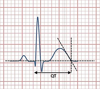
The QT interval is a measurement made on an electrocardiogram used to assess some of the electrical properties of the heart. It is calculated as the time from the start of the Q wave to the end of the T wave, and approximates to the time taken from when the cardiac ventricles start to contract to when they finish relaxing. An abnormally long or abnormally short QT interval is associated with an increased risk of developing abnormal heart rhythms and sudden cardiac death. Abnormalities in the QT interval can be caused by genetic conditions such as long QT syndrome, by certain medications such as sotalol or pitolisant, by disturbances in the concentrations of certain salts within the blood such as hypokalaemia, or by hormonal imbalances such as hypothyroidism.

Sotalol

Sotalol, sold under the brand name Betapace among others, is a medication used to treat and prevent abnormal heart rhythms. It is only recommended in those with significant abnormal heart rhythms due to potentially serious side effects. Evidence does not support a decreased risk of death with long term use. It is taken by mouth or injection into a vein.

Jervell and Lange-Nielsen syndrome Medical condition

Jervell and Lange-Nielsen syndrome (JLNS) is a rare type of long QT syndrome associated with severe, bilateral sensorineural hearing loss. Those with JLNS are at risk of abnormal heart rhythms called arrhythmias, which can lead to fainting, seizures, or sudden death. JLNS, like other forms of long QT syndrome, causes the cardiac muscle to take longer than usual to recharge between beats. It is caused by genetic variants responsible for producing ion channels that carry transport potassium out of cells. The condition is usually diagnosed using an electrocardiogram, but genetic testing can also be used. Treatment includes lifestyle measures, beta blockers, and implantation of a defibrillator in some cases. It was first described by Anton Jervell and Fred Lange-Nielsen in 1957.

Romano–Ward syndrome Medical condition

Romano–Ward syndrome is the most common form of congenital Long QT syndrome (LQTS), a genetic heart condition that affects the electrical properties of heart muscle cells. Those affected are at risk of abnormal heart rhythms which can lead to fainting, seizures, or sudden death. Romano–Ward syndrome can be distinguished clinically from other forms of inherited LQTS as it affects only the electrical properties of the heart, while other forms of LQTS can also affect other parts of the body.

Andersen–Tawil syndrome Rare autosomal dominant genetic disorder

Andersen–Tawil syndrome, also called Andersen syndrome and long QT syndrome 7, is a rare genetic disorder affecting several parts of the body. The three predominant features of Andersen–Tawil syndrome include disturbances of the electrical function of the heart characterised by an abnormality seen on an electrocardiogram and a tendency to abnormal heart rhythms, physical characteristics including low-set ears and a small lower jaw, and intermittent periods of muscle weakness known as hypokalaemic periodic paralysis.

Azimilide

Azimilide is a class ΙΙΙ antiarrhythmic drug. The agents from this heterogeneous group have an effect on the repolarization, they prolong the duration of the action potential and the refractory period. Also they slow down the spontaneous discharge frequency of automatic pacemakers by depressing the slope of diastolic depolarization. They shift the threshold towards zero or hyperpolarize the membrane potential. Although each agent has its own properties and will have thus a different function.

Lorcainide

Lorcainide is a Class 1c antiarrhythmic agent that is used to help restore normal heart rhythm and conduction in patients with premature ventricular contractions, ventricular tachycardiac and Wolff-Parkinson-White syndrome. Lorcainide was developed by Janssen Pharmaceutica (Belgium) in 1968 under the commercial name Remivox and is designated by code numbers R-15889 or Ro 13-1042/001. It has a half-life of 8.9 +- 2.3 hrs which may be prolonged to 66 hrs in people with cardiac disease.

Arrhythmia Group of conditions in which the heartbeat is irregular, too fast, or too slow

Arrhythmia, also known as cardiac arrhythmia or heart arrhythmia, is a group of conditions in which the heartbeat is irregular, too fast, or too slow. The heart rate that is too fast – above 100 beats per minute in adults – is called tachycardia, and a heart rate that is too slow – below 60 beats per minute – is called bradycardia. Some types of arrhythmias have no symptoms. Symptoms, when present, may include palpitations or feeling a pause between heartbeats. In more serious cases, there may be lightheadedness, passing out, shortness of breath or chest pain. While most types of arrhythmia are not serious, some predispose a person to complications such as stroke or heart failure. Others may result in sudden death.

Celivarone Experimental drug being tested for use in pharmacological antiarrhythmic therapy

Celivarone is an experimental drug being tested for use in pharmacological antiarrhythmic therapy. Cardiac arrhythmia is any abnormality in the electrical activity of the heart. Arrhythmias range from mild to severe, sometimes causing symptoms like palpitations, dizziness, fainting, and even death. They can manifest as slow (bradycardia) or fast (tachycardia) heart rate, and may have a regular or irregular rhythm.

Illnesses and injuries during space missions are a range of medical conditions and injuries that may occur during space flights. Some of these medical conditions occur due to the changes withstood by the human body during space flight itself, while others are injuries that could have occurred on Earth's surface. A non-exhaustive list of these conditions and their probability of occurrence can be found in the following sources:

Skeletal muscles, particularly postural muscles of the lower limb, undergo atrophy and structural and metabolic alterations during space flight. The relationships between in-flight exercise, muscle changes and performance are not well understood. Efforts should be made to try to understand the current status of in-flight and post-flight exercise performance capacity and what the goals/target areas for protection are with the current in flight exercise program.

QT prolongation is a measure of delayed ventricular repolarisation, which means the heart muscle takes longer than normal to recharge between beats. It is an electrical disturbance which can be seen on an electrocardiogram (ECG). Excessive QT prolongation can trigger tachycardias such as torsades de pointes (TdP). QT prolongation is an established side effect of anti-arrhythmic medicines, but can also be caused by a wide range of non-cardiac medicines, including antibiotics, antihistamines, opioid analgesics and complementary medicines. On an EKG, the QT interval represents the summation of action potentials in cardiac muscle cells, which can be caused by an increase in inward current through sodium or calcium channels, or a decrease in outward current through potassium channels. By binding to and inhibiting the “rapid” delayed rectifier potassium current protein, certain drugs are able to decrease the outward flow of potassium ions and extend the length of phase 3 myocardial repolarization, resulting in QT prolongation.

References

  1. Myerburg, RJ; Kessler, KM; Bassett, AL; Castellanos, A (15 June 1989). "A biological approach to sudden cardiac death: structure, function and cause". The American Journal of Cardiology. 63 (20): 1512–6. doi:10.1016/0002-9149(89)90017-9. PMID   2524961.
  2. Lerman, BB; Stein, KM; Markowitz, SM (June 1996). "Adenosine-sensitive ventricular tachycardia: a conceptual approach". Journal of Cardiovascular Electrophysiology. 7 (6): 559–69. doi:10.1111/j.1540-8167.1996.tb00563.x. PMID   8743762. S2CID   39409797.
  3. 1 2 3 4 5 Fritsch-Yelle, JM; Leuenberger, UA; D'Aunno, DS; Rossum, AC; Brown, TE; Wood, ML; Josephson, ME; Goldberger, AL (1 June 1998). "An episode of ventricular tachycardia during long-duration spaceflight" (PDF). The American Journal of Cardiology. 81 (11): 1391–2. doi:10.1016/s0002-9149(98)00179-9. PMID   9631987.
  4. 1 2 3 Fritsch-Yelle, JM; Charles, JB; Jones, MM; Wood, ML (March 1996). "Microgravity decreases heart rate and arterial pressure in humans". Journal of Applied Physiology. 80 (3): 910–4. doi:10.1152/jappl.1996.80.3.910. PMID   8964756.
  5. 1 2 3 4 Rossum, AC; Wood, ML; Bishop, SL; Deblock, H; Charles, JB (15 April 1997). "Evaluation of cardiac rhythm disturbances during extravehicular activity". The American Journal of Cardiology. 79 (8): 1153–5. doi:10.1016/s0002-9149(97)00071-4. PMID   9114789.
  6. Goldberger, AL; Bungo, MW; Baevsky, RM; Bennett, BS; Rigney, DR; Mietus, JE; Nikulina, GA; Charles, JB (July 1994). "Heart rate dynamics during long-term space flight: report on Mir cosmonauts". American Heart Journal. 128 (1): 202–4. doi:10.1016/0002-8703(94)90033-7. PMID   8017279.
  7. 1 2 D'Aunno, DS; Dougherty, AH; DeBlock, HF; Meck, JV (15 February 2003). "Effect of short- and long-duration spaceflight on QTc intervals in healthy astronauts". The American Journal of Cardiology. 91 (4): 494–7. doi:10.1016/S0002-9149(02)03259-9. PMID   12586278.
  8. 1 2 3 Summers, RL; Martin, DS; Meck, JV; Coleman, TG (1 May 2005). "Mechanism of spaceflight-induced changes in left ventricular mass". The American Journal of Cardiology. 95 (9): 1128–30. doi:10.1016/j.amjcard.2005.01.033. PMID   15842991.
  9. 1 2 3 4 5 6 Perhonen, MA; Franco, F; Lane, LD; Buckey, JC; Blomqvist, CG; Zerwekh, JE; Peshock, RM; Weatherall, PT; Levine, BD (August 2001). "Cardiac atrophy after bed rest and spaceflight". Journal of Applied Physiology. 91 (2): 645–53. doi:10.1152/jappl.2001.91.2.645. PMID   11457776.
  10. 1 2 3 Leguay, G; Seigneuric, A (July 1981). "Cardiac arrhythmias in space. Role of vagotonia". Acta Astronautica. 8 (7): 795–801. Bibcode:1981AcAau...8..795L. doi:10.1016/0094-5765(81)90019-9. PMID   11542963.
  11. Baevsky, R.M.; Funtova, I.I.; Diedrich, A.; Pashchenko, A.V.; Chernikova, A.G.; Drescher, J.; Baranov, V.M.; Tank, J. (1 October 2007). "Autonomic function testing on board the ISS—update on "Pneumocard"". Acta Astronautica. 61 (7–8): 672–675. Bibcode:2007AcAau..61..672B. doi:10.1016/j.actaastro.2006.11.017.
  12. Colucci, WS (17 October 1996). "Apoptosis in the heart". The New England Journal of Medicine. 335 (16): 1224–6. doi:10.1056/nejm199610173351610. PMID   8815947.
  13. James, TN (1998). "Normal and abnormal consequences of apoptosis in the human heart". Annual Review of Physiology. 60: 309–25. doi:10.1146/annurev.physiol.60.1.309. PMID   9558466.
  14. Mallat, Ziad; Tedgui, Alain; Fontaliran, Fabrice; Frank, Robert; Durigon, Michel; Fontaine, Guy (17 October 1996). "Evidence of Apoptosis in Arrhythmogenic Right Ventricular Dysplasia". New England Journal of Medicine. 335 (16): 1190–1197. doi:10.1056/NEJM199610173351604. PMID   8815941.
  15. Basso, C; Thiene, G; Corrado, D; Angelini, A; Nava, A; Valente, M (1 September 1996). "Arrhythmogenic right ventricular cardiomyopathy. Dysplasia, dystrophy, or myocarditis?". Circulation. 94 (5): 983–91. doi:10.1161/01.cir.94.5.983. PMID   8790036.
  16. 1 2 Dorfman, TA; Levine, BD; Tillery, T; Peshock, RM; Hastings, JL; Schneider, SM; Macias, BR; Biolo, G; Hargens, AR (July 2007). "Cardiac atrophy in women following bed rest". Journal of Applied Physiology. 103 (1): 8–16. doi:10.1152/japplphysiol.01162.2006. PMID   17379748.
  17. El-Sherif, N (August 2001). "Mechanism of ventricular arrhythmias in the long QT syndrome: on hermeneutics". Journal of Cardiovascular Electrophysiology. 12 (8): 973–6. doi:10.1046/j.1540-8167.2001.00973.x. PMID   11513452. S2CID   3198696.
  18. Lux, RL; Hilbel, T; Brockmeier, K (2001). "Electrocardiographic measures of repolarization revisited: why? what? how?". Journal of Electrocardiology . 34 Suppl (4): 259–64. doi:10.1054/jelc.2001.28909. PMID   11781965.
  19. Shusterman, V; Goldberg, A; London, B (27 June 2006). "Upsurge in T-wave alternans and nonalternating repolarization instability precedes spontaneous initiation of ventricular tachyarrhythmias in humans". Circulation. 113 (25): 2880–7. doi: 10.1161/circulationaha.105.607895 . PMID   16785339.
  20. Ishida, S; Takahashi, N; Nakagawa, M; Fujino, T; Saikawa, T; Ito, M (August 1995). "Relation between QT and RR intervals in patients with bradyarrhythmias". British Heart Journal. 74 (2): 159–62. doi:10.1136/hrt.74.2.159. PMC   483992 . PMID   7546995.
  21. Khan, IA (January 2002). "Clinical and therapeutic aspects of congenital and acquired long QT syndrome". The American Journal of Medicine. 112 (1): 58–66. doi:10.1016/S0002-9343(01)01011-7. PMID   11812408.
  22. Lo, SS; Mathias, CJ; Sutton, MS (May 1996). "QT interval and dispersion in primary autonomic failure". Heart. 75 (5): 498–501. doi:10.1136/hrt.75.5.498. PMC   484349 . PMID   8665344.
  23. Savelieva, I; Yap, YG; Yi, G; Guo, XH; Hnatkova, K; Camm, AJ; Malik, M (October 1999). "Relation of ventricular repolarization to cardiac cycle length in normal subjects, hypertrophic cardiomyopathy, and patients with myocardial infarction". Clinical Cardiology. 22 (10): 649–54. doi:10.1002/clc.4960221011. PMC   6655915 . PMID   10526689.
  24. Fritsch-Yelle, JM; Whitson, PA; Bondar, RL; Brown, TE (November 1996). "Subnormal norepinephrine release relates to presyncope in astronauts after spaceflight". Journal of Applied Physiology. 81 (5): 2134–41. doi:10.1152/jappl.1996.81.5.2134. PMID   8941538.
  25. Fritsch, JM; Charles, JB; Bennett, BS; Jones, MM; Eckberg, DL (August 1992). "Short-duration spaceflight impairs human carotid baroreceptor-cardiac reflex responses". Journal of Applied Physiology. 73 (2): 664–71. Bibcode:1992JAPh...73..664F. doi:10.1152/jappl.1992.73.2.664. PMID   1399995.
  26. 1 2 Meck, JV; Reyes, CJ; Perez, SA; Goldberger, AL; Ziegler, MG (November–December 2001). "Marked exacerbation of orthostatic intolerance after long- vs. short-duration spaceflight in veteran astronauts". Psychosomatic Medicine. 63 (6): 865–73. doi:10.1097/00006842-200111000-00003. PMID   11719623. S2CID   18756171.
  27. Rossum, AC; Ziegler, MG; Meck, JV (1 December 2001). "Effect of spaceflight on cardiovascular responses to upright posture in a 77-year-old astronaut". The American Journal of Cardiology. 88 (11): 1335–7. doi:10.1016/s0002-9149(01)02104-x. PMID   11728371. Archived from the original on 24 June 2013.
  28. Levine, BD; Zuckerman, JH; Pawelczyk, JA (15 July 1997). "Cardiac atrophy after bed-rest deconditioning: a nonneural mechanism for orthostatic intolerance". Circulation. 96 (2): 517–25. doi:10.1161/01.cir.96.2.517. PMID   9244220.
  29. Arbeille, P; Fomina, G; Roumy, J; Alferova, I; Tobal, N; Herault, S (December 2001). "Adaptation of the left heart, cerebral and femoral arteries, and jugular and femoral veins during short- and long-term head-down tilt and spaceflights". European Journal of Applied Physiology. 86 (2): 157–68. doi:10.1007/s004210100473. PMID   11822475. S2CID   31653815.
  30. Ray, CA; Vasques, M; Miller, TA; Wilkerson, MK; Delp, MD (September 2001). "Effect of short-term microgravity and long-term hindlimb unloading on rat cardiac mass and function". Journal of Applied Physiology. 91 (3): 1207–13. doi:10.1152/jappl.2001.91.3.1207. PMID   11509517.
  31. Bao, JX; Zhang, LF; Shang, HH; Yu, ZB; Qian, YQ (April 1999). "[Echocardiographic assessment of left ventricular structure and function after simulated weightlessness in rats]". Hang Tian Yi Xue Yu Yi Xue Gong Cheng = Space Medicine & Medical Engineering. 12 (2): 88–91. PMID   12428650.
  32. Rowell, Loring B. (1993). Human cardiovascular control. New York: Oxford University Press. ISBN   978-0195073621.
  33. Summers, RL; Martin, DS; Meck, JV; Coleman, TG (March 2007). "Computer systems analysis of spaceflight induced changes in left ventricular mass". Computers in Biology and Medicine. 37 (3): 358–63. doi:10.1016/j.compbiomed.2006.04.003. PMID   16808910.
  34. Mitchell, BM; Meck, JV (15 April 2004). "Short-duration spaceflight does not prolong QTc intervals in male astronauts". The American Journal of Cardiology. 93 (8): 1051–2. doi:10.1016/j.amjcard.2003.12.060. PMID   15081456.

PD-icon.svg This article incorporates  public domain material from the National Aeronautics and Space Administration document: "Human Health and Performance Risks of Space Exploration Missions" (PDF).(NASA SP-2009-3405)