Crown-rump length

Last updated
An ultrasound showing an embryo measured to have a crown-rump length of 1.67 cm and estimated to have a gestational age of 8 weeks and 1 day Ultrasound of human fetus, 8 weeks and 1 day.jpg
An ultrasound showing an embryo measured to have a crown-rump length of 1.67 cm and estimated to have a gestational age of 8 weeks and 1 day

Crown-rump length (CRL) is the measurement of the length of human embryos and fetuses from the top of the head (crown) to the bottom of the buttocks (rump). It is typically determined from ultrasound imagery and can be used to estimate gestational age.

Contents

Introduction

Diagram showing crown-rump length by gestational age. The blue line is the mean and the green area delimits the 3rd versus the 97th percentile. Crown-rump length by gestational age.png
Diagram showing crown-rump length by gestational age. The blue line is the mean and the green area delimits the 3rd versus the 97th percentile.
Ultrasound image of the foetus at 12 weeks of pregnancy in a sagittal scan. Measurements of fetal Crown Rump Length (CRL). CRL Crown rump length 12 weeks ecografia Dr. Wolfgang Moroder.jpg
Ultrasound image of the foetus at 12 weeks of pregnancy in a sagittal scan. Measurements of fetal Crown Rump Length (CRL).

The embryo and fetus float in the amniotic fluid inside the uterus of the mother usually in a curved posture resembling the letter C. The measurement can actually vary slightly if the fetus is temporarily stretching (straightening) its body. The measurement needs to be in the natural state with an unstretched body which is actually C shaped. The measurement of CRL is useful in determining the gestational age (menstrual age starting from the first day of the last menstrual period) and thus the expected date of delivery (EDD). Different human fetuses grow at different rates and thus the gestational age is an approximation. Recent evidence has indicated that CRL growth (and thus the approximation of gestational age) may be influenced by maternal factors such as age, smoking, and folic acid intake. Early in pregnancy gestational age 8 weeks, it is accurate within about +/- 5 days but later in pregnancy due to different growth rates, the accuracy is less. In that situation, other parameters can be used in addition to CRL. The length of the umbilical cord is approximately equal to the CRL throughout pregnancy.

Gestational age is not the same as fertilization age. It takes about 14 days from the first day of the last menstrual period for conception to take place and thus for the conceptus to form. The age from this point in time (conception) is called the fertilization age and is thus 2 weeks shorter than the gestational age. Thus a 6-week gestational age would be a 4-week fertilization age. Some authorities however casually interchange these terms [ citation needed ] and the reader is advised to be cautious. An average gestational period (duration of pregnancy from the first day of the last menstrual period up to delivery) is 280 days. On average, this is 9 months and 6 days.

Gestational age estimation

A commonly used estimate of gestational age in weeks is (as described by Verburg et al. [2] ):

Crown–rump length (CRL) in relation to gestational age [2]
CRL (mm)Gestational age (weeks + days)
5th centileMedian95th centile
55 + 66 + 26 + 6
107 + 07 + 48 + 1
157 + 58 + 29 + 0
208 + 29 + 09 + 5
258 + 69 + 410 + 2
309 + 210 + 010 + 6
359 + 510 + 311 + 2
4010 + 110 + 611 + 5
4510 + 311 + 212 + 1
5010 + 611 + 512 + 4
5511 + 112 + 013 + 0
6011 + 312 + 313 + 3
6511 + 612 + 513 + 5
7012 + 113 + 014 + 0
7512 + 313 + 314 + 3
8012 + 513 + 514 + 5
8513 + 014 + 015 + 1
9013 + 214 + 215 + 3
9513 + 414 + 415 + 5
10013 + 615 + 016 + 1

Gestational age estimation in days is carried out according to the equations:

 ; and SD of GA = 2.39102 + (0.0193474 × CRL).

See also

Related Research Articles

<span class="mw-page-title-main">Pregnancy (mammals)</span> Period of reproduction

In mammals, pregnancy is the period of reproduction during which a female carries one or more live offspring from implantation in the uterus through gestation. It begins when a fertilized zygote implants in the female's uterus, and ends once it leaves the uterus.

<span class="mw-page-title-main">Ectopic pregnancy</span> Female reproductive system health issue

Ectopic pregnancy is a complication of pregnancy in which the embryo attaches outside the uterus. Signs and symptoms classically include abdominal pain and vaginal bleeding, but fewer than 50 percent of affected women have both of these symptoms. The pain may be described as sharp, dull, or crampy. Pain may also spread to the shoulder if bleeding into the abdomen has occurred. Severe bleeding may result in a fast heart rate, fainting, or shock. With very rare exceptions, the fetus is unable to survive.

<span class="mw-page-title-main">Obstetric ultrasonography</span> Use of medical ultrasonography in pregnancy

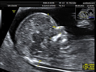
Obstetric ultrasonography, or prenatal ultrasound, is the use of medical ultrasonography in pregnancy, in which sound waves are used to create real-time visual images of the developing embryo or fetus in the uterus (womb). The procedure is a standard part of prenatal care in many countries, as it can provide a variety of information about the health of the mother, the timing and progress of the pregnancy, and the health and development of the embryo or fetus.

<span class="mw-page-title-main">Gestational sac</span> Cavity of fluid surrounding an embryo

The gestational sac is the large cavity of fluid surrounding the embryo. During early embryogenesis it consists of the extraembryonic coelom, also called the chorionic cavity. The gestational sac is normally contained within the uterus. It is the only available structure that can be used to determine if an intrauterine pregnancy exists until the embryo can be identified.

The fetal pole is a thickening on the margin of the yolk sac of a fetus during pregnancy. It is usually identified at six weeks with vaginal ultrasound and at six and a half weeks with abdominal ultrasound. However, it is not unheard of for the fetal pole to not be visible until about 9 weeks. The fetal pole may be seen at 2–4 mm crown-rump length (CRL).

<span class="mw-page-title-main">Blighted ovum</span> Medical condition

A blighted ovum is a pregnancy in which the embryo never develops or develops and is reabsorbed. In a normal pregnancy, an embryo would be visible on an ultrasound by six weeks after the woman's last menstrual period. Anembryonic gestation is one of the causes of miscarriage of a pregnancy.

In obstetrics, gestational age is a measure of the age of a pregnancy taken from the beginning of the woman's last menstrual period (LMP), or the corresponding age of the gestation as estimated by a more accurate method if available. Such methods include adding 14 days to a known duration since fertilization, or by obstetric ultrasonography. The popularity of using this measure of pregnancy is due to the fact that menstrual periods are usually noticed, while there is generally no convenient way to discern when fertilization or implantation occurred. Gestational age is contrasted with fertilization age which takes the date of fertilization as the start date of gestation, and pregnancy which begins with implantation.

<span class="mw-page-title-main">Pregnancy</span> Time of offspring development in mothers body

Pregnancy is the time during which one or more offspring develops (gestates) inside a woman's uterus (womb). A multiple pregnancy involves more than one offspring, such as with twins.

Prenatal development includes the development of the embryo and of the fetus during a viviparous animal's gestation. Prenatal development starts with fertilization, in the germinal stage of embryonic development, and continues in fetal development until birth.
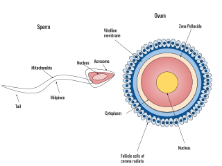
<span class="mw-page-title-main">Human fertilization</span> Union of a human egg and sperm

Human fertilization is the union of a human egg and sperm, occurring primarily in the ampulla of the fallopian tube. The result of this union leads to the production of a fertilized egg called a zygote, initiating embryonic development. Scientists discovered the dynamics of human fertilization in the nineteenth century.

<span class="mw-page-title-main">Human reproduction</span> Procreative biological processes of humanity

Human reproduction is sexual reproduction that results in human fertilization to produce a human offspring. It typically involves sexual intercourse between a sexually mature human male and female. During sexual intercourse, the interaction between the male and female reproductive systems results in fertilization of the ovum by the sperm to form a zygote. These specialized reproductive cells are called gametes, which are created in a process called gametogenesis. While normal cells contain 46 chromosomes, gamete cells only contain 23 single chromosomes, and it is when these two cells merge into one zygote cell that genetic recombination occurs and the new zygote contains 23 chromosomes from each parent, giving it 46 chromosomes. The zygote then undergoes a defined development process that is known as human embryogenesis, and this starts the typical 9-month gestation period that is followed by childbirth. The fertilization of the ovum may be achieved by artificial insemination methods, which do not involve sexual intercourse. Assisted reproductive technology also exists.

Postterm pregnancy is when a woman has not yet delivered her baby after 42 weeks of gestation, two weeks beyond the typical 40-week duration of pregnancy. Postmature births carry risks for both the mother and the baby, including fetal malnutrition, meconium aspiration syndrome, and stillbirths. After the 42nd week of gestation, the placenta, which supplies the baby with nutrients and oxygen from the mother, starts aging and will eventually fail. Postterm pregnancy is a reason to induce labor.

<span class="mw-page-title-main">Nuchal scan</span> Routine ultrasound done between 11 and 14 weeks pregnancy

A nuchal scan or nuchal translucency (NT) scan/procedure is a sonographic prenatal screening scan (ultrasound) to detect chromosomal abnormalities in a fetus, though altered extracellular matrix composition and limited lymphatic drainage can also be detected.

A fetus or foetus is the unborn offspring that develops from an animal embryo. Following embryonic development the fetal stage of development takes place. In human prenatal development, fetal development begins from the ninth week after fertilization and continues until birth. Prenatal development is a continuum, with no clear defining feature distinguishing an embryo from a fetus. However, a fetus is characterized by the presence of all the major body organs, though they will not yet be fully developed and functional and some not yet situated in their final anatomical location.

Pregnancy rate is the success rate for getting pregnant. It is the percentage of all attempts that leads to pregnancy, with attempts generally referring to menstrual cycles where insemination or any artificial equivalent is used, which may be simple artificial insemination (AI) or AI with additional in vitro fertilization (IVF).

The estimated date of delivery (EDD), also known as expected date of confinement, and estimated due date or simply due date, is a term describing the estimated delivery date for a pregnant person. Normal pregnancies last between 38 and 42 weeks. Children are delivered on their expected due date about 4% of the time.

Post-maturity syndrome develops in about 20% of human pregnancies continuing past the expected dates. Ten years ago it was generally held that the postmature fetus ran some risk of dying in the uterus before the onset of labour because of degeneration and calcification of the placenta. Features of post-maturity syndrome include oligohydramnios, meconium aspiration, macrosomia and fetal problems such as dry peeling skin, overgrown nails, abundant scalp hair, visible creases on palms and soles, minimal fat deposition and skin colour become green or yellow due to meconium staining. Post-maturity refers to any baby born after 42 weeks gestation or 294 days past the first day of the mother's last menstrual period. Less than 6 percent of all babies are born at 42 weeks or later. In most cases, continued fetal growth between 39 and 43 wk gestation results in a macrosomic infant. However, sometimes the placenta involutes, and multiple infarcts and villous degeneration cause placental insufficiency syndrome. In this syndrome, the fetus receives inadequate nutrients and oxygen from the mother, resulting in a thin, small-for-gestational-age, undernourished infant with depleted glycogen stores. Post term, the amniotic fluid volume eventually decreases, leading to oligohydramnios. Although pregnancy is said to last nine months, health care providers track pregnancy by weeks and days. The estimated delivery date, also called the estimated due date or EDD, is calculated as 40 weeks or 280 days from the first day of the last menstrual period. Only 4 percent women will deliver on their due date. The terms Post-maturity or "Post-term" are both words used to describe babies born after 42 weeks. The terms "post-maturity" and "post-term" are interchangeable. As there are many definitions for prolonged pregnancy the incidence varies from 2 to 10%.When incidence is taken as delivery beyond 42 weeks it is 10%, if it is taken according to the delivered baby's weight and length it is 2%. The baby may have birth weight of 4 kg and length of 54 cm but these findings are variable, even the baby may have underweight. Post-maturity is more likely to happen when a mother has had a post-term pregnancy before. After one post-term pregnancy, the risk of a second post-term birth increases by 2 to 3 times. Other, minor risk factors include an older or obese mother, a white mother, male baby, or a family history of post-maturity. Maternal risks include obstructed labor, perennial damage, instrumental vaginal delivery, a Cesarean section, infection, and post postpartum hemorrhage. Accurate pregnancy due dates can help identify babies at risk for post-maturity. Ultrasound examinations early in pregnancy help establish more accurate dating by measurements taken of the fetus. Pregnancies complicated by gestational diabetes, hypertension, or other high-risk conditions should be managed according to guidelines for those conditions.

<span class="mw-page-title-main">Prenatal nutrition</span>

Prenatal nutrition addresses nutrient recommendations before and during pregnancy. Nutrition and weight management before and during pregnancy has a profound effect on the development of infants. This is a rather critical time for healthy development since infants rely heavily on maternal stores and nutrient for optimal growth and health outcome later in life.

Anthropometry is defined as the scientific study of the human body measurements and proportions. These studies are generally used by clinicians and pathologists for adequate assessments of the growth and development of the fetus at any specific point of gestational maturity. Fetal height, fetal weight, head circumference (HC), crown to rump length (CR), dermatological observations like skin thickness etc. are measured individually to assess the growth and development of the organs and the fetus as a whole and can be a parameter for normal or abnormal development also including adaptation of the fetus to its newer environment.

References

  1. Ohuma EO, Papageorghiou AT, Villar J, Altman DG (December 2013). "Estimation of gestational age in early pregnancy from crown-rump length when gestational age range is truncated: the case study of the INTERGROWTH-21st Project". BMC Medical Research Methodology. 13: 151. doi:10.1186/1471-2288-13-151. PMC   4029763 . PMID   24314232.
    Crown rump length before a gestational age of 9 weeks is extrapolated by assuming a crown rump length of 0 at a gestational age of 2 weeks, corresponding to an embryonic age of 0 weeks. This extrapolated part is blurred because of uncertain values.
  2. 1 2 Verburg BO, Steegers EA, De Ridder M, Snijders RJ, Smith E, Hofman A, et al. (April 2008). "New charts for ultrasound dating of pregnancy and assessment of fetal growth: longitudinal data from a population-based cohort study". Ultrasound in Obstetrics & Gynecology. 31 (4): 388–96. doi: 10.1002/uog.5225 . PMID   18348183. S2CID   349860.