Crown-to-root ratio

Last updated
This X-ray film reveals a poor crown-to-root ratio for tooth #21 (right), the lower left first premolar. The tooth exhibits 50% bone loss, adding roughly 5-7 mm to the clinical crown of what is actually anatomical root. The fulcrum, existing somewhere immediately apical to the height of the bone, does not allow for any adjacent bone to avoid compression or tension, resulting in virtually complete widening of the PDL and a grim prognosis, due to secondary occlusal trauma. Periodontalboneloss.JPG
This X-ray film reveals a poor crown-to-root ratio for tooth #21 (right), the lower left first premolar. The tooth exhibits 50% bone loss, adding roughly 5-7 mm to the clinical crown of what is actually anatomical root. The fulcrum, existing somewhere immediately apical to the height of the bone, does not allow for any adjacent bone to avoid compression or tension, resulting in virtually complete widening of the PDL and a grim prognosis, due to secondary occlusal trauma.

Crown-to-root-ratio is the ratio of the length of the part of a tooth that appears above the alveolar bone versus what lies below it. [1] It is an important consideration in the diagnosis, treatment planning and restoration of teeth, one that hopefully guides the plan of treatment to the proper end result.

Ratio relationship between two numbers of the same kind

In mathematics, a ratio is a relationship between two numbers indicating how many times the first number contains the second. For example, if a bowl of fruit contains eight oranges and six lemons, then the ratio of oranges to lemons is eight to six. Similarly, the ratio of lemons to oranges is 6:8 and the ratio of oranges to the total amount of fruit is 8:14.

Alveolar process thickened ridge of bone that contains the tooth sockets (dental alveoli) on bones that hold teeth

The alveolar process is the thickened ridge of bone that contains the tooth sockets on the jaw bones that hold teeth. In humans, the tooth-bearing bones are the maxilla and the mandible. The curved part of each alveolar process on the jaw is called the alveolar arch.

Contents

Perspectives of different terminologies

Anatomical terms

All teeth have two general sections, the crown and the root. Because the crown is covered with enamel and the root is covered with cementum, the line at which these two sections join is called the cementoenamel junction. In health, the roots of teeth are entirely surrounded by and submerged in the alveolar bone of either the maxilla or the mandible, depending on whether the tooth in question is an upper or a lower tooth, up until the cementoenamel junction. Overlying the bone is the gingival soft tissue, which is, on average, about 1 millimeter in thickness. [2] Because of this gingiva, the crown, which exists entirely outside of the surrounding bone, is somewhat obscured at the apical millimeter or so. Thus, crown and root can be used as anatomical terms, defining the actual parts of a tooth. Gingival recession is a related condition to decreased crown-to-root ratio, but referring to gingiva.

Tooth enamel outer tooth tissue

Tooth enamel is one of the four major tissues that make up the tooth in humans and many other animals, including some species of fish. It makes up the normally visible part of the tooth, covering the crown. The other major tissues are dentin, cementum, and dental pulp. It is a very hard, white to off-white, highly mineralised substance that acts as a barrier to protect the tooth but can become susceptible to degradation, especially by acids from food and drink. In rare circumstances enamel fails to form, leaving the underlying dentine exposed on the surface.

Cementum specialized calcified substance covering the root of a tooth; part of the periodontium that attaches the teeth to the alveolar bone by anchoring the periodontal ligament

Cementum is a specialized calcified substance covering the root of a tooth. The cementum is the part of the periodontium that attaches the teeth to the alveolar bone by anchoring the periodontal ligament.

Cementoenamel junction

The cementoenamel junction, frequently abbreviated as the CEJ, is a slightly visible anatomical border identified on a tooth. It is the location where the enamel, which covers the anatomical crown of a tooth, and the cementum, which covers the anatomical root of a tooth, meet. Informally it is known as the neck of the tooth. The border created by these two dental tissues has much significance as it is usually the location where the gingiva attaches to a healthy tooth by fibers called the gingival fibers.

Clinical terms

These terms can also be used to refer to the clinical sense of the words, as relative crown and relative root, by describing how much tooth remains surrounded by bone. As the inflammation associated with periodontal disease causes the bone to resorb and disappear, revealing more of the root structure, the effective crown height in relation to the effective root height is reduced and the prognosis shifts from favorable to unfavorable.

Periodontal disease human disease of the tissues surrounding the teeth

Periodontal disease, also known as gum disease, is a set of inflammatory conditions affecting the tissues surrounding the teeth. In its early stage, called gingivitis, the gums become swollen, red, and may bleed. In its more serious form, called periodontitis, the gums can pull away from the tooth, bone can be lost, and the teeth may loosen or fall out. Bad breath may also occur.

Prognosis is a medical term for predicting the likely or expected development of a disease, including whether the signs and symptoms will improve or worsen or remain stable over time; expectations of quality of life, such as the ability to carry out daily activities; the potential for complications and associated health issues; and the likelihood of survival. A prognosis is made on the basis of the normal course of the diagnosed disease, the individual's physical and mental condition, the available treatments, and additional factors. A complete prognosis includes the expected duration, function, and description of the course of the disease, such as progressive decline, intermittent crisis, or sudden, unpredictable crisis.

Clinical importance

This X-ray film displays horizontal bone loss of the mandible, in the lower right quadrant. Although the two premolars and the molar exhibit moderate to severe bone loss, there was no tooth mobility at the time this film was taken. Mand Right Premolar PA.JPG
This X-ray film displays horizontal bone loss of the mandible, in the lower right quadrant. Although the two premolars and the molar exhibit moderate to severe bone loss, there was no tooth mobility at the time this film was taken.

Clinically, the anatomical definitions don't really matter; what is important in terms of support for the teeth within the bone is how much of the teeth remain embedded; this is where the crown-to-root ratio becomes important.

Naturally, the cementoenamel junction exists much closer to the incisal or occlusal surface of a tooth than to the tip of the root or roots. Because of this fact, root length is considerably longer than crown length, and this helps allow for proper support of the teeth during normal function, not unlike a tree that has a root system hidden below ground which is more often than not considerably larger and more elaborate than the form taken by the growing branches. If a towering tree doesn't have enough support provided by its root system, it will easily be knocked over in even a slight wind; similarly, a tooth requires a sturdy root system encased in bone to protect it from being knocked out of the mouth.

The natural crown-to-root ratio is thus termed a favorable crown-to-root ratio, because the root system existing within the surrounding bone is more than sufficient to support the tooth under normal physiologic stresses. After some bone loss is incurred, though, and more root structure is visible outside of the supporting bone, not only is there less effective root support, but there is an increased height of revealed tooth structure that this now diminished submerged root is responsible for supporting. In a way, then, each millimeter of lost bone contributes, for example, to a millimeter of less support and a millimeter of more structure to support. As can be extrapolated, this can easily become a very serious problem once three, four or five millimeters of bone have been lost due to periodontal disease. [3] The minimum crown-to-root ratio necessary is 1:1; any less support provided by the roots drastically reduces the prognosis of the tooth and its restoration.

Physiology science of the function of living systems

Physiology is the scientific study of the functions and mechanisms which work within a living system.

Models of human teeth as they exist within the alveolar bone. The roots, as they can be seen through the transparent "bone", exist naturally in greater inciso-apical lengths than the crowns. Jointtypodonts.jpg
Models of human teeth as they exist within the alveolar bone. The roots, as they can be seen through the transparent "bone", exist naturally in greater inciso-apical lengths than the crowns.

Teeth are constantly subject to both horizontal and vertical occlusal forces. With the center of rotation of the tooth acting as a fulcrum, the surface of bone adjacent to the pressured side of the tooth will undergo resorption and disappear, while the surface of bone adjacent to the tensioned side of the tooth will undergo apposition and increase in volume. [4] When the amount of root remaining in the bone is so short that the entire surface of bone adjacent to the root surface is constantly under compression or tension (with no middle section acting as a stabilizer for the fulcrum), the prognosis for the tooth is deemed highly unfavorable. This is usually the outcome associated with untreated secondary occlusal trauma.

Occlusal trauma

Occlusal trauma is the damage to teeth when they are not properly aligned when the jaw is closed.

See also

Related Research Articles

Gums mucosal tissue that lies over the mandible and maxilla inside the mouth

The gums or gingiva, consist of the mucosal tissue that lies over the mandible and maxilla inside the mouth. Gum health and disease can have an effect on general health.

Bridge (dentistry) fixed dental restoration

A bridge is a fixed dental restoration used to replace one or more missing teeth by joining an artificial tooth definitively to adjacent teeth or dental implants.

Periodontium the structures surrounding and supporting the tooth

The periodontium is the specialized tissues that both surround and support the teeth, maintaining them in the maxillary and mandibular bones. The word comes from the Greek terms περί peri-, meaning "around" and -odont, meaning "tooth". Literally taken, it means that which is "around the tooth". Periodontics is the dental specialty that relates specifically to the care and maintenance of these tissues. It provides the support necessary to maintain teeth in function. It consists of four principal components, namely:

Periodontal fiber group of specialized connective tissue fibers that essentially attach a tooth to the alveolar bone within which it sits

The periodontal ligament, commonly abbreviated as the PDL, is a group of specialized connective tissue fibers that essentially attach a tooth to the alveolar bone within which it sits. It inserts into root cementum one side and onto alveolar bone on the other.

Crown lengthening

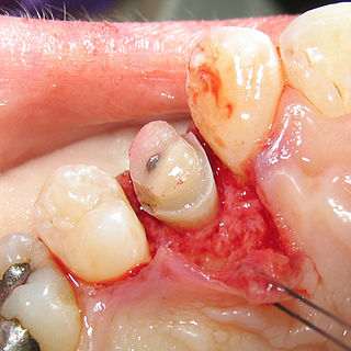
Crown lengthening is a surgical procedure performed by a dentist, or more frequently a specialist periodontist. There are a number of reasons for considering crown lengthening in a treatment plan. Commonly, the procedure is used to expose a greater amount of tooth structure for the purpose of subsequently restoring the tooth prosthetically. However, other indications include accessing subgingival caries, accessing perforations and to treat aesthetic disproportions such as a gummy smile. There are a number of procedures used to achieve an increase in crown length.

Gingival and periodontal pocket

Gingival and periodontal pockets are dental terms indicating the presence of an abnormal depth of the gingival sulcus near the point at which the gingival tissue contacts the tooth.

The junctional epithelium (JE) is that epithelium which lies at, and in health also defines, the base of the gingival sulcus. The probing depth of the gingival sulcus is measured by a calibrated periodontal probe. In a healthy-case scenario, the probe is gently inserted, slides by the sulcular epithelium (SE), and is stopped by the epithelial attachment (EA). However, the probing depth of the gingival sulcus may be considerably different from the true histological gingival sulcus depth.

A mucogingival junction is an anatomical feature found on the intraoral mucosa. The mucosa of the cheeks and floor of the mouth are freely moveable and fragile, whereas the mucosa around the teeth and on the palate are firm and keratinized. Where the two tissue types meet is known as a mucogingival junction.

The gingival fibers are the connective tissue fibers that inhabit the gingival tissue adjacent to teeth and help hold the tissue firmly against the teeth. They are primarily composed of type I collagen, although type III fibers are also involved.

Gingivectomy is a dental procedure in which a dentist or oral surgeon cuts away part of the gums in the mouth.

Periodontal scaler

Periodontal scalers are dental instruments used in the prophylactic and periodontal care of teeth, including scaling and root planing. The working ends come in a variety of shapes and sizes, but they are always narrow at the tip, so as to allow for access to narrow embrasure spaces between teeth. They differ from periodontal curettes, which possess a blunt tip.

Debridement (dental) removal of plaque and calculus

In dentistry, debridement refers to the removal by dental cleaning of accumulations of plaque and calculus (tartar) in order to maintain dental health.

Furcation defect

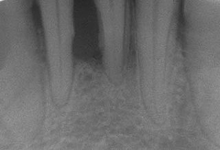
In dentistry, a furcation defect is bone loss, usually a result of periodontal disease, affecting the base of the root trunk of a tooth where two or more roots meet. The extent and configuration of the defect are factors in both diagnosis and treatment planning.

Periodontal abscess

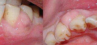
A periodontal abscess, is a localized collection of pus within the tissues of the periodontium. It is a type of dental abscess. A periodontal abscess occurs alongside a tooth, and is different from the more common periapical abscess, which represents the spread of infection from a dead tooth. To reflect this, sometimes the term "lateral (periodontal) abscess" is used. In contrast to a periapical abscess, periodontal abscesses are usually associated with a vital (living) tooth. Abscesses of the periodontium are acute bacterial infections classified primarily by location.

Clinical attachment loss (CAL) is the predominant clinical manifestation and determinant of periodontal disease.

Tooth mobility

Tooth mobility is the medical term for loose tooth.

References

  1. Page 114 in: Czochrowska, E.; Stenvik, A.; Bjercke, B.; Zachrisson, B. (2002). "Outcome of tooth transplantation: Survival and success rates 17-41 years posttreatment". American Journal of Orthodontics and Dentofacial Orthopedics. 121 (2): 110–119. doi:10.1067/mod.2002.119979. PMID   11840123. "Archived copy" (PDF). Archived from the original (PDF) on 2011-08-15. Retrieved 2010-08-16.
  2. Fermin A. Carranza. CARRANZA'S CLINICAL PERIODONTOLOGY, 9th edition, 2002. page 16
  3. Fermin A. Carranza. CARRANZA'S CLINICAL PERIODONTOLOGY, 9th edition, 2002. page 481
  4. Trauma from Occlusion Handout, Dr. Michael Deasy, Department of Periodontics, NJDS 2007. page 4