The lumbricals are intrinsic muscles of the hand that flex the metacarpophalangeal joints and extend the interphalangeal joints.

The anatomical snuff box or snuffbox is a triangular deepening on the radial, dorsal aspect of the hand—at the level of the carpal bones, specifically, the scaphoid and trapezium bones forming the floor. The name originates from the use of this surface for placing and then sniffing powdered tobacco, or "snuff." It is sometimes referred to by its French name tabatière.

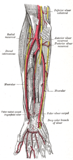
In human anatomy, the radial artery is the main artery of the lateral aspect of the forearm.
The ulnar artery is the main blood vessel, with oxygenated blood, of the medial aspect of the forearm. It arises from the brachial artery and terminates in the superficial palmar arch, which joins with the superficial branch of the radial artery. It is palpable on the anterior and medial aspect of the wrist.

In human anatomy, the adductor pollicis muscle is a muscle in the hand that functions to adduct the thumb. It has two heads: transverse and oblique.

Palmaris brevis is a thin, quadrilateral muscle, placed beneath the integument of the ulnar side of the hand. It acts to fold the skin of the hypothenar eminence transversally.

The palmar aponeurosis invests the muscles of the palm, and consists of central, lateral, and medial portions.
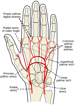
The superficial palmar arch is formed predominantly by the ulnar artery, with a contribution from the superficial palmar branch of the radial artery. However, in some individuals the contribution from the radial artery might be absent, and instead anastomoses with either the princeps pollicis artery, the radialis indicis artery, or the median artery, the former two of which are branches from the radial artery.
The palmar metacarpal arteries, three or four in number, arise from the convexity of the deep volar arch
The radialis indicis artery is a branch of the radial artery that provides blood to the index finger.

The dorsal carpal arch is an anatomical term for the combination (anastomosis) of dorsal carpal branch of the radial artery and the dorsal carpal branch of the ulnar artery near the back of the wrist.
Most of the dorsal metacarpal arteries arise from the dorsal carpal arch and run downward on the second, third, and fourth dorsal interossei of the hand and bifurcate into the dorsal digital arteries. Near their origin, they anastomose with the deep palmar arch by perforating arteries. They also anastomose with common palmar digital arteries, also via perforating arteries.
The palmar carpal arch is the combination (anastomosis) of two arteries: the palmar carpal branch of radial artery and the palmar carpal branch of ulnar artery.
A circulatory anastomosis is a connection between two blood vessels, such as between arteries, between veins or between an artery and a vein. Anastomoses between arteries and between veins result in a multitude of arteries and veins, respectively, serving the same volume of tissue. Such anastomoses occur normally in the body in the circulatory system, serving as backup routes for blood to flow if one link is blocked or otherwise compromised, but may also occur pathologically.
The deep palmar branch of ulnar artery passes between the Abductor digiti minimi and Flexor digiti minimi brevis and through the origin of the Opponens digiti minimi; it anastomoses with the radial artery, and completes the deep volar arch.

The deep branch of the ulnar nerve is a terminal, primarily motor branch of the ulnar nerve. It is accompanied by the deep palmar branch of ulnar artery.
The palmar digital veins on each finger are connected to the dorsal digital veins by oblique intercapitular veins.
The superficial palmar arch is accompanied by a pair of venae comitantes which constitute the superficial palmar venous arch. It receives the veins corresponding to the branches of the superficial arterial arch: the common palmar digital veins.
The public domain consists of all the creative works to which no exclusive intellectual property rights apply. Those rights may have expired, been forfeited, expressly waived, or may be inapplicable.

Gray's Anatomy is an English language textbook of human anatomy originally written by Henry Gray and illustrated by Henry Vandyke Carter. Earlier editions were called Anatomy: Descriptive and Surgical, Anatomy of the Human Body and Gray's Anatomy: Descriptive and Applied, but the book's name is commonly shortened to, and later editions are titled, Gray's Anatomy. The book is widely regarded as an extremely influential work on the subject, and has continued to be revised and republished from its initial publication in 1858 to the present day. The latest edition of the book, the 41st, was published in September 2015.