Dinophysis acuminata

Last updated

Dinophysis acuminata
Dinophysis acuminata.jpg
Formalin fixed sample, collected from sampling station 7, North Sea
Scientific classification OOjs UI icon edit-ltr.svg
Domain: Eukaryota
Clade: Diaphoretickes
Clade: SAR
Clade: Alveolata
Phylum: Myzozoa
Superclass: Dinoflagellata
Class: Dinophyceae
Order: Dinophysiales
Family: Dinophysaceae
Genus: Dinophysis
Species:
D. acuminata
Binomial name
Dinophysis acuminata
Claparède & Lachmann

Dinophysis acuminata is a marine plankton species of dinoflagellates that is found in coastal waters of the north Atlantic and Pacific oceans. [1] The genus Dinophysis includes both phototrophic and heterotrophic species. D. acuminata is one of several phototrophic species of Dinophysis classed as toxic, as they produce okadaic acid which can cause diarrhetic shellfish poisoning (DSP). Okadiac acid is taken up by shellfish and has been found in the soft tissue of mussels and the liver of flounder species. When contaminated animals are consumed, they cause severe diarrhoea. D. acuminata blooms are constant threat to and indication of diarrhoeatic shellfish poisoning outbreaks. [2] [3] [4]

Contents

Dinophysis acuminata is a photosynthesising Dinophysis species by acquiring secondary plastids from consuming the ciliate Myrionecta rubra , [5] which in turn had ingested them from the alga Teleaulax amphioxeia . [6] [7] Thus, D. acuminata is a mixotroph, primarily a heterotroph, but autotroph once it acquires plastids. This is also an example of cell organelle stealing, the concept called kleptoplasty, and endosymbiosis. Dinophysis acuminata reproduces sexually and asexually. [8]

Description

Dinophysis acuminata is an oval-shaped protist. It measures 30-35 μm in length and 38-58 μm in diameter. The body is reddish-brown in colour and is covered with an armor-like covering called theca, which is made up of grass. The anterior end has a crown-like platform, which is the smaller epitheca; while the posterior is simply rounded constituting a larger hypotheca. The cell has two flagella for locomotion. Reproduction is by simple binary fission. In lateral view D. acuminata cells are irregularly egg-shaped, dorsally convex and have large hypothecal plates with a more or less oval shape. The dorsal contour is always more strongly convex than the ventral one. Compared to other species of Dinophysis, D. acuminata has a more straight ventral margin and larger left sulcal lists with more prominent ribs. The nucleus is prominently at the centre of the cell. The unusual feature of the cell is that it contains reddish-brown chloroplast. [1]

The taxonomic identification of Dinophysis species is largely based on cell contouring, size and shape of their large hypothecal plates and the shape of their left sulcal lists and ribs. When viewed laterally species in the Dinophysis are laterally compressed with a cap-like epitheca and a much larger hypotheca although the size and shape of these species varies greatly due to their polymorphic life cycle. Due to the morphological variability of Dinophysis species identification can be hard, especially when two species (D. acuminata and D. sacculus ) co-exist. For this reason the term "D. acuminata complex" was coined to label a group of co-existing species difficult to discriminate. [9]

Dinophysis morphology

Dinophysis acuminata can be very hard to identify, and requires careful observations[3]. It can be identified by its midsection[1]. It is very large (38-55μm) and wide (30-38μm) in the middle unlike D. norvegica that is 34-50μm long and 36-43μm wide[2]. The Dinophysis norvegica is smaller and widest in the middle region[1]. D. norvegica and D. acuminata are a very similar species as a result, they exhibits similar behaviors and are commonly misidentified[1]. Furthermore, other ways to identify D. acuminata from other Dinophysis species can be done by comparison of the left sulcal list (cellulose extensions of the cell[5]) and LSL identification in ribs[1].Cells have convex dorsal margins and small oval shaped cells and their thecal plates are covered with areolae (circular depression on the cellulose wall of a dinoflagellate[5]) each with a pore[4]. Continuously, the knob-shaped protrusions and round antapex (botton end of a dinoflagellate[5]) are ways to identify D.acuminata[4].

Feeding and endosymbiosis

Dinophysis acuminata is basically a heterotroph feeding on the ciliate Mesodinium rubrum . M. rubrum in turn feeds on green algae that contain plastids. (The endosymbiont is used by the ciliate for its own photosynthesis.) [10] Microscopic observations of live cells using established cultures revealed that D. acuminata uses a peduncle, extending from the flagellar pore, to extract the cell contents of the marine ciliate M. rubrum. After about 1 minute the trapped M. rubrum becomes immobile after which the D. acuminata slowly consumes the ciliate, over 1–2 hours, filling its vacuoles with the ciliate's cytoplasm. [9] The algal plastids are not destroyed by D. acuminata but use it for its own photosynthesis, thereby becoming an autotroph. However, unlike its prey M. rubum, it is not clear whether D. acuminata uses the plastids permanently or temporarily. [11] [12] Food vacuoles found in the vacuoles of this primitive genus indicates that organisms in this genus are mixotrophs especially D. norvegica[1]. Mixotrophy is the ability of an organism to use different sources of carbon and energy instead of having a single mode of feeding (autotroph or heterotroph). However, certain species related to Dinophysis acuminata prefer one mode of feeding over another. Dinophysis rotundata uses myzocytosis to feed [1]. Their mixotrophic conditions and size are influenced by prey populations and hydrographic conditions [2].

Ecology and current threats of Dinophysis

Dinophysis acuminata has caused several problems in oceanic ecosystems. The main cause of DSP, diarrhetic shellfish poisoning, outbreaks in waters along Europe has been due to this species[3]. Likewise, the close knit sisters of Dinophysis acuminata called Dinophysis norvegica, a photosynthetic organism with yellow chloroplast and posterior nucleus, Dinophysis acuta, and Dinophysis fortii have also been known to cause the same problems as D. acuminata[1]. Dinophysis norvegica is a marine plankton dinoflagellate that is found in neritic waters[1]. This species of the Dinophysis genus is a bloom-forming toxic species[1]. Both species reproduce asexually by binary fission to make identical copies of itself[1]. Speculations of sexual dimorphism that is the difference between the female and male counterparts have allowed researchers to draw conclusion that species of this genus can undergo sexual reproduction[1]. The first record of DSP with Dinophysis acuminata and Dinophysis fortii was in 1980 in the Patagonian coast[4]. A year after the report, another occurrence of DSP unleashed in December 1993 and November 1994[4]. Dinophysis acuminata releases lipophilic shellfish toxins (LSTs) and have been found to cause trouble to ecological marines and aquaculture farmers[2]'. The lipophilic toxins accumulate in shellfishes and causes diarrhea and shellfish poisoning to consumers. Dinophysis acuminata is the cause of DSP in Brazil and creates a disturbing impact due to its long and early blooming species[4]. However, their presence is restricted around the spring and summer[4], but in higher concentrations in December. DSP is a particular kind food poisoning that causes severe gastrointestinal illness in humans and this is related to the ingestion of toxin contaminated shellfishes from contaminated water[4]. Some of the symptoms of DSP include diarrhea, stomach pain, vomiting, nausea and fever; reported human ingestion shows that the toxins are capable of causing stomach tumors and chronic problems to consumers[4]. Government involvement as a result of high concentrations of toxins in the Dinophysis toxic shellfish epidemic has caused economic crisis in Europe and the aquaculture industry'[2]'.

Related Research Articles

<span class="mw-page-title-main">Toxin</span> Naturally occurring organic poison

A toxin is a naturally occurring organic poison produced by metabolic activities of living cells or organisms. They occur especially as proteins, often conjugated. The term was first used by organic chemist Ludwig Brieger (1849–1919) and is derived from the word "toxic".

<span class="mw-page-title-main">Dinoflagellate</span> Unicellular algae with two flagella

The dinoflagellates are a monophyletic group of single-celled eukaryotes constituting the phylum Dinoflagellata and are usually considered protists. Dinoflagellates are mostly marine plankton, but they also are common in freshwater habitats. Their populations vary with sea surface temperature, salinity, and depth. Many dinoflagellates are photosynthetic, but a large fraction of these are in fact mixotrophic, combining photosynthesis with ingestion of prey.

<span class="mw-page-title-main">Alveolate</span> Superphylum of protists

The alveolates are a group of protists, considered a major clade and superphylum within Eukarya. They are currently grouped with the stramenopiles and Rhizaria among the protists with tubulocristate mitochondria into the SAR supergroup.

<span class="mw-page-title-main">Kleptoplasty</span> Form of algae symbiosis

Kleptoplasty or kleptoplastidy is a process in symbiotic relationships whereby plastids, notably chloroplasts from algae, are sequestered by the host. The word is derived from Kleptes (κλέπτης) which is Greek for thief. The alga is eaten normally and partially digested, leaving the plastid intact. The plastids are maintained within the host, temporarily continuing photosynthesis and benefiting the host.

Okadaic acid, C44H68O13, is a toxin produced by several species of dinoflagellates, and is known to accumulate in both marine sponges and shellfish. One of the primary causes of diarrhetic shellfish poisoning, okadaic acid is a potent inhibitor of specific protein phosphatases and is known to have a variety of negative effects on cells. A polyketide, polyether derivative of a C38 fatty acid, okadaic acid and other members of its family have shined light upon many biological processes both with respect to dinoflagellete polyketide synthesis as well as the role of protein phosphatases in cell growth.

<i>Noctiluca scintillans</i> Bioluminescent, marine dinoflagellate

Noctiluca scintillans is a marine species of dinoflagellate that can exist in a green or red form, depending on the pigmentation in its vacuoles. It can be found worldwide, but its geographical distribution varies depending on whether it is green or red. This unicellular microorganism is known for its ability to bioluminesce, giving the water a bright blue glow seen at night. However, blooms of this species can be responsible for environmental hazards, such as toxic red tides. They may also be an indicator of anthropogenic eutrophication.

<span class="mw-page-title-main">Paralytic shellfish poisoning</span> Syndrome of shellfish poisoning

Paralytic shellfish poisoning (PSP) is one of the four recognized syndromes of shellfish poisoning, which share some common features and are primarily associated with bivalve mollusks. These shellfish are filter feeders and accumulate neurotoxins, chiefly saxitoxin, produced by microscopic algae, such as dinoflagellates, diatoms, and cyanobacteria. Dinoflagellates of the genus Alexandrium are the most numerous and widespread saxitoxin producers and are responsible for PSP blooms in subarctic, temperate, and tropical locations. The majority of toxic blooms have been caused by the morphospecies Alexandrium catenella, Alexandrium tamarense, Gonyaulax catenella and Alexandrium fundyense, which together comprise the A. tamarense species complex. In Asia, PSP is mostly associated with the occurrence of the species Pyrodinium bahamense.

Diarrheic shellfish poisoning (DSP) is one of the four recognized symptom types of shellfish poisoning, alongside paralytic shellfish poisoning, neurotoxic shellfish poisoning and amnesic shellfish poisoning. As the name suggests, it mainly manifests as diarrhea. Abdominal pain, nausea and vomiting may also occur.

<i>Karenia</i> (dinoflagellate) Genus of single-celled organisms

Karenia is a genus that consists of unicellular, photosynthetic, planktonic organisms found in marine environments. The genus currently consists of 12 described species. They are best known for their dense toxic algal blooms and red tides that cause considerable ecological and economical damage; some Karenia species cause severe animal mortality. One species, Karenia brevis, is known to cause respiratory distress and neurotoxic shellfish poisoning (NSP) in humans.

<i>Ornithocercus</i> Genus of single-celled organisms

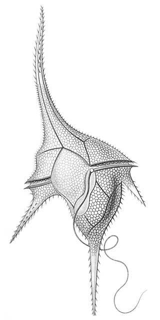
Ornithocercus is a genus of planktonic dinoflagellate that is known for its complex morphology that features considerable lists growing from its thecal plates, giving an attractive appearance. Discovered in 1883, this genus has a small number of species currently categorized but is widespread in tropical and sub-tropical oceans. The genus is marked by exosymbiotic bacteria gardens under its lists, the inter-organismal dynamics of which are a current field of research. As they reside only in warm water, the genus has been used as a proxy for climate change and has potential to be an indicator species for environmental change if found in novel environments.

<i>Dinophysis</i> Genus of single-celled organisms

Dinophysis is a genus of dinoflagellates common in tropical, temperate, coastal and oceanic waters. It was first described in 1839 by Christian Gottfried Ehrenberg.

<span class="mw-page-title-main">Yessotoxin</span> Chemical compound

Yessotoxins are a group of lipophilic, sulfur bearing polyether toxins that are related to ciguatoxins. They are produced by a variety of dinoflagellates, most notably Lingulodinium polyedrum and Gonyaulax spinifera.

<i>Dinophysis norvegica</i> Species of single-celled organism

Dinophysis norvegica is a species of dinoflagellate most commonly associated with diarrheal shellfish poisoning.

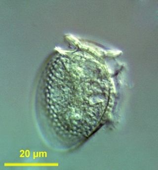
Mesodinium chamaeleon is a ciliate of the genus Mesodinium. It is known for being able to consume and maintain algae endosymbiotically for days before digesting the algae. It has the ability to eat red and green algae, and afterwards using the chlorophyll granules from the algae to generate energy, turning itself from being a heterotroph into an autotroph. The species was discovered in January 2012 outside the coast of Nivå, Denmark by professor Øjvind Moestrup.

<i>Alexandrium</i> (dinoflagellate) Genus of single-celled organisms

Alexandrium is a genus of dinoflagellates. It contains some of the dinoflagellate species most harmful to humans, because it produces toxic harmful algal blooms (HAB) that cause paralytic shellfish poisoning (PSP) in humans. There are about 30 species of Alexandrium that form a clade, defined primarily on morphological characters in their thecal plates.

Dinotoxins are a group of toxins which are produced by flagellate, aquatic, unicellular protists called dinoflagellates. Dinotoxin was coined by Hardy and Wallace in 2012 as a general term for the variety of toxins produced by dinoflagellates. Dinoflagellates are an enormous group of marine life, with much diversity. With great diversity comes many different toxins, however, there are a few toxins that multiple species have in common.

<i>Mesodinium rubrum</i> Species of single-celled organism

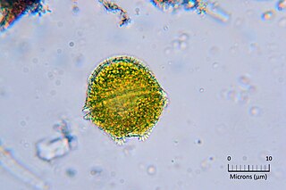
Mesodinium rubrum is a species of ciliates. It constitutes a plankton community and is found throughout the year, most abundantly in spring and fall, in coastal areas. Although discovered in 1908, its scientific importance came into light in the late 1960s when it attracted scientists by the recurrent red colouration it caused by forming massive blooms, that cause red tides in the oceans.

<i>Dinophysis acuta</i> Species of dinoflagellate

Dinophysis acuta is a species of flagellated planktons belonging to the genus Dinophysis. It is one of the few unusual photosynthetic protists that acquire plastids from algae by endosymbiosis. By forming massive blooms, particularly in late summer and spring, it causes red tides. It produces toxic substances and the red tides cause widespread infection of seafood, particularly crabs and mussels. When infected animals are consumed, severe diarrhoea occurs. The clinical symptom is called diarrhetic shellfish poisoning. The main chemical toxins were identified in 2006 as okadaic acid and pectenotoxins. They can produce non-fatal or fatal amounts of toxins in their predators, which can become toxic to humans.

<span class="mw-page-title-main">Mixotrophic dinoflagellate</span> Plankton

Dinoflagellates are eukaryotic plankton, existing in marine and freshwater environments. Previously, dinoflagellates had been grouped into two categories, phagotrophs and phototrophs. Mixotrophs, however include a combination of phagotrophy and phototrophy. Mixotrophic dinoflagellates are a sub-type of planktonic dinoflagellates and are part of the phylum Dinoflagellata. They are flagellated eukaryotes that combine photoautotrophy when light is available, and heterotrophy via phagocytosis. Dinoflagellates are one of the most diverse and numerous species of phytoplankton, second to diatoms.

<i>Mesodinium</i> Genus of single-celled organisms

Mesodinium is a genus of ciliates that are widely distributed and are abundant in marine and brackish waters.

References

  1. 1 2 Setälä, Outi; Autio, Riitta; Kuosa, Harri; Rintala, Janne; Ylöstalo, Pasi (2005). "Survival and photosynthetic activity of different Dinophysis acuminata populations in the northern Baltic Sea". Harmful Algae. 4 (2): 337–350. doi:10.1016/j.hal.2004.06.017. ISSN   1568-9883.
  2. Díaz, Patricio; Reguera, Beatriz; Ruiz-Villarreal, Manuel; Pazos, Yolanda; Velo-Suárez, Lourdes; Berger, Henrick; Sourisseau, Marc (2013). "Climate variability and oceanographic settings associated with interannual variability in the initiation of Dinophysis acuminata blooms". Marine Drugs. 11 (8): 2964–2981. doi: 10.3390/md11082964 . PMC   3766876 . PMID   23959151.
  3. Lee, Ka Jeong; Mok, Jong Soo; Song, Ki Cheol; Yu, Hongsik; Jung, Jee Hyung; Kim, Ji Hoe (2011). "Geographical and annual variation in lipophilic shellfish toxins from oysters and mussels along the south coast of Korea". Journal of Food Protection. 74 (12): 2127–2133. doi: 10.4315/0362-028X.JFP-11-148 . PMID   22186054.
  4. Naustvoll, L.-J.; Gustad, E.; Dahl, E. (2012). "Monitoring of Dinophysis species and diarrhetic shellfish toxins in Flødevigen Bay, Norway: inter-annual variability over a 25-year time-series". Food Additives & Contaminants: Part A. 29 (10): 1605–1615. doi:10.1080/19440049.2012.714908. PMID   22891979.
  5. Johnson, Matthew D.; Oldach, David; Delwiche, Charles F.; Stoecker, Diane K. (2007). "Retention of transcriptionally active cryptophyte nuclei by the ciliate Myrionecta rubra". Nature. 445 (7126): 426–428. doi:10.1038/nature05496. PMID   17251979.
  6. Janson, Sven (2004). "Molecular evidence that plastids in the toxin-producing dinoflagellate genus Dinophysis originate from the free-living cryptophyte Teleaulax amphioxeia". Environmental Microbiology. 6 (10): 1102–1106. doi:10.1111/j.1462-2920.2004.00646.x. PMID   15344936.
  7. Nishitani, G.; Nagai, S.; Baba, K.; Kiyokawa, S.; Kosaka, Y.; Miyamura, K.; Nishikawa, T.; Sakurada, K.; Shinada, A.; Kamiyama, T. (2010). "High-level congruence of Myrionecta rubra prey and Dinophysis species plastid identities as revealed by genetic analyses of isolates from Japanese coastal waters". Applied and Environmental Microbiology. 76 (9): 2791–2798. doi:10.1128/AEM.02566-09. PMC   2863437 . PMID   20305031.
  8. "WoRMS - World Register of Marine Species - Dinophysis acuminata Claparède & Lachmann, 1859". www.marinespecies.org. Retrieved 2016-09-28.
  9. 1 2 Raho, Nicolás; Pizarro, Gemita; Escalera, Laura; Reguera, Beatriz; Marín, Irma (2008). "Morphology, toxin composition and molecular analysis of Dinophysis ovum Schütt, a dinoflagellate of the "Dinophysis acuminata complex"". Harmful Algae. 7 (6): 839–848. doi:10.1016/j.hal.2008.04.006. ISSN   1568-9883.
  10. Dorrell, R. G.; Howe, C. J. (2012). "What makes a chloroplast? Reconstructing the establishment of photosynthetic symbioses". Journal of Cell Science. 125 (8): 1865–1875. doi: 10.1242/jcs.102285 . PMID   22547565.
  11. Takishita, K; Koike, K; Maruyama, T; Ogata, T (2002). "Molecular evidence for plastid robbery (Kleptoplastidy) in Dinophysis, a dinoflagellate causing diarrhetic shellfish poisoning". Protist. 153 (3): 293–302. doi:10.1078/1434-4610-00106. PMID   12389818.
  12. Wisecaver, Jennifer H; Hackett, Jeremiah D (2010). "Transcriptome analysis reveals nuclear-encoded proteins for the maintenance of temporary plastids in the dinoflagellate Dinophysis acuminata". BMC Genomics. 11 (1): 366. doi: 10.1186/1471-2164-11-366 . PMC   3017763 . PMID   20537123.