This article is an orphan, as no other articles link to it . Please introduce links to this page from related articles ; try the Find link tool for suggestions. (June 2024) |
Endogenous regeneration in the brain is the ability of cells to engage in the repair and regeneration process. While the brain has a limited capacity for regeneration, endogenous neural stem cells, as well as numerous pro-regenerative molecules, can participate in replacing and repairing damaged or diseased neurons and glial cells. Another benefit that can be achieved by using endogenous regeneration could be avoiding an immune response from the host. [1]
During the early development of a human, neural stem cells lie in the germinal layer of the developing brain, ventricular and subventricular zones. In brain development, multipotent stem cells (those that can generate different types of cells) are present in these regions, and all of these cells differentiate into neural cell forms, such as neurons, oligodendrocytes and astrocytes. A long-held belief states that the multipotency of neural stem cells would be lost in the adult human brain. [2] However, it is only in vitro , using neurosphere and adherent monolayer cultures, that stem cells from the adult mammalian brain have shown multipotent capacity, while the in vivo study is not convincing. Therefore, the term "neural progenitor" is used instead of "stem cell" to describe limited regeneration ability in the adult brain stem cell. [3]
Neural stem cells (NSC) reside in the subventricular zone (SVZ) of the adult human brain and the dentate gyrus of the adult mammalian hippocampus. Newly formed neurons from these regions participate in learning, memory, olfaction and mood modulation. [3] It has not been definitively determined whether or not these stem cells are multipotents. NSC from the hippocampus of rodents, which can differentiate into dentate granule cells, have developed into many cell types when studied in culture. [4] However, another in vivo study, using NSCs in the postnatal SVZ, showed that the stem cell is restricted to developing into different neuronal sub-type cells in the olfactory bulb. It is believed that the various spatial location niches regulate the differentiation of the neural stem cell. [5]
Santiago Ramon y Cajal, a neuroscience pioneer, concluded that the generation of neurons occurs only in the prenatal phase of human development, not after birth. This theory had long been the fundamental principle of neuroscience. [4] However, in the mid-20th century, evidence of adult mammalian neurogenesis was found in rodent hippocampus and other region of the brain. [6] In the intact adult mammalian brain, neuroregeneration maintains the function and structure of the central nervous system (CNS). The most adult stem cells in the brain are found in the subventricular zone at the lateral walls of the lateral ventricle. Another region where neurogenesis takes place in the adult brain is the subgranular zone (SGZ) of the dentate gyrus in the hippocampus. While the exact mechanism that maintains functional NSCs in these regions is still unknown, NSCs have shown an ability to restore neurons and glia in response to certain pathological conditions. However, so far, this regeneration by NSCs is insufficient to restore the full function and structure of an injured brain. However, endogenous neuroregeneration, unlike using embryonic stem cell implantation, is anticipated to treat damaged CNS without immunogenesis or tumorigenesis. [7]
Progenitor cells in the dentate gyrus of the hippocampus migrate to the nearby location and differentiate into granule cells. As a part of the limbic system, new neurons of the hippocampus maintain the function of controlling mood, learning and memory. In the dentate gyrus, putative stem cells, called type 1 cells, proliferate into type 2 and type 3 cells, which are transiently amplifying, lineage-determined progenitor cells. Type 1 cells in the hippocampus are multipotent in vitro. However, although there is evidence that both new neurons and glia are generated in the hippocampus in vivo, no exact relationship of neurogenesis to type 1 cells is shown. [8]
In the hippocampus, newly formed neurons contribute only a small portion to the entire neuron population. These new neurons have different electrophysiology compared to the rest of the existing neurons. This may be evidence that generating new neurons in the SGZ is part of learning and memorizing activity of mammals. Several studies have been performed to explain the relationship between neurogenesis and learning. In the case of learning, that related to the hippocampal function, a significantly increased number of neurons are generated in the SGZ and survival of the new neurons is increased if they are required for retention of memory. [9] [10] In addition to learning and memorizing, neurogenesis in the SGZ is also affected by mood and emotion. With constant, inescapable stress, which usually results in emotional depression, there is a significant decrease in neurogenesis, the effect of which can be reversed by treatment with fluoxetine. [11]
The largest NSC population in the brain is found in the SVZ. The SVZ is considered a micro-environment called a "stem cell niche" that retains the NSC's capacity of self-renewing and multipotency. Basic fibroblast growth factor (FGF2), hepatocyte growth factor (HGF), Notch-1, sonic hedgehog (SHH), noggin, ciliary neurotrophic factor (CNTF), and a soluble carbohydrate-binding protein, Galectin-1, are reported as factors that maintain such properties of NSC in stem cell niche. Like stem cells in SGZ, progenitor cells in SVZ also differentiate into neurons and form an intermediate cell called a transiently amplifying cell (TAC). A recent study revealed that beta-catenin signaling, Wnt β-catenin, regulates the differentiation of TAC. [12]
NSCs in the SVZ have a distinct capacity to migrate into the olfactory bulb in the anterior tip of the telencephalon by a pathway called the rostral migratory stream (RMS). This migration is unique to new neurons in the SVZ that embryonic neurogenesis and neurogenesis at other region of the brain are not able to perform. Another unique neurogenesis in the SVZ is neurogenesis by astrocytes. A study done by Doetsch (1999) showed that astrocytes in the SVZ can be dedifferentiate and differentiate into neurons in the olfactory bulb. Among four types of cells in the SVZ (migrating neuroblasts, immature precursors, astrocytes, and ependymal cells), migrating neuroblasts and immature precursors are silenced with the anti-mitotic agent and astrocytes are infected with a retrovirus. In the result, neurons that have the retrovirus are found in the olfactory bulb. [13]
Neurogenesis in the adult mammalian brain is affected by various factors, including exercise, stroke, brain insult and pharmacological treatments. For example, kainic acid-induced seizures, antidepressant (fluoxetine), neurotransmitters such as GABA and growth factors (fibroblast growth factors (FGFs), epidermal growth factor (EGF), neuregulins (NRGs), vascular endothelial growth factor (VEGF), and pigment epithelium-derived factor (PEDF) induce formation of neuroblasts. The final destination of NSCs is determined by "niche" signals. Wnt signaling drives NSCs to the formation of new neurons in the SGZ, whereas bone morphogenic proteins (BMPs) promote NSC differentiation into glia cells in the SVZ. [5]
However, in the case of brain injury, neurogenesis seems insufficient to repair damaged neurons. Thus, Cajal's theory was accepted for a long time. In actuality, in the intercranial physiological condition, many neurogenesis inhibitors are present (for example, axon growth-inhibitory ligands expressed in oligodendrocytes, myelin, NG2-glia, and reactive astrocytes in the lesion and degenerating tracts, and fibroblasts in scar tissue). The inhibitory ligands bind to growth cone receptors on a damaged neuron, which causes repulsion and collapse of the growth cone in the damaged regions. Among inhibitory factors, oligodendrocyte and myelin-derived inhibitory ligands are membrane-bound, meaning that, in the case of injury, those factors are not upregulated or overexpressed, rather it is from direct contact between intact or degraded myelin (or oligodendrocytes) and newly forming neurons.
Nevertheless, with scar formation, many cell types in the brain release growth-inhibitory ligands such as basal lamina components, inhibitory axon guidance molecules and chondroitin sulfate proteoglycans. [14] Inhibitory action of such factors may be a protection of the brain from inflammation. Okano and Sawamoto used an astrocyte-selective conditional Stat3-deficient mice model to examine the role of reactive astrocytes. The result was increased widespread CD11b-positive inflammatory cell invasion and demyelination. [15]
Brain damage itself can induce endogenous regeneration. Many studies have proven endogenous regeneration as a possible treatment of brain damage. However, the inhibitory reaction of the surrounding tissue of damaged region must be overcome before the treatment produces significant improvement.
In the study of the endogenous regeneration of the brain done by Scharff and co-researchers, damaged neurons in a songbird brain are regenerated with the same neuronal types where regeneration occurs (in the case of the study, the hippocampus). However, in places where normal regeneration of the neuron does not occur, there was no replacement of damaged neurons. [16] Thus, recovering brain function after a brain injury was supposed to have limitations. However, a current study revealed that neurons are repaired to some degree after damage, from the SVZ.
The migrating ability of progenitor cells in the SVZ form chain-like structures and laterally move progenitor cells towards the injured region. Along with progenitor cells, thin astrocytic processes and blood vessels also play an important role in the migration of neuroblasts, suggesting that the blood vessels may act as a scaffold. Other factors that contribute of the migration are slit proteins (produced at the choroid plexus) and their gradient (generated by the flow of cerebrospinal fluid). However, only 0.2% of new neurons survived and functioned in this study. Enhancing neurogenesis can be done by injecting growth factors such as fibroblast growth factor-2 (FGF-2) and epidermal growth factor (EGF). However, enhanced neurogenesis also have the possibility of epilepsy resulting in prolonged seizures. [17]
Although endogenous regeneration methods are showing some promising evidence in treating brain ischemia, the current body of knowledge regarding promoting and inhibiting endogenous regeneration is not sufficient to treat Parkinson's disease. Both extrinsic and intrinsic modulation of pathological and physiological stimulation prevent the progenitor cell from differentiating into dopamine cells. Further research must be done to understand factors that affect progenitor cell differentiation in order to treat Parkinson's disease. [18]
Despite the difficulties in replacing compromised dopamine neurons through endogenous sources, recent work suggests that pharmacological activation of endogenous neural stem cells or neural precursor cells results in powerful neuronal rescue and motor skill improvements through a signal transduction pathway that involves the phosphorylation of STAT3 on the serine residue and subsequent Hes3 expression increase (STAT3-Ser/Hes3 Signaling Axis). [19] [20]
Adult neurogenesis is the process in which neurons are generated from neural stem cells in the adult. This process differs from prenatal neurogenesis.
In vertebrates, a neuroblast or primitive nerve cell is a postmitotic cell that does not divide further, and which will develop into a neuron after a migration phase. In invertebrates such as Drosophila, neuroblasts are neural progenitor cells which divide asymmetrically to produce a neuroblast, and a daughter cell of varying potency depending on the type of neuroblast. Vertebrate neuroblasts differentiate from radial glial cells and are committed to becoming neurons. Neural stem cells, which only divide symmetrically to produce more neural stem cells, transition gradually into radial glial cells. Radial glial cells, also called radial glial progenitor cells, divide asymmetrically to produce a neuroblast and another radial glial cell that will re-enter the cell cycle.
Oligodendrocyte progenitor cells (OPCs), also known as oligodendrocyte precursor cells, NG2-glia, O2A cells, or polydendrocytes, are a subtype of glia in the central nervous system named for their essential role as precursors to oligodendrocytes. They are typically identified in the human by co-expression of PDGFRA and CSPG4.
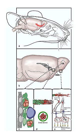
The rostral migratory stream (RMS) is a specialized migratory route found in the brain of some animals along which neuronal precursors that originated in the subventricular zone (SVZ) of the brain migrate to reach the main olfactory bulb (OB). The importance of the RMS lies in its ability to refine and even change an animal's sensitivity to smells, which explains its importance and larger size in the rodent brain as compared to the human brain, as our olfactory sense is not as developed. This pathway has been studied in the rodent, rabbit, and both the squirrel monkey and rhesus monkey. When the neurons reach the OB they differentiate into GABAergic interneurons as they are integrated into either the granule cell layer or periglomerular layer.
Neuroepithelial cells, or neuroectodermal cells, form the wall of the closed neural tube in early embryonic development. The neuroepithelial cells span the thickness of the tube's wall, connecting with the pial surface and with the ventricular or lumenal surface. They are joined at the lumen of the tube by junctional complexes, where they form a pseudostratified layer of epithelium called neuroepithelium.
Neural stem cells (NSCs) are self-renewing, multipotent cells that firstly generate the radial glial progenitor cells that generate the neurons and glia of the nervous system of all animals during embryonic development. Some neural progenitor stem cells persist in highly restricted regions in the adult vertebrate brain and continue to produce neurons throughout life. Differences in the size of the central nervous system are among the most important distinctions between the species and thus mutations in the genes that regulate the size of the neural stem cell compartment are among the most important drivers of vertebrate evolution.
Radial glial cells, or radial glial progenitor cells (RGPs), are bipolar-shaped progenitor cells that are responsible for producing all of the neurons in the cerebral cortex. RGPs also produce certain lineages of glia, including astrocytes and oligodendrocytes. Their cell bodies (somata) reside in the embryonic ventricular zone, which lies next to the developing ventricular system.
Neuropoiesis is the process by which neural stem cells differentiate to form mature neurons, astrocytes, and oligodendrocytes in the adult mammal. This process is also referred to as adult neurogenesis.
The subventricular zone (SVZ) is a region situated on the outside wall of each lateral ventricle of the vertebrate brain. It is present in both the embryonic and adult brain. In embryonic life, the SVZ refers to a secondary proliferative zone containing neural progenitor cells, which divide to produce neurons in the process of neurogenesis. The primary neural stem cells of the brain and spinal cord, termed radial glial cells, instead reside in the ventricular zone (VZ).
A neurosphere is a culture system composed of free-floating clusters of neural stem cells. Neurospheres provide a method to investigate neural precursor cells in vitro. Putative neural stem cells are suspended in a medium lacking adherent substrates but containing necessary growth factors, such as epidermal growth factor and fibroblast growth factor. This allows the neural stem cells to form into characteristic 3-D clusters. However, neurospheres are not identical to stem cells; rather, they only contain a small percentage of neural stem cells.
The subgranular zone (SGZ) is a brain region in the hippocampus where adult neurogenesis occurs. The other major site of adult neurogenesis is the subventricular zone (SVZ) in the brain.
Gliogenesis is the generation of non-neuronal glia populations derived from multipotent neural stem cells.
Eomesodermin also known as T-box brain protein 2 (Tbr2) is a protein that in humans is encoded by the EOMES gene.
The STAT3-Ser/Hes3 signaling axis is a specific type of intracellular signaling pathway that regulates several fundamental properties of cells.
Epigenetic regulation of neurogenesis is the role that epigenetics plays in the regulation of neurogenesis.
A neuronal lineage marker is an endogenous tag that is expressed in different cells along neurogenesis and differentiated cells such as neurons. It allows detection and identification of cells by using different techniques. A neuronal lineage marker can be either DNA, mRNA or RNA expressed in a cell of interest. It can also be a protein tag, as a partial protein, a protein or an epitope that discriminates between different cell types or different states of a common cell. An ideal marker is specific to a given cell type in normal conditions and/or during injury. Cell markers are very valuable tools for examining the function of cells in normal conditions as well as during disease. The discovery of various proteins specific to certain cells led to the production of cell-type-specific antibodies that have been used to identify cells.
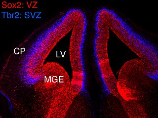
In vertebrates, the ventricular zone (VZ) is a transient embryonic layer of tissue containing neural stem cells, principally radial glial cells, of the central nervous system (CNS). The VZ is so named because it lines the ventricular system, which contains cerebrospinal fluid (CSF). The embryonic ventricular system contains growth factors and other nutrients needed for the proper function of neural stem cells. Neurogenesis, or the generation of neurons, occurs in the VZ during embryonic and fetal development as a function of the Notch pathway, and the newborn neurons must migrate substantial distances to their final destination in the developing brain or spinal cord where they will establish neural circuits. A secondary proliferative zone, the subventricular zone (SVZ), lies adjacent to the VZ. In the embryonic cerebral cortex, the SVZ contains intermediate neuronal progenitors that continue to divide into post-mitotic neurons. Through the process of neurogenesis, the parent neural stem cell pool is depleted and the VZ disappears. The balance between the rates of stem cell proliferation and neurogenesis changes during development, and species from mouse to human show large differences in the number of cell cycles, cell cycle length, and other parameters, which is thought to give rise to the large diversity in brain size and structure.
Neurogenesis is the process by which nervous system cells, the neurons, are produced by neural stem cells (NSCs). This occurs in all species of animals except the porifera (sponges) and placozoans. Types of NSCs include neuroepithelial cells (NECs), radial glial cells (RGCs), basal progenitors (BPs), intermediate neuronal precursors (INPs), subventricular zone astrocytes, and subgranular zone radial astrocytes, among others.
Intermediate progenitor cells (IPCs) are a type of progenitor cell in the developing cerebral cortex. They are multipolar cells produced by radial glial cells who have undergone asymmetric division. IPCs can produce neuron cells via neurogenesis and are responsible for ensuring the proper quantity of cortical neurons are produced. In mammals, neural stem cells are the primary progenitors during embryogenesis whereas intermediate progenitor cells are the secondary progenitors.
Arturo Álvarez-Buylla Roces is a professor and endowed chair in neurological surgery, and a researcher in neurobiology at the University of California, San Francisco.