Myxozoa is a subphylum of aquatic cnidarian animals – all obligate parasites. It contains the smallest animals ever known to have lived. Over 2,180 species have been described and some estimates have suggested at least 30,000 undiscovered species. Many have a two-host lifecycle, involving a fish and an annelid worm or a bryozoan. The average size of a myxosporean spore usually ranges from 10 μm to 20 μm, whereas that of a malacosporean spore can be up to 2 mm. Myxozoans can live in both freshwater and marine habitats.

Myxosporea is a class of microscopic parasites, belonging to the Myxozoa clade within Cnidaria. They have a complex life cycle which comprises vegetative forms in two hosts, an aquatic invertebrate and an ectothermic vertebrate, usually a fish. Each host releases a different type of spore. The two forms of spore are so different that until relatively recently they were treated as belonging to different classes within the Myxozoa.
Myxobolus cerebralis is a myxosporean parasite of salmonids that causes whirling disease in farmed salmon and trout and also in wild fish populations. It was first described in rainbow trout in Germany in 1893, but its range has spread and it has appeared in most of Europe, the United States, South Africa, Canada and other countries from shipments of cultured and wild fish. In the 1980s, M. cerebralis was found to require a tubificid oligochaete to complete its life cycle. The parasite infects its hosts with its cells after piercing them with polar filaments ejected from nematocyst-like capsules. This infects the cartilage and possibly the nervous tissue of salmonids, causing a potentially lethal infection in which the host develops a black tail, spinal deformities, and possibly more deformities in the anterior part of the fish.
Tetracapsuloides bryosalmonae is a myxozoan parasite of salmonid fish. It is the only species currently recognized in the monotypic genus Tetracapsuloides. It is the cause of proliferative kidney disease (PKD), one of the most serious parasitic diseases of salmonid populations in Europe and North America that can result in losses of up to 90% in infected populations.
Kudoa thyrsites is a myxosporean parasite of marine fishes. It has a worldwide distribution, and infects a wide range of host species. This parasite is responsible for causing economic losses to the fisheries sector, by causing post-mortem "myoliquefaction", a softening of the flesh to such an extent that the fish becomes unmarketable. It is not infective to humans.
Enteric redmouth disease, or simply redmouth disease is a bacterial infection of freshwater and marine fish caused by the pathogen Yersinia ruckeri. It is primarily found in rainbow trout and other cultured salmonids. The disease is characterized by subcutaneous hemorrhaging of the mouth, fins, and eyes. It is most commonly seen in fish farms with poor water quality. Redmouth disease was first discovered in Idaho rainbow trout in the 1950s. The disease does not infect humans.
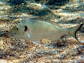
The gilt-head (sea) bream is a fish of the bream family Sparidae found in the Mediterranean Sea and the eastern coastal regions of the North Atlantic Ocean. It commonly reaches about 35 centimetres (1.15 ft) in length, but may reach 70 cm (2.3 ft) and weigh up to about 7.36 kilograms (16.2 lb).
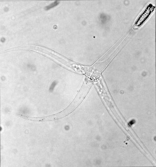
Ceratomyxa is a genus of myxozoan.
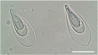
Gadimyxa arctica is a species of parasitic myxozoan. Together with G. atlantica and G. sphaerica, they infect Gadus morhua and Arctogadus glacialis by developing coelozoically in bisporic plasmodia in their urinary systems. These 3 species' spores exhibit two morphological forms: wide and subspherical, being both types bilaterally symmetrical along the suture line. The wide spores have a mean width ranging from 7.5-10μm, respectively, while the subspherical ones range from 5.3-8μm in mean width. The subspherical forms of Gadimyxa are similar to Ortholinea, differing in the development of the spores and in the arrangement of the polar capsules.
Tenacibaculum is a Gram-negative and motile bacterial genus from the family of Flavobacteriaceae.

Philasterides dicentrarchi is a marine protozoan ciliate that was first identified in 1995 after being isolated from infected European sea bass reared in France. The species was also identified as the causative agent of outbreaks of scuticociliatosis that occurred between summer 1999 and spring 2000 in turbot cultivated in the Atlantic Ocean. Infections caused by P. dicentrarchi have since been observed in turbot reared in both open flow and recirculating production systems. In addition, the ciliate has also been reported to cause infections in other flatfishes, such as the olive flounder in Korea and the fine flounder in Peru, as well as in seadragons, seahorses, and several species of sharks in other parts of the world.
Scuticociliatosis is a severe and often fatal parasitic infection of several groups of marine organisms. Species known to be susceptible include a broad range of teleosts, seahorses, sharks, and some crustaceans. The disease can be caused by any one of about 20 distinct species of unicellular eukaryotes known as scuticociliates, which are free-living marine microorganisms that are opportunistic or facultative parasites. Scuticociliatosis has been described in the wild, in captive animals in aquariums, and in aquaculture. It is best studied in fish species that are commonly farmed, in which typical effects of infection include skin ulceration, hemorrhage, and necrosis, with post-mortem examination identifying ciliates in the skin, gills, blood, and internal organs including the brain.
Bothriocephalus gregarius is a tapeworm that parasitises the turbot. It has a complex life cycle including two intermediate hosts, a copepod and a small fish.

Polypodium is a genus of cnidarians that parasitizes in the eggs of sturgeon and similar fishes. It is one of the few metazoans (animals) that live inside the cells of other animals.
Enteromyxum is a genus of myxozoans.

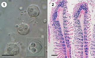
Enteromyxum leei is a species of myxozoan, histozoic parasite that infects the intestinal tract and sometimes associated organs, like gall bladder and liver, of several teleostean fish species. Myxozoans are microscopic metazoans, with an obligate parasitic life-style. The parasite stages of this species live in the paracelullar space between fish enterocytes. It is the causative agent of enteromyxosis, or emaciative disease, also known as "razor blade syndrome" in sparid fish. E. leei has a wide host and geographical range within marine fish, and even freshwater fish have been infected experimentally. E. leei initially emerged in the Mediterranean in the late 1980s and it is believed to have been unintentionally introduced into the Red Sea. Its pathogenicity and economic impact depend on the host species. In the gilt-head seabream, it is manifested as a chronic disease that provokes anorexia, delayed growth with weight loss, cachexia, reduced marketability and increased mortality. In other species, it has no clinical signs. In sharpsnout seabream, infection results in very high mortality rates, which have pushed fish farmers to abandon the culture of this fish species.
Sphaerospora molnari is a microscopic endoparasite of carp in pond cultures and natural freshwater habitats in Central and Eastern Europe. In natural infections, S. molnari invades the epithelia of gills and surrounding skin regions. It then forms spores in between epithelial cells, causing sphaerosporosis, a pathological condition of the skin and gill tissues. Affected tissues show marked dystrophic changes and necrosis, causing secondary bacterial infections and resulting in osmoregulatory and respiratory failure. Mortalities can reach 100% but little is known about the overall distribution of the parasite species in European carp ponds or its economic impact on carp aquaculture.
Thelohanellus kitauei is a myxozoan endoparasite identified as the agent of intestinal giant-cystic disease (IGCD) of common carp Cyprinus carpio. The species was first identified in Japan, in 1980 and later formally described by Egusa & Nakajima. Fan subsequently reported the parasite in China, and several other reports from carp and Koi carp in China and Korea followed. Reports referred to an intestinal infection, swelling and emaciation of fish due to blockage of the intestinal tract by giant cysts. The intestine of carp was believed to be the only infection site of T. kitauei until Zhai et al. reported large cysts of T. kitauei in the skin, with morphologically similar and molecularly identical spores. T. kitauei has been recognized as the most detrimental disease of farmed carp in Asia with around 20% of farmed carp killed annually. In 2014, the genome of T. kitauei was sequenced, and in 2016, its life cycle was found to include the oligochaete Branchiura sowerbyi. Infected oligochaete worms were first discovered in Hungary and raised concerns of the introduction of T. kitauei into European carp culture ponds, since it was believed to be endemic to Asia. However, the related disease (IGCD) has not yet been reported in Europe.

Enterospora nucleophila is a microsporidian infecting the gilt-head bream. It develops primarily within the nuclei of rodlet cells and enterocytes, at the intestinal epithelium. It can also be found in cytoplasmic position within other cell types, including phagocytes, at subepithelial layers. It is the causative agent of emaciative microsporidiosis of gilthead sea bream, a chronic condition manifested as a severe growth arrestment, normally accompanied by trickling mortality.
Ellipsomyxa is a genus of cnidarian that is part of the family Ceratomyxidae.