Facilitated diffusion

Last updated
Facilitated diffusion in cell membrane, showing ion channels and carrier proteins Scheme facilitated diffusion in cell membrane-en.svg
Facilitated diffusion in cell membrane, showing ion channels and carrier proteins

Facilitated diffusion (also known as facilitated transport or passive-mediated transport) is the process of spontaneous passive transport (as opposed to active transport) of molecules or ions across a biological membrane via specific transmembrane integral proteins. [1] Being passive, facilitated transport does not directly require chemical energy from ATP hydrolysis in the transport step itself; rather, molecules and ions move down their concentration gradient reflecting its diffusive nature.

Contents

Insoluble molecules diffusing through an integral protein. Facilitated Diffusion.svg
Insoluble molecules diffusing through an integral protein.

Facilitated diffusion differs from simple diffusion in several ways:

  1. The transport relies on molecular binding between the cargo and the membrane-embedded channel or carrier protein.
  2. The rate of facilitated diffusion is saturable with respect to the concentration difference between the two phases; unlike free diffusion which is linear in the concentration difference.
  3. The temperature dependence of facilitated transport is substantially different due to the presence of an activated binding event, as compared to free diffusion where the dependence on temperature is mild. [2]
3D rendering of facilitated diffusion Blausen 0394 Facilitated Diffusion.png
3D rendering of facilitated diffusion

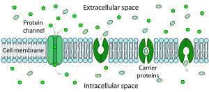
Polar molecules and large ions dissolved in water cannot diffuse freely across the plasma membrane due to the hydrophobic nature of the fatty acid tails of the phospholipids that comprise the lipid bilayer. Only small, non-polar molecules, such as oxygen and carbon dioxide, can diffuse easily across the membrane. Hence, small polar molecules are transported by proteins in the form of transmembrane channels. These channels are gated, meaning that they open and close, and thus deregulate the flow of ions or small polar molecules across membranes, sometimes against the osmotic gradient. Larger molecules are transported by transmembrane carrier proteins, such as permeases, that change their conformation as the molecules are carried across (e.g. glucose or amino acids). Non-polar molecules, such as retinol or lipids, are poorly soluble in water. They are transported through aqueous compartments of cells or through extracellular space by water-soluble carriers (e.g. retinol binding protein). The metabolites are not altered because no energy is required for facilitated diffusion. Only permease changes its shape in order to transport metabolites. The form of transport through a cell membrane in which a metabolite is modified is called group translocation transportation.

Glucose, sodium ions, and chloride ions are just a few examples of molecules and ions that must efficiently cross the plasma membrane but to which the lipid bilayer of the membrane is virtually impermeable. Their transport must therefore be "facilitated" by proteins that span the membrane and provide an alternative route or bypass mechanism. Some examples of proteins that mediate this process are glucose transporters, organic cation transport proteins, urea transporter, monocarboxylate transporter 8 and monocarboxylate transporter 10.

In vivo model of facilitated diffusion

Many physical and biochemical processes are regulated by diffusion. [3] Facilitated diffusion is one form of diffusion and it is important in several metabolic processes. Facilitated diffusion is the main mechanism behind the binding of Transcription Factors (TFs) to designated target sites on the DNA molecule. The in vitro model, which is a very well known method of facilitated diffusion, that takes place outside of a living cell, explains the 3-dimensional pattern of diffusion in the cytosol and the 1-dimensional diffusion along the DNA contour. [4] After carrying out extensive research on processes occurring out of the cell, this mechanism was generally accepted but there was a need to verify that this mechanism could take place in vivo or inside of living cells. Bauer & Metzler (2013) [4] therefore carried out an experiment using a bacterial genome in which they investigated the average time for TF – DNA binding to occur. After analyzing the process for the time it takes for TF's to diffuse across the contour and cytoplasm of the bacteria's DNA, it was concluded that in vitro and in vivo are similar in that the association and dissociation rates of TF's to and from the DNA are similar in both. Also, on the DNA contour, the motion is slower and target sites are easy to localize while in the cytoplasm, the motion is faster but the TF's are not sensitive to their targets and so binding is restricted.

Intracellular facilitated diffusion

Single-molecule imaging is an imaging technique which provides an ideal resolution necessary for the study of the Transcription factor binding mechanism in living cells. [5] In prokaryotic bacteria cells such as E. coli, facilitated diffusion is required in order for regulatory proteins to locate and bind to target sites on DNA base pairs. [3] [5] [6] There are 2 main steps involved: the protein binds to a non-specific site on the DNA and then it diffuses along the DNA chain until it locates a target site, a process referred to as sliding. [3] According to Brackley et al. (2013), during the process of protein sliding, the protein searches the entire length of the DNA chain using 3-D and 1-D diffusion patterns. During 3-D diffusion, the high incidence of Crowder proteins creates an osmotic pressure which brings searcher proteins (e.g. Lac Repressor) closer to the DNA to increase their attraction and enable them to bind, as well as steric effect which exclude the Crowder proteins from this region (Lac operator region). Blocker proteins participate in 1-D diffusion only i.e. bind to and diffuse along the DNA contour and not in the cytosol.

Facilitated diffusion of proteins on Chromatin

The in vivo model mentioned above clearly explains 3-D and 1-D diffusion along the DNA strand and the binding of proteins to target sites on the chain. Just like prokaryotic cells, in eukaryotes, facilitated diffusion occurs in the nucleoplasm on chromatin filaments, accounted for by the switching dynamics of a protein when it is either bound to a chromatin thread or when freely diffusing in the nucleoplasm. [7] In addition, given that the chromatin molecule is fragmented, its fractal properties need to be considered. After calculating the search time for a target protein, alternating between the 3-D and 1-D diffusion phases on the chromatin fractal structure, it was deduced that facilitated diffusion in eukaryotes precipitates the searching process and minimizes the searching time by increasing the DNA-protein affinity. [7]

For oxygen

The oxygen affinity with hemoglobin on red blood cell surfaces enhances this bonding ability. [8] In a system of facilitated diffusion of oxygen, there is a tight relationship between the ligand which is oxygen and the carrier which is either hemoglobin or myoglobin. [9] This mechanism of facilitated diffusion of oxygen by hemoglobin or myoglobin was discovered and initiated by Wittenberg and Scholander. [10] They carried out experiments to test for the steady-state of diffusion of oxygen at various pressures. Oxygen-facilitated diffusion occurs in a homogeneous environment where oxygen pressure can be relatively controlled. [11] [12] For oxygen diffusion to occur, there must be a full saturation pressure (more) on one side of the membrane and full reduced pressure (less) on the other side of the membrane i.e. one side of the membrane must be of higher concentration. During facilitated diffusion, hemoglobin increases the rate of constant diffusion of oxygen and facilitated diffusion occurs when oxyhemoglobin molecule is randomly displaced.

For carbon monoxide

Facilitated diffusion of carbon monoxide is similar to that of oxygen. Carbon monoxide also combines with hemoglobin and myoglobin, [12] but carbon monoxide has a dissociation velocity that 100 times less than that of oxygen. Its affinity for myoglobin is 40 times higher and 250 times higher for hemoglobin, compared to oxygen. [13]

For glucose

Since glucose is a large molecule, its diffusion across a membrane is difficult. [14] Hence, it diffuses across membranes through facilitated diffusion, down the concentration gradient. The carrier protein at the membrane binds to the glucose and alters its shape such that it can easily to be transported. [15] Movement of glucose into the cell could be rapid or slow depending on the number of membrane-spanning protein. It is transported against the concentration gradient by a dependent glucose symporter which provides a driving force to other glucose molecules in the cells. Facilitated diffusion helps in the release of accumulated glucose into the extracellular space adjacent to the blood capillary. [15]

See also

Related Research Articles

<span class="mw-page-title-main">Hemoglobin</span> Metalloprotein that binds with oxygen

Hemoglobin is a protein containing iron that facilitates the transport of oxygen in red blood cells. Almost all vertebrates contain hemoglobin, with the exception of the fish family Channichthyidae and the tissues of some invertebrate animals. Hemoglobin in the blood carries oxygen from the respiratory organs to the other tissues of the body, where it releases the oxygen to enable aerobic respiration which powers the animal's metabolism. A healthy human has 12 to 20 grams of hemoglobin in every 100 mL of blood. Hemoglobin is a metalloprotein, a chromoprotein, and globulin.

<span class="mw-page-title-main">Red blood cell</span> Oxygen-delivering blood cell and the most common type of blood cell

Red blood cells (RBCs), scientific name erythrocytes (from Greek erythros 'red' and kytos 'hollow vessel', with -cyte translated as 'cell' in modern usage), also referred to as red cells, red blood corpuscles (in humans or other animals not having nucleus in red blood cells) or haematids, are the most common type of blood cell and the vertebrate's principal means of delivering oxygen (O2) to the body tissues—via blood flow through the circulatory system. Erythrocytes take up oxygen in the lungs, or in fish the gills, and release it into tissues while squeezing through the body's capillaries.

<span class="mw-page-title-main">Myoglobin</span> Iron and oxygen-binding protein

Myoglobin is an iron- and oxygen-binding protein found in the cardiac and skeletal muscle tissue of vertebrates in general and in almost all mammals. Myoglobin is distantly related to hemoglobin. Compared to hemoglobin, myoglobin has a higher affinity for oxygen and does not have cooperative binding with oxygen like hemoglobin does. Myoglobin consists of non-polar amino acids at the core of the globulin, where the heme group is non-covalently bounded with the surrounding polypeptide of myoglobin. In humans, myoglobin is only found in the bloodstream after muscle injury.

<span class="mw-page-title-main">Hemoprotein</span> Protein containing a heme prosthetic group

A hemeprotein, or heme protein, is a protein that contains a heme prosthetic group. They are a very large class of metalloproteins. The heme group confers functionality, which can include oxygen carrying, oxygen reduction, electron transfer, and other processes. Heme is bound to the protein either covalently or noncovalently or both.

In cellular biology, active transport is the movement of molecules or ions across a cell membrane from a region of lower concentration to a region of higher concentration—against the concentration gradient. Active transport requires cellular energy to achieve this movement. There are two types of active transport: primary active transport that uses adenosine triphosphate (ATP), and secondary active transport that uses an electrochemical gradient. This process is in contrast to passive transport, which allows molecules or ions to move down their concentration gradient, from an area of high concentration to an area of low concentration, without energy.

<span class="mw-page-title-main">Passive transport</span> Transport that does not require energy

Passive transport is a type of membrane transport that does not require energy to move substances across cell membranes. Instead of using cellular energy, like active transport, passive transport relies on the second law of thermodynamics to drive the movement of substances across cell membranes. Fundamentally, substances follow Fick's first law, and move from an area of high concentration to an area of low concentration because this movement increases the entropy of the overall system. The rate of passive transport depends on the permeability of the cell membrane, which, in turn, depends on the organization and characteristics of the membrane lipids and proteins. The four main kinds of passive transport are simple diffusion, facilitated diffusion, filtration, and/or osmosis.

<span class="mw-page-title-main">Mediated transport</span> Transportation of substances via membrane

Mediated transport refers to transport mediated by a membrane transport protein. Substances in the human body may be hydrophobic, electrophilic, contain a positively or negatively charge, or have another property. As such there are times when those substances may not be able to pass over the cell membrane using protein-independent movement. The cell membrane is imbedded with many membrane transport proteins that allow such molecules to travel in and out of the cell. There are three types of mediated transporters: uniport, symport, and antiport. Things that can be transported are nutrients, ions, glucose, etc, all depending on the needs of the cell. One example of a uniport mediated transport protein is GLUT1. GLUT1 is a transmembrane protein, which means it spans the entire width of the cell membrane, connecting the extracellular and intracellular region. It is a uniport system because it specifically transports glucose in only one direction, down its concentration gradient across the cell membrane.

A membrane transport protein is a membrane protein involved in the movement of ions, small molecules, and macromolecules, such as another protein, across a biological membrane. Transport proteins are integral transmembrane proteins; that is they exist permanently within and span the membrane across which they transport substances. The proteins may assist in the movement of substances by facilitated diffusion, active transport, osmosis, or reverse diffusion. The two main types of proteins involved in such transport are broadly categorized as either channels or carriers. Examples of channel/carrier proteins include the GLUT 1 uniporter, sodium channels, and potassium channels. The solute carriers and atypical SLCs are secondary active or facilitative transporters in humans. Collectively membrane transporters and channels are known as the transportome. Transportomes govern cellular influx and efflux of not only ions and nutrients but drugs as well.

In cellular biology, membrane transport refers to the collection of mechanisms that regulate the passage of solutes such as ions and small molecules through biological membranes, which are lipid bilayers that contain proteins embedded in them. The regulation of passage through the membrane is due to selective membrane permeability – a characteristic of biological membranes which allows them to separate substances of distinct chemical nature. In other words, they can be permeable to certain substances but not to others.

<span class="mw-page-title-main">Uniporter</span>

Uniporters, also known as solute carriers or facilitated transporters, are a type of membrane transport protein that passively transports solutes across a cell membrane. It uses facilitated diffusion for the movement of solutes down their concentration gradient from an area of high concentration to an area of low concentration. Unlike active transport, it does not require energy in the form of ATP to function. Uniporters are specialized to carry one specific ion or molecule and can be categorized as either channels or carriers. Facilitated diffusion may occur through three mechanisms: uniport, symport, or antiport. The difference between each mechanism depends on the direction of transport, in which uniport is the only transport not coupled to the transport of another solute.

<span class="mw-page-title-main">Cotransporter</span> Type of membrane transport proteins

Cotransporters are a subcategory of membrane transport proteins (transporters) that couple the favorable movement of one molecule with its concentration gradient and unfavorable movement of another molecule against its concentration gradient. They enable coupled or cotransport and include antiporters and symporters. In general, cotransporters consist of two out of the three classes of integral membrane proteins known as transporters that move molecules and ions across biomembranes. Uniporters are also transporters but move only one type of molecule down its concentration gradient and are not classified as cotransporters.

<span class="mw-page-title-main">Glucose transporter</span> Family of monosaccharide transport proteins

Glucose transporters are a wide group of membrane proteins that facilitate the transport of glucose across the plasma membrane, a process known as facilitated diffusion. Because glucose is a vital source of energy for all life, these transporters are present in all phyla. The GLUT or SLC2A family are a protein family that is found in most mammalian cells. 14 GLUTS are encoded by the human genome. GLUT is a type of uniporter transporter protein.

Carbaminohemoglobin (carbaminohaemoglobin BrE) (CO2Hb, also known as carbhemoglobin and carbohemoglobin) is a compound of hemoglobin and carbon dioxide, and is one of the forms in which carbon dioxide exists in the blood. Twenty-three percent of carbon dioxide is carried in blood this way (70% is converted into bicarbonate by carbonic anhydrase and then carried in plasma, 7% carried as free CO2, dissolved in plasma).

<span class="mw-page-title-main">Ion transporter</span> Transmembrane protein that moves ions across a biological membrane

In biology, a transporter is a transmembrane protein that moves ions across a biological membrane to accomplish many different biological functions, including cellular communication, maintaining homeostasis, energy production, etc. There are different types of transporters including pumps, uniporters, antiporters, and symporters. Active transporters or ion pumps are transporters that convert energy from various sources—including adenosine triphosphate (ATP), sunlight, and other redox reactions—to potential energy by pumping an ion up its concentration gradient. This potential energy could then be used by secondary transporters, including ion carriers and ion channels, to drive vital cellular processes, such as ATP synthesis.

Method of glucose uptake differs throughout tissues depending on two factors; the metabolic needs of the tissue and availability of glucose. The two ways in which glucose uptake can take place are facilitated diffusion and secondary active transport. Active transport is the movement of ions or molecules going against the concentration gradient.

<span class="mw-page-title-main">Iron in biology</span> Use of Iron by organisms

Iron is an important biological element. It is used in both the ubiquitous iron-sulfur proteins and in vertebrates it is used in hemoglobin which is essential for blood and oxygen transport.

<span class="mw-page-title-main">Single-particle tracking</span> AliAlamerr

Single-particle tracking (SPT) is the observation of the motion of individual particles within a medium. The coordinates time series, which can be either in two dimensions (x, y) or in three dimensions (x, y, z), is referred to as a trajectory. The trajectory is typically analyzed using statistical methods to extract information about the underlying dynamics of the particle. These dynamics can reveal information about the type of transport being observed (e.g., thermal or active), the medium where the particle is moving, and interactions with other particles. In the case of random motion, trajectory analysis can be used to measure the diffusion coefficient.

Transcellular transport involves the transportation of solutes by a cell through a cell. Transcellular transport can occur in three different ways active transport, passive transport, and transcytosis.

The insulin transduction pathway is a biochemical pathway by which insulin increases the uptake of glucose into fat and muscle cells and reduces the synthesis of glucose in the liver and hence is involved in maintaining glucose homeostasis. This pathway is also influenced by fed versus fasting states, stress levels, and a variety of other hormones.

<span class="mw-page-title-main">Cell membrane</span> Biological membrane that separates the interior of a cell from its outside environment

The cell membrane is a biological membrane that separates and protects the interior of a cell from the outside environment. The cell membrane consists of a lipid bilayer, made up of two layers of phospholipids with cholesterols interspersed between them, maintaining appropriate membrane fluidity at various temperatures. The membrane also contains membrane proteins, including integral proteins that span the membrane and serve as membrane transporters, and peripheral proteins that loosely attach to the outer (peripheral) side of the cell membrane, acting as enzymes to facilitate interaction with the cell's environment. Glycolipids embedded in the outer lipid layer serve a similar purpose. The cell membrane controls the movement of substances in and out of a cell, being selectively permeable to ions and organic molecules. In addition, cell membranes are involved in a variety of cellular processes such as cell adhesion, ion conductivity, and cell signalling and serve as the attachment surface for several extracellular structures, including the cell wall and the carbohydrate layer called the glycocalyx, as well as the intracellular network of protein fibers called the cytoskeleton. In the field of synthetic biology, cell membranes can be artificially reassembled.

References

  1. Pratt CA, Voet D, Voet JG (2002). Fundamentals of biochemistry upgrade. New York: Wiley. pp. 264–266. ISBN   0-471-41759-9.
  2. Friedman, Morton (2008). Principles and models of biological transport. Springer. ISBN   978-0387-79239-2.
  3. 1 2 3 Klenin, Konstantin V.; Merlitz, Holger; Langowski, Jörg; Wu, Chen-Xu (2006). "Facilitated Diffusion of DNA-Binding Proteins". Physical Review Letters. 96 (1): 018104. arXiv: physics/0507056 . Bibcode:2006PhRvL..96a8104K. doi:10.1103/PhysRevLett.96.018104. ISSN   0031-9007. PMID   16486524. S2CID   8937433.
  4. 1 2 Bauer M, Metzler R (2013). "In vivo facilitated diffusion model". PLOS ONE. 8 (1): e53956. arXiv: 1301.5502 . Bibcode:2013PLoSO...853956B. doi: 10.1371/journal.pone.0053956 . PMC   3548819 . PMID   23349772.
  5. 1 2 Hammar, P.; Leroy, P.; Mahmutovic, A.; Marklund, E. G.; Berg, O. G.; Elf, J. (2012). "The lac Repressor Displays Facilitated Diffusion in Living Cells". Science. 336 (6088): 1595–1598. Bibcode:2012Sci...336.1595H. doi:10.1126/science.1221648. ISSN   0036-8075. PMID   22723426. S2CID   21351861.
  6. Brackley CA, Cates ME, Marenduzzo D (September 2013). "Intracellular facilitated diffusion: searchers, crowders, and blockers". Phys. Rev. Lett. 111 (10): 108101. arXiv: 1309.1010 . Bibcode:2013PhRvL.111j8101B. doi:10.1103/PhysRevLett.111.108101. PMID   25166711. S2CID   13220767.
  7. 1 2 Bénichou O, Chevalier C, Meyer B, Voituriez R (January 2011). "Facilitated diffusion of proteins on chromatin". Phys. Rev. Lett. 106 (3): 038102. arXiv: 1006.4758 . Bibcode:2011PhRvL.106c8102B. doi:10.1103/PhysRevLett.106.038102. PMID   21405302. S2CID   15977456.
  8. Kreuzer, F. (1970). "Facilitated diffusion of oxygen and its possible significance; a review". Respiration Physiology. 9 (1): 1–30. doi:10.1016/0034-5687(70)90002-2. ISSN   0034-5687. PMID   4910215.
  9. Jacquez JA, Kutchai H, Daniels E (June 1972). "Hemoglobin-facilitated diffusion of oxygen: interfacial and thickness effects" (PDF). Respir Physiol. 15 (2): 166–81. doi:10.1016/0034-5687(72)90096-5. hdl: 2027.42/34087 . PMID   5042165.
  10. Rubinow SI, Dembo M (April 1977). "The facilitated diffusion of oxygen by hemoglobin and myoglobin". Biophys. J. 18 (1): 29–42. Bibcode:1977BpJ....18...29R. doi:10.1016/S0006-3495(77)85594-X. PMC   1473276 . PMID   856316.
  11. Kreuzer F, Hoofd LJ (May 1972). "Factors influencing facilitated diffusion of oxygen in the presence of hemoglobin and myoglobin". Respir Physiol. 15 (1): 104–24. doi:10.1016/0034-5687(72)90008-4. PMID   5079218.
  12. 1 2 Wittenberg JB (January 1966). "The molecular mechanism of hemoglobin-facilitated oxygen diffusion". J. Biol. Chem. 241 (1): 104–14. doi: 10.1016/S0021-9258(18)96964-4 . PMID   5901041.
  13. Murray JD, Wyman J (October 1971). "Facilitated diffusion. The case of carbon monoxide". J. Biol. Chem. 246 (19): 5903–6. doi: 10.1016/S0021-9258(18)61811-3 . PMID   5116656.
  14. Thorens B (1993). "Facilitated glucose transporters in eithelial cells". Annu. Rev. Physiol. 55: 591–608. doi:10.1146/annurev.ph.55.030193.003111. PMID   8466187.
  15. 1 2 Carruthers, A. (1990). "Facilitated diffusion of glucose". Physiological Reviews. 70 (4): 1135–1176. doi:10.1152/physrev.1990.70.4.1135. ISSN   0031-9333. PMID   2217557.