Farber disease

Last updated
Farber disease
Other namesAcid ceramidase deficiency [1]
Specialty Endocrinology   OOjs UI icon edit-ltr-progressive.svg

Farber disease (also known as Farber's lipogranulomatosis, acid ceramidase deficiency, "Lipogranulomatosis", [2] and ASAH1-related disorders) is an extremely rare, progressive, autosomal recessive lysosomal storage disease caused by a deficiency of the acid ceramidase enzyme. Acid ceramidase is responsible for breaking down ceramide into sphingosine and fatty acid. [3] When the enzyme is deficient, this leads to an accumulation of fatty material (called ceramide) in the lysosomes of the cells, leading to the signs and symptoms of this disorder. [4]

Contents

Signs and symptoms

Hands in Farber disease Lipogranulomatosis 1.jpg
Hands in Farber disease

The symptoms of Farber disease develop over time. The onset of symptoms and how quickly they progress vary from person to person. [5]

The most common symptoms include: [4]

Other symptoms observed in some individuals with Farber disease include: [5] [3]

Genetics

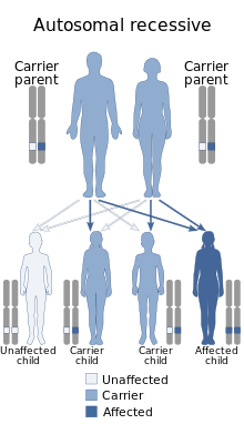
This condition is inherited in an autosomal recessive manner Autosomal recessive - en.svg
This condition is inherited in an autosomal recessive manner

Farber disease is caused by variants in the ASAH1 gene. This gene codes for the acid ceramidase enzyme. Individuals with Farber disease have two copies of this gene that are not functioning properly leading to the enzyme deficiency. Over 73 different gene variants have been reported to cause Farber disease. No definitive genotype-phenotype correlations are known. [6]

Farber disease is inherited in an autosomal recessive manner. Affected individuals inherit one copy of the gene that is not functioning properly from each parent. Each parent is a called a carrier and has one copy of the gene that is functioning properly and one that is not. Siblings of individuals with Farber disease have a 25% chance to also have Farber disease, a 50% chance to be a carrier like the parents, and a 25% chance of being unaffected and not a carrier. [4]

The ASAH1 gene is also known to cause a condition called spinal muscular atrophy with progressive myoclonic epilepsy (SMA-PME). [4] Farber disease and SMA-PME have been classified as separate disorders, however more recent case reports have identified some individuals with overlapping symptoms of both disorders. [7] [8] Some individuals with SMA symptoms without seizures have also been described. [9]

Diagnosis

Disease onset is typically in early infancy but may occur later in life. Children who have the classic form of Farber disease develop symptoms within the first few weeks to months of life. [4] Individuals with moderate or attenuated forms may develop symptoms at any time in childhood. Sometimes it is difficult to diagnose Farber disease because the symptoms can be misdiagnosed as Juvenile Idiopathic Arthritis (JIA). [10] [11] Diagnosis is confirmed by molecular genetic testing of the ASAH1 gene or by measuring acid ceramidase enzyme activity. [4]

Treatment

There is no disease specific treatment for Farber disease. Anti-inflammatory medications, specifically tocilizumab (an interleukin-6 receptor inhibitor), has been shown to improve inflammation and pain in some patients. [12] Bone marrow transplant may improve granulomas (small masses of inflamed tissue) and inflammation in patients with little or no lung or nervous system complications. [13] Supportive therapies such as physical therapy, respiratory support, and mobility aids may be required.[ citation needed ]

Studies in cells and mice have shown proof-of-concept for enzyme replacement therapy for Farber disease. [14] Aceragen, a biopharmaceutical company, is currently developing an investigational enzyme replacement therapy with a clinical study planned for late 2022.[ citation needed ]

Prognosis

Children with the most severe forms of Farber disease generally die by age 2–3 years. [4] The life expectancy of individuals with moderate or attenuated forms is unknown. The oldest reported individuals living with Farber disease were in their 50s and 60s. [15]

Prevalence

To date, there have been approximately 200 reported cases of Farber disease and SMA-PME in the literature. [3] The disorders are ultra-rare and estimated to occur in fewer than one per million. [4]

Eponym

It is named for Sidney Farber. [16] [17]

Related Research Articles

<span class="mw-page-title-main">Occipital horn syndrome</span> Medical condition

Occipital horn syndrome (OHS), formerly considered a variant of Ehlers–Danlos syndrome, is an X-linked recessive mitochondrial and connective tissue disorder. It is caused by a deficiency in the transport of the essential mineral copper, associated with mutations in the ATP7A gene.

<span class="mw-page-title-main">Lysosomal storage disease</span> Medical condition

Lysosomal storage diseases are a group of over 70 rare inherited metabolic disorders that result from defects in lysosomal function. Lysosomes are sacs of enzymes within cells that digest large molecules and pass the fragments on to other parts of the cell for recycling. This process requires several critical enzymes. If one of these enzymes is defective due to a mutation, the large molecules accumulate within the cell, eventually killing it.

<span class="mw-page-title-main">Niemann–Pick disease</span> Medical condition

Niemann–Pick disease (NP), also known as acid sphingomyelinase deficiency, is a group of rare genetic diseases of varying severity. These are inherited metabolic disorders in which sphingomyelin accumulates in lysosomes in cells of many organs. NP types A, A/B, and B are caused by mutations in the SMPD1 gene, which causes a deficiency of an acid sphingomyelinase (ASM). NP type C is now considered a separate disease, as SMPD1 is not involved, and there is no deficiency in ASM.

Hypotonia is a state of low muscle tone, often involving reduced muscle strength. Hypotonia is not a specific medical disorder, but a potential manifestation of many different diseases and disorders that affect motor nerve control by the brain or muscle strength. Hypotonia is a lack of resistance to passive movement, whereas muscle weakness results in impaired active movement. Central hypotonia originates from the central nervous system, while peripheral hypotonia is related to problems within the spinal cord, peripheral nerves and/or skeletal muscles. Severe hypotonia in infancy is commonly known as floppy baby syndrome. Recognizing hypotonia, even in early infancy, is usually relatively straightforward, but diagnosing the underlying cause can be difficult and often unsuccessful. The long-term effects of hypotonia on a child's development and later life depend primarily on the severity of the muscle weakness and the nature of the cause. Some disorders have a specific treatment but the principal treatment for most hypotonia of idiopathic or neurologic cause is physical therapy and/or occupational therapy for remediation.

<span class="mw-page-title-main">Mitochondrial myopathy</span> Muscle disorders caused by mitochondrial dysfunction

Mitochondrial myopathies are types of myopathies associated with mitochondrial disease. Adenosine triphosphate (ATP), the chemical used to provide energy for the cell, cannot be produced sufficiently by oxidative phosphorylation when the mitochondrion is either damaged or missing necessary enzymes or transport proteins. With ATP production deficient in mitochondria, there is an over-reliance on anaerobic glycolysis which leads to lactic acidosis either at rest or exercise-induced.

Pyruvate dehydrogenase deficiency is a rare neurodegenerative disorder associated with abnormal mitochondrial metabolism. PDCD is a genetic disease resulting from mutations in one of the components of the pyruvate dehydrogenase complex (PDC). The PDC is a multi-enzyme complex that plays a vital role as a key regulatory step in the central pathways of energy metabolism in the mitochondria. The disorder shows heterogeneous characteristics in both clinical presentation and biochemical abnormality.

<span class="mw-page-title-main">Spinal muscular atrophy</span> Rare congenital neuromuscular disorder

Spinal muscular atrophy (SMA) is a rare neuromuscular disorder that results in the loss of motor neurons and progressive muscle wasting. It is usually diagnosed in infancy or early childhood and if left untreated it is the most common genetic cause of infant death. It may also appear later in life and then have a milder course of the disease. The common feature is progressive weakness of voluntary muscles, with arm, leg and respiratory muscles being affected first. Associated problems may include poor head control, difficulties swallowing, scoliosis, and joint contractures.

<span class="mw-page-title-main">Ubiquitin-activating enzyme</span> Class of enzymes

Ubiquitin-activating enzymes, also known as E1 enzymes, catalyze the first step in the ubiquitination reaction, which can target a protein for degradation via a proteasome. This covalent bond of ubiquitin or ubiquitin-like proteins to targeted proteins is a major mechanism for regulating protein function in eukaryotic organisms. Many processes such as cell division, immune responses and embryonic development are also regulated by post-translational modification by ubiquitin and ubiquitin-like proteins.

Ceramidase is an enzyme which cleaves fatty acids from ceramide, producing sphingosine (SPH) which in turn is phosphorylated by a sphingosine kinase to form sphingosine-1-phosphate (S1P).

Progressive Myoclonic Epilepsies (PME) are a rare group of inherited neurodegenerative diseases characterized by myoclonus, resistance to treatment, and neurological deterioration. The cause of PME depends largely on the type of PME. Most PMEs are caused by autosomal dominant or recessive and mitochondrial mutations. The location of the mutation also affects the inheritance and treatment of PME. Diagnosing PME is difficult due to their genetic heterogeneity and the lack of a genetic mutation identified in some patients. The prognosis depends largely on the worsening symptoms and failure to respond to treatment. There is no current cure for PME and treatment focuses on managing myoclonus and seizures through antiepileptic medication (AED).

<span class="mw-page-title-main">Glycine—tRNA ligase</span> Protein-coding gene in the species Homo sapiens

Glycine—tRNA ligase also known as glycyl–tRNA synthetase is an enzyme that in humans is encoded by the GARS1 gene.

<span class="mw-page-title-main">ASAH1</span> Protein-coding gene in the species Homo sapiens

The ASAH1 gene encodes in humans the acid ceramidase enzyme.

Mitochondrially encoded tRNA threonine also known as MT-TT is a transfer RNA which in humans is encoded by the mitochondrial MT-TT gene.

<span class="mw-page-title-main">Spinal disease</span> Disease involving the vertebral column

Spinal disease refers to a condition impairing the backbone. These include various diseases of the back or spine ("dorso-"), such as kyphosis. Dorsalgia refers to back pain. Some other spinal diseases include spinal muscular atrophy, ankylosing spondylitis, scoliosis, lumbar spinal stenosis, spina bifida, spinal tumors, osteoporosis and cauda equina syndrome.

<span class="mw-page-title-main">X-linked spinal muscular atrophy type 2</span> Medical condition

X-linked spinal muscular atrophy type 2, also known as arthrogryposis multiplex congenita X-linked type 1 (AMCX1), is a rare neurological disorder involving death of motor neurons in the anterior horn of spinal cord resulting in generalised muscle wasting (atrophy). The disease is caused by a mutation in UBA1 gene and is passed in an X-linked recessive manner by carrier mothers to affected sons.

<span class="mw-page-title-main">Distal spinal muscular atrophy type 1</span> Medical condition

Distal spinal muscular atrophy type 1 (DSMA1), also known as spinal muscular atrophy with respiratory distress type 1 (SMARD1), is a rare neuromuscular disorder involving death of motor neurons in the spinal cord which leads to a generalised progressive atrophy of body muscles.

<span class="mw-page-title-main">Robert J. Desnick</span> American geneticist

Robert J. Desnick is an American human geneticist whose basic and translational research accomplishments include significant discoveries in genomics, pharmacogenetics, gene therapy, personalized medicine, and the treatment of genetic diseases. His translational research has led to the development of the enzyme replacement therapy (ERT) and the chaperone therapy for Fabry disease, ERT for Niemann–Pick disease type B, and the RNA Interference Therapy for the Acute Hepatic Porphyrias.

<span class="mw-page-title-main">Spinal muscular atrophy with progressive myoclonic epilepsy</span> Rare neurodegenerative disease whose symptoms include slowly progressive muscle wasting

Spinal muscular atrophy with progressive myoclonic epilepsy (SMA-PME), sometimes called Jankovic–Rivera syndrome, is a very rare neurodegenerative disease whose symptoms include slowly progressive muscle loss (atrophy), predominantly affecting proximal muscles, combined with denervation and myoclonic seizures. Only 12 known human families are described in scientific literature to have SMA-PME.

Congenital distal spinal muscular atrophy (cDSMA), also known as distal hereditary motor neuropathytype VIII (dHMN8), is a hereditary medical condition characterized by muscle wasting (atrophy), particularly of distal muscles in legs and hands, and by early-onset contractures of the hip, knee, and ankle. Affected individuals often have shorter lower limbs relative to the trunk and upper limbs. The condition is a result of a loss of anterior horn cells localized to lumbar and cervical regions of the spinal cord early in infancy, which in turn is caused by a mutation of the TRPV4 gene. The disorder is inherited in an autosomal dominant manner. Arm muscle and function, as well as cardiac and respiratory functions are typically well preserved.

<span class="mw-page-title-main">Nusinersen</span> Medication used for spinal muscular atrophy

Nusinersen, marketed as Spinraza, is a medication used in treating spinal muscular atrophy (SMA), a rare neuromuscular disorder. In December 2016, it became the first approved drug used in treating this disorder.

References

  1. RESERVED, INSERM US14-- ALL RIGHTS. "Orphanet: Farber disease". www.orpha.net. Retrieved 17 April 2019.{{cite web}}: CS1 maint: numeric names: authors list (link)
  2. James, William D.; Berger, Timothy G.; et al. (2006). Andrews' Diseases of the Skin: clinical Dermatology. Saunders Elsevier. ISBN   978-0-7216-2921-6.
  3. 1 2 3 Yu, FPS; Amintas, S; Levade, T; Medin, JA (20 July 2018). "Acid ceramidase deficiency: Farber disease and SMA-PME". Orphanet Journal of Rare Diseases. 13 (121): 20. doi: 10.1186/s13023-018-0845-z . PMC   6053731 . PMID   30029679.
  4. 1 2 3 4 5 6 7 8 Dyment, DA; Bennett, SAL; Medin, JA; Levade, T; Adam, MP; Ardinger, HH; Pagon, RA; Wallace, SE; Bean, LJH; Mirzaa, G; Amemiya, A (2018). "ASAH1-Related Disorders". GeneReviews. PMID   29595935.
  5. 1 2 "Farber disease". rarediseases.info.nih.gov. Genetic and Rare Diseases Information Center (GARD) – an NCATS Program. Archived from the original on 2021-08-23. Retrieved 2021-08-17.
  6. Elsea, SH; Solyom, A; Martin, K; Harmatz, P; Mitchell, J; Lampe, C; Grant, C; Selim, L; Mungan, NO; Guelbert, N; Magnusson, B; Sundberg, E; Puri, R; Kapoor, S; Arslan, N; DiRocco, M; Zaki, M; Ozen, S; Mahmoud, IG; Ehlert, K; Hahn, A; Gokcay, G; Torcoletti, M; Ferreira, CR (September 2020). "ASAH1 pathogenic variants associated with acid ceramidase deficiency: Farber disease and spinal muscular atrophy with progressive myoclonic epilepsy". Human Mutation. 41 (9): 1469–1487. doi: 10.1002/humu.24056 . PMID   32449975. S2CID   218895424.
  7. Teoh, HL; Solyom, A; Schuchman, EH; Mowat, D; Roscioli, T; Farrar, M; Sampaio, H (October 2016). "Polyarticular Arthritis and Spinal Muscular Atrophy in Acid Ceramidase Deficiency". Pediatrics. 138 (4): e20161068. doi: 10.1542/peds.2016-1068 . PMID   27650050. S2CID   2152446.
  8. Lee, BH; Mongiovi, P; Levade, T; Marston, B; Mountain, J; Ciafaloni, E (October 2020). "Spinal muscular atrophy and Farber disease due to ASAH1 variants: A case report". American Journal of Medical Genetics. Part A. 182 (10): 2369–2371. doi:10.1002/ajmg.a.61764. PMID   32627310. S2CID   220369745.
  9. Filosto, M; Aureli, M; Castellotti, B; Rinaldi, F; Schiumarini, D; Valsecchi, M; Lualdi, S; Mazzotti, R; Pensato, V; Rota, S; Gellera, C; Filocamo, M; Padovani, A (November 2016). "ASAH1 variant causing a mild SMA phenotype with no myoclonic epilepsy: a clinical, biochemical and molecular study". European Journal of Human Genetics. 24 (11): 1578–1583. doi:10.1038/ejhg.2016.28. PMC   5110045 . PMID   27026573.
  10. Schuchman, Edward H.; Mitchell, John; Solyom, Alex (2 September 2017). "Morbidity and mortality associated with Farber disease and prospects for therapy". Expert Opinion on Orphan Drugs. 5 (9): 717–726. doi:10.1080/21678707.2017.1359086. S2CID   79980666.
  11. Moghadam, SH; Tavasoli, AR; Modaresi, M; Ziaee, V (1 December 2019). "Farber disease: report of three cases with joint involvement mimicking juvenile idiopathic arthritis". Journal of Musculoskeletal & Neuronal Interactions. 19 (4): 521–525. PMC   6944811 . PMID   31789304.
  12. Mitchell, John; Solyom, Alexander; Makay, Balahan; Arslan, Nur; Batu, Ezgi Deniz; Ozen, Seza; Hügle, Boris; Schuchman, Edward; Magnusson, Bo (February 2016). "Farber disease: Implications of anti-inflammatory treatment". Molecular Genetics and Metabolism. 117 (2): S81–S82. doi:10.1016/j.ymgme.2015.12.364.
  13. Ehlert, K; Levade, T; Di Rocco, M; Lanino, E; Albert, MH; Führer, M; Jarisch, A; Güngör, T; Ayuk, F; Vormoor, J (March 2019). "Allogeneic hematopoietic cell transplantation in Farber disease". Journal of Inherited Metabolic Disease. 42 (2): 286–294. doi:10.1002/jimd.12043. PMID   30815900. S2CID   73478264.
  14. He, X; Dworski, S; Zhu, C; DeAngelis, V; Solyom, A; Medin, JA; Simonaro, CM; Schuchman, EH (June 2017). "Enzyme replacement therapy for Farber disease: Proof-of-concept studies in cells and mice". BBA Clinical. 7: 85–96. doi:10.1016/j.bbacli.2017.02.001. PMC   5338723 . PMID   28275553.
  15. Bonafé, L; Kariminejad, A; Li, J; Royer-Bertrand, B; Garcia, V; Mahdavi, S; Bozorgmehr, B; Lachman, RL; Mittaz-Crettol, L; Campos-Xavier, B; Nampoothiri, S; Unger, S; Rivolta, C; Levade, T; Superti-Furga, A (September 2016). "Brief Report: Peripheral Osteolysis in Adults Linked to ASAH1 (Acid Ceramidase) Mutations: A New Presentation of Farber's Disease". Arthritis & Rheumatology. 68 (9): 2323–7. doi: 10.1002/art.39659 . PMID   26945816. S2CID   37749661.
  16. synd/453 at Who Named It?
  17. Farber S (1952). "A lipid metabolic disorder: disseminated lipogranulomatosis; a syndrome with similarity to, and important difference from, Niemann-Pick and Hand-Schüller-Christian disease". American Journal of Diseases of Children. 84 (4): 499–500. PMID   12975849.