Immunodiffusion

Last updated
Immunodiffusion
MeSH D005779

Immunodiffusion is a laboratory technique used to detect and quantify antigens and antibodies by observing their interactions within a gel medium. [1] This technique involves the diffusion of antigens and antibodies through a gel, usually agar, resulting in the formation of a visible precipitate when they interact. [1] [2]

Contents

Applications

Immunodiffusion techniques are widely used in immunology for various purposes, including [1] [2] :

Types of Immunodiffusion

Single Immunodiffusion (Radial Immunodiffusion)

In this method, antibodies are uniformly distributed in an agar gel, and the antigen sample is placed in wells cut into the gel. As the antigen diffuses radially, it forms a precipitation ring with the antibody. The diameter of this ring corresponds to the concentration of the antigen in the solution. [3] [2]

Double Immunodiffusion (Ouchterlony Technique)

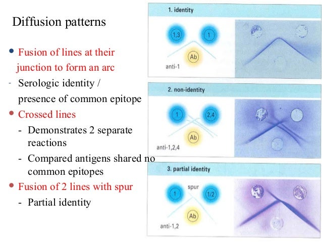
This method involves both antigen and antibody diffusing through the gel from separate wells, forming precipitation lines where they meet and react. [4]

Other types:

  1. Single diffusion in one dimension (Oudin procedure)
  2. Double diffusion in one dimension (Oakley Fulthorpe procedure)

Advantages

Limitations

Notes

  1. 1 2 3 4 "Immunodiffusion - Protocol & Troubleshooting - Creative Biolabs". www.creativebiolabs.net. Retrieved 2024-07-25.
  2. 1 2 3 Aryal, Sagar (2022-07-04). "Radial Immunodiffusion- Principle, Procedure, Results, Uses". microbenotes.com. Retrieved 2024-07-25.
  3. "Radial Immunodiffusion". Edvotek, Inc. 2017. Archived from the original (photograph) on 2017-08-07. Retrieved 2017-08-07. Photograph of precipitin circles in a Petri dish during radial immunodiffusion.
  4. "Diffusion Patterns". Immunodiffusion principles and application. Archived from the original on 2019-12-11. Retrieved 2017-05-19. Photographs of Ouchterlony immunodiffusion patterns showing stained precipitin lines of full identity, partial identity and non-identity.
  5. Mujtaba, Mustafa G.; Baliban, Tara; Bhagu, Jamini; Herrera, Michael. "A Laboratory Exercise Simulating Antibody and Antigen Reactions of the Ouchterlony Double Immunodiffusion Assay Using Inorganic Salts". Journal of Microbiology & Biology Education. 22 (2): e00103–21. doi:10.1128/jmbe.00103-21. ISSN   1935-7877. PMC   8442017 . PMID   34594450.

Related Research Articles

<span class="mw-page-title-main">Agar</span> Thickening agent used in microbiology and food

Agar, or agar-agar, is a jelly-like substance consisting of polysaccharides obtained from the cell walls of some species of red algae, primarily from "ogonori" (Gracilaria) and "tengusa" (Gelidiaceae). As found in nature, agar is a mixture of two components, the linear polysaccharide agarose and a heterogeneous mixture of smaller molecules called agaropectin. It forms the supporting structure in the cell walls of certain species of algae and is released on boiling. These algae are known as agarophytes, belonging to the Rhodophyta phylum. The processing of food-grade agar removes the agaropectin, and the commercial product is essentially pure agarose.

<span class="mw-page-title-main">ELISA</span> Method to detect an antigen using an antibody and enzyme

The enzyme-linked immunosorbent assay (ELISA) is a commonly used analytical biochemistry assay, first described by Eva Engvall and Peter Perlmann in 1971. The assay is a solid-phase type of enzyme immunoassay (EIA) to detect the presence of a ligand in a liquid sample using antibodies directed against the ligand to be measured. ELISA has been used as a diagnostic tool in medicine, plant pathology, and biotechnology, as well as a quality control check in various industries.

<span class="mw-page-title-main">Western blot</span> Analytical technique used in molecular biology

The western blot, or western blotting, is a widely used analytical technique in molecular biology and immunogenetics to detect specific proteins in a sample of tissue homogenate or extract. Besides detecting the proteins, this technique is also utilized to visualize, distinguish, and quantify the different proteins in a complicated protein combination.

An assay is an investigative (analytic) procedure in laboratory medicine, mining, pharmacology, environmental biology and molecular biology for qualitatively assessing or quantitatively measuring the presence, amount, or functional activity of a target entity. The measured entity is often called the analyte, the measurand, or the target of the assay. The analyte can be a drug, biochemical substance, chemical element or compound, or cell in an organism or organic sample. An assay usually aims to measure an analyte's intensive property and express it in the relevant measurement unit.

<span class="mw-page-title-main">Immunostaining</span> Biochemical technique

In biochemistry, immunostaining is any use of an antibody-based method to detect a specific protein in a sample. The term "immunostaining" was originally used to refer to the immunohistochemical staining of tissue sections, as first described by Albert Coons in 1941. However, immunostaining now encompasses a broad range of techniques used in histology, cell biology, and molecular biology that use antibody-based staining methods.

<span class="mw-page-title-main">Hemagglutination assay</span> Measure of virus or bacteria concentration

The hemagglutination assay or haemagglutination assay (HA) and the hemagglutination inhibition assay were developed in 1941–42 by American virologist George Hirst as methods for quantifying the relative concentration of viruses, bacteria, or antibodies.

<span class="mw-page-title-main">Hypersensitivity pneumonitis</span> Medical condition

Hypersensitivity pneumonitis (HP) or extrinsic allergic alveolitis (EAA) is a syndrome caused by the repetitive inhalation of antigens from the environment in susceptible or sensitized people. Common antigens include molds, bacteria, bird droppings, bird feathers, agricultural dusts, bioaerosols and chemicals from paints or plastics. People affected by this type of lung inflammation (pneumonitis) are commonly exposed to the antigens by their occupations, hobbies, the environment and animals. The inhaled antigens produce a hypersensitivity immune reaction causing inflammation of the airspaces (alveoli) and small airways (bronchioles) within the lung. Hypersensitivity pneumonitis may eventually lead to interstitial lung disease.

<span class="mw-page-title-main">Immunoelectrophoresis</span> Biochemical methods of separation and characterization of proteins

Immunoelectrophoresis is a general name for a number of biochemical methods for separation and characterization of proteins based on electrophoresis and reaction with antibodies. All variants of immunoelectrophoresis require immunoglobulins, also known as antibodies, reacting with the proteins to be separated or characterized. The methods were developed and used extensively during the second half of the 20th century. In somewhat chronological order: Immunoelectrophoretic analysis, crossed immunoelectrophoresis, rocket-immunoelectrophoresis, fused rocket immunoelectrophoresis ad modum Svendsen and Harboe, affinity immunoelectrophoresis ad modum Bøg-Hansen.

Protein methods are the techniques used to study proteins. There are experimental methods for studying proteins. Computational methods typically use computer programs to analyze proteins. However, many experimental methods require computational analysis of the raw data.

<span class="mw-page-title-main">Antibiotic sensitivity testing</span> Microbiology test used in medicine

Antibiotic sensitivity testing or antibiotic susceptibility testing is the measurement of the susceptibility of bacteria to antibiotics. It is used because bacteria may have resistance to some antibiotics. Sensitivity testing results can allow a clinician to change the choice of antibiotics from empiric therapy, which is when an antibiotic is selected based on clinical suspicion about the site of an infection and common causative bacteria, to directed therapy, in which the choice of antibiotic is based on knowledge of the organism and its sensitivities.

<span class="mw-page-title-main">Ouchterlony double immunodiffusion</span> Biomedical technique

Ouchterlony double immunodiffusion is an immunological technique used in the detection, identification and quantification of antibodies and antigens, such as immunoglobulins and extractable nuclear antigens. The technique is named after Örjan Ouchterlony, the Swedish physician who developed the test in 1948 to evaluate the production of diphtheria toxins from isolated bacteria.

Extractable nuclear antigens (ENAs) are over 100 different soluble cytoplasmic and nuclear antigens. They are known as "extractable" because they can be removed from cell nuclei using saline and represent six main proteins: Ro, La, Sm, RNP, Scl-70, Jo1. Most ENAs are part of spliceosomes or nucleosomes complexes and are a type of small nuclear ribonucleoprotein (snRNPS). The location in the nucleus and association with spliceosomes or nucleosomes results in these ENAs being associated with additional RNA and proteins such as polymerases. This quality of ENAs often makes it difficult to purify and quantify their presence for clinical use.

<span class="mw-page-title-main">Immunoproteomics</span> Study of large set of protein

Immunoproteomics is the study of large sets of proteins (proteomics) involved in the immune response.

<span class="mw-page-title-main">Disk diffusion test</span> Microbiology assay used in diagnostic and drug discovery laboratories

The disk diffusion test is a culture-based microbiology assay used in diagnostic and drug discovery laboratories. In diagnostic labs, the assay is used to determine the susceptibility of bacteria isolated from a patient's infection to clinically approved antibiotics. This allows physicians to prescribe the most appropriate antibiotic treatment. In drug discovery labs, especially bioprospecting labs, the assay is used to screen biological material and drug candidates for antibacterial activity. When bioprospecting, the assay can be performed with paired strains of bacteria to achieve dereplication and provisionally identify antibacterial mechanism of action.

<span class="mw-page-title-main">Counterimmunoelectrophoresis</span>

Counterimmunoelectrophoresis is a laboratory technique used to evaluate the binding of an antibody to its antigen, it is similar to immunodiffusion, but with the addition of an applied electrical field across the diffusion medium, usually an agar or polyacrylamide gel. The effect is rapid migration of the antibody and antigen out of their respective wells towards one another to form a line of precipitation, or a precipitin line, indicating binding.

Örjan Thomas Ouchterlony was a Swedish bacteriologist and immunologist who is credited with the creation of the Ouchterlony double immuno diffusion test in the 1940s. He was trained at Karolinska Institute, where his received his medical doctorate. He worked at Sweden's State Bacteriology Laboratory from 1935 to 1952. Ouchterlony was a professor of bacteriology at the Medical Faculty of Gothenburg University from 1952 to 1980 and was elected a member of the Royal Swedish Academy of Sciences in 1968. In addition to his laboratory work, he did research in field epidemiology of infectious diseases and worked and lectured in Africa and the United States, as well as in several countries in Europe. Upon his retirement in 1980, the successor to his professorial chair was Jan Holmgren.

The Ouchterlony plate is one of the more frequently used techniques for the identification of antigens and antibodies, by measurement of diffusion gradients in gel. Among its many applications has been the search for tumor-specific antigens. The technique was introduced by Örjan Ouchterlony of Sweden, in 1948, initially for the in vitro testing of the toxin-producing capacity of diphtheria bacteria. The technique was proved well suited to the analysis of complex serological systems, including analysis that helped to elucidate the structural heterogeneity of immunoglobulins. Ouchterlony reviewed the history of the developments, which extends back to the late Nineteenth Century.

Radial immunodiffusion (RID), Mancini immunodiffusion or single radial immunodiffusion assay, is an immunodiffusion technique used in immunology to determine the quantity or concentration of an antigen in a sample.

Virus quantification is counting or calculating the number of virus particles (virions) in a sample to determine the virus concentration. It is used in both research and development (R&D) in academic and commercial laboratories as well as in production situations where the quantity of virus at various steps is an important variable that must be monitored. For example, the production of virus-based vaccines, recombinant proteins using viral vectors, and viral antigens all require virus quantification to continually monitor and/or modify the process in order to optimize product quality and production yields and to respond to ever changing demands and applications. Other examples of specific instances where viruses need to be quantified include clone screening, multiplicity of infection (MOI) optimization, and adaptation of methods to cell culture.

Antigen-antibody interaction, or antigen-antibody reaction, is a specific chemical interaction between antibodies produced by B cells of the white blood cells and antigens during immune reaction. The antigens and antibodies combine by a process called agglutination. It is the fundamental reaction in the body by which the body is protected from complex foreign molecules, such as pathogens and their chemical toxins. In the blood, the antigens are specifically and with high affinity bound by antibodies to form an antigen-antibody complex. The immune complex is then transported to cellular systems where it can be destroyed or deactivated.

Diagnostic microbiology is the study of microbial identification. Since the discovery of the germ theory of disease, scientists have been finding ways to harvest specific organisms. Using methods such as differential media or genome sequencing, physicians and scientists can observe novel functions in organisms for more effective and accurate diagnosis of organisms. Methods used in diagnostic microbiology are often used to take advantage of a particular difference in organisms and attain information about what species it can be identified as, which is often through a reference of previous studies. New studies provide information that others can reference so that scientists can attain a basic understanding of the organism they are examining.