Chromatin immunoprecipitation

Last updated
ChIP-sequencing workflow Chromatin immunoprecipitation sequencing.svg
ChIP-sequencing workflow

Chromatin immunoprecipitation (ChIP) is a type of immunoprecipitation experimental technique used to investigate the interaction between proteins and DNA in the cell. It aims to determine whether specific proteins are associated with specific genomic regions, such as transcription factors on promoters or other DNA binding sites, and possibly define cistromes. ChIP also aims to determine the specific location in the genome that various histone modifications are associated with, indicating the target of the histone modifiers. [1] ChIP is crucial for the advancements in the field of epigenomics and learning more about epigenetic phenomena. [2]

Contents

Briefly, the conventional method is as follows:

  1. DNA and associated proteins on chromatin in living cells or tissues are crosslinked (this step is omitted in Native ChIP).
  2. The DNA-protein complexes (chromatin-protein) are then sheared into ~500 bp DNA fragments by sonication or nuclease digestion.
  3. Cross-linked DNA fragments associated with the protein(s) of interest are selectively immunoprecipitated from the cell debris using an appropriate protein-specific antibody.
  4. The associated DNA fragments are purified and their sequence is determined. Enrichment of specific DNA sequences represents regions on the genome that the protein of interest is associated with in vivo.

Typical ChIP

There are mainly two types of ChIP, primarily differing in the starting chromatin preparation. The first uses reversibly cross-linked chromatin sheared by sonication called cross-linked ChIP (XChIP). Native ChIP (NChIP) uses native chromatin sheared by micrococcal nuclease digestion.[ citation needed ]

Cross-linked ChIP (XChIP)

Cross-linked ChIP is mainly suited for mapping the DNA target of transcription factors or other chromatin-associated proteins, and uses reversibly cross-linked chromatin as starting material. The agent for reversible cross-linking could be formaldehyde [3] or UV light. [4] Then the cross-linked chromatin is usually sheared by sonication, providing fragments of 300 - 1000 base pairs (bp) in length. Mild formaldehyde crosslinking followed by nuclease digestion has been used to shear the chromatin. [5] Chromatin fragments of 400 - 500bp have proven to be suitable for ChIP assays as they cover two to three nucleosomes.

Cell debris in the sheared lysate is then cleared by sedimentation and protein–DNA complexes are selectively immunoprecipitated using specific antibodies to the protein(s) of interest. The antibodies are commonly coupled to agarose, sepharose, or magnetic beads. Alternatively, chromatin-antibody complexes can be selectively retained and eluted by inert polymer discs. [6] [7] The immunoprecipitated complexes (i.e., the bead–antibody–protein–target DNA sequence complex) are then collected and washed to remove non-specifically bound chromatin, the protein–DNA cross-link is reversed and proteins are removed by digestion with proteinase K. An epitope-tagged version of the protein of interest, or in vivo biotinylation [8] can be used instead of antibodies to the native protein of interest.

The DNA associated with the complex is then purified and identified by polymerase chain reaction (PCR), microarrays (ChIP-on-chip), molecular cloning and sequencing, or direct high-throughput sequencing (ChIP-Seq).[ citation needed ]

Native ChIP (NChIP)

Native ChIP is mainly suited for mapping the DNA target of histone modifiers. Generally, native chromatin is used as starting chromatin. As histones wrap around DNA to form nucleosomes, they are naturally linked. Then the chromatin is sheared by micrococcal nuclease digestion, which cuts DNA at the length of the linker, leaving nucleosomes intact and providing DNA fragments of one nucleosome (200bp) to five nucleosomes (1000bp) in length. Thereafter, methods similar to XChIP are used for clearing the cell debris, immunoprecipitating the protein of interest, removing protein from the immunoprecipitated complex, and purifying and analyzing the complex-associated DNA.[ citation needed ]

Comparison of XChIP and NChIP

The major advantage of NChIP is antibody specificity. It is important to note that most antibodies to modified histones are raised against unfixed, synthetic peptide antigens and that the epitopes they need to recognize in the XChIP may be disrupted or destroyed by formaldehyde cross-linking, particularly as the cross-links are likely to involve lysine e-amino groups in the N-terminals, disrupting the epitopes. This is likely to explain the consistently low efficiency of XChIP protocols compared to NChIP.

But XChIP and NChIP have different aims and advantages relative to each other. XChIP is for mapping target sites of transcription factors and other chromatin-associated proteins; NChIP is for mapping target sites of histone modifiers (see Table 1).

Comparison of ChIP-seq and ChIP-chip

Chromatin Immunoprecipitation sequencing, also known as ChIP-seq, is an experimental technique used to identify transcription factor binding events throughout an entire genome. Knowing how the proteins in the human body interact with DNA to regulate gene expression is a key component of our knowledge of human diseases and biological processes. ChIP-seq is the primary technique to complete this task, as it has proven to be extremely effective in resolving how proteins and transcription factors influence phenotypical mechanisms. Overall ChIP-seq has risen to be a very efficient method for determining these factors, but there is a rivaling method known as ChIP-on-chip.

ChIP-on-chip, also known as ChIP-chip, is an experimental technique used to isolate and identify genomic sites occupied by specific DNA-binding proteins in living cells. ChIP-on-chip is a relatively newer technique, as it was introduced in 2001 by Peggy Farnham and Michael Zhang. ChIP-on-chip gets its name by combining the methods of Chromatin Immunoprecipitation and DNA microarray, thus creating ChIP-on-chip.

The two methods seek similar results, as they both strive to find protein binding sites that can help identify elements in the human genome. Those elements in the human genome are important for the advancement of knowledge in human diseases and biological processes. The difference between ChIP-seq and ChIP-chip is established by the specific site of the protein binding identification. The main difference comes from the efficacy of the two techniques, ChIP-seq produces results with higher sensitivity and spatial resolution because of the wide range of genomic coverage. Even though ChIP-seq has proven to be more efficient than ChIP-chip, ChIP-seq is not always the first choice for scientists. The cost and accessibility of ChIP-seq is a major disadvantage, which has led to the more predominant use of ChIP-chip in laboratories across the world. [2]

This photo compares the efficacy of the two experimental techniques, ChIP-seq and ChIP-chip. ChIP-seq vs ChIP-chip.png
This photo compares the efficacy of the two experimental techniques, ChIP-seq and ChIP-chip.

Table 1 Advantages and disadvantages of NChIP and XChIP

XChIPNChIP
AdvantagesSuitable for transcriptional factors, or any other weakly binding chromatin associated proteins.
Applicable to any organisms where native protein is hard to prepare
Testable antibody specificity
Better antibody specificity as target protein naturally intact

Better chromatin and protein revery efficiency due to better antibody specificity

DisadvantagesInefficient chromatin recovery due to antibody target protein epitope disruption
May cause false positive result due to fixation of transient proteins to chromatin
Wide range of chromatin shearing size due to random cut by sonication.
Usually not suitable for non-histone proteins
Nucleosomes may rearrange during digestion

History and New ChIP methods

In 1984 John T. Lis and David Gilmour, at the time a graduate student in the Lis lab, used UV irradiation, a zero-length protein-nucleic acid crosslinking agent, to covalently cross-link proteins bound to DNA in living bacterial cells. Following lysis of cross-linked cells and immunoprecipitation of bacterial RNA polymerase, DNA associated with enriched RNA polymerase was hybridized to probes corresponding to different regions of known genes to determine the in vivo distribution and density of RNA polymerase at these genes. A year later they used the same methodology to study the distribution of eukaryotic RNA polymerase II on fruit fly heat shock genes. These reports are considered the pioneering studies in the field of chromatin immunoprecipitation. [9] [10] XChIP was further modified and developed by Alexander Varshavsky and co-workers, who examined the distribution of histone H4 on heat shock genes using formaldehyde cross-linking. [11] [12] This technique was extensively developed and refined thereafter. [13] NChIP approach was first described by Hebbes et al., 1988, [14] and has also been developed and refined quickly. [15] The typical ChIP assay usually takes 4–5 days and requires 106~ 107 cells at least. Now new techniques on ChIP could be achieved as few as 100~1000 cells and completed within one day.

ChIP has also been applied for genome-wide analysis by combining with microarray technology (ChIP-on-chip) or second-generation DNA-sequencing technology (Chip-Sequencing). ChIP can also combine with paired-end tags sequencing in Chromatin Interaction Analysis using Paired End Tag sequencing (ChIA-PET), a technique developed for large-scale, de novo analysis of higher-order chromatin structures. [25] [26] [27]

Limitations

See also

Related Research Articles

<span class="mw-page-title-main">ChIP-on-chip</span> Molecular biology method

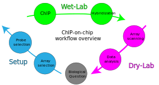
ChIP-on-chip is a technology that combines chromatin immunoprecipitation ('ChIP') with DNA microarray ("chip"). Like regular ChIP, ChIP-on-chip is used to investigate interactions between proteins and DNA in vivo. Specifically, it allows the identification of the cistrome, the sum of binding sites, for DNA-binding proteins on a genome-wide basis. Whole-genome analysis can be performed to determine the locations of binding sites for almost any protein of interest. As the name of the technique suggests, such proteins are generally those operating in the context of chromatin. The most prominent representatives of this class are transcription factors, replication-related proteins, like origin recognition complex protein (ORC), histones, their variants, and histone modifications.

ChIP-sequencing, also known as ChIP-seq, is a method used to analyze protein interactions with DNA. ChIP-seq combines chromatin immunoprecipitation (ChIP) with massively parallel DNA sequencing to identify the binding sites of DNA-associated proteins. It can be used to map global binding sites precisely for any protein of interest. Previously, ChIP-on-chip was the most common technique utilized to study these protein–DNA relations.

Epigenomics is the study of the complete set of epigenetic modifications on the genetic material of a cell, known as the epigenome. The field is analogous to genomics and proteomics, which are the study of the genome and proteome of a cell. Epigenetic modifications are reversible modifications on a cell's DNA or histones that affect gene expression without altering the DNA sequence. Epigenomic maintenance is a continuous process and plays an important role in stability of eukaryotic genomes by taking part in crucial biological mechanisms like DNA repair. Plant flavones are said to be inhibiting epigenomic marks that cause cancers. Two of the most characterized epigenetic modifications are DNA methylation and histone modification. Epigenetic modifications play an important role in gene expression and regulation, and are involved in numerous cellular processes such as in differentiation/development and tumorigenesis. The study of epigenetics on a global level has been made possible only recently through the adaptation of genomic high-throughput assays.

<span class="mw-page-title-main">ChIP-exo</span>

ChIP-exo is a chromatin immunoprecipitation based method for mapping the locations at which a protein of interest binds to the genome. It is a modification of the ChIP-seq protocol, improving the resolution of binding sites from hundreds of base pairs to almost one base pair. It employs the use of exonucleases to degrade strands of the protein-bound DNA in the 5'-3' direction to within a small number of nucleotides of the protein binding site. The nucleotides of the exonuclease-treated ends are determined using some combination of DNA sequencing, microarrays, and PCR. These sequences are then mapped to the genome to identify the locations on the genome at which the protein binds.

H3K27ac is an epigenetic modification to the DNA packaging protein histone H3. It is a mark that indicates acetylation of the lysine residue at N-terminal position 27 of the histone H3 protein.

H3K9me3 is an epigenetic modification to the DNA packaging protein Histone H3. It is a mark that indicates the tri-methylation at the 9th lysine residue of the histone H3 protein and is often associated with heterochromatin.

H3K4me1 is an epigenetic modification to the DNA packaging protein Histone H3. It is a mark that indicates the mono-methylation at the 4th lysine residue of the histone H3 protein and often associated with gene enhancers.

H3K79me2 is an epigenetic modification to the DNA packaging protein Histone H3. It is a mark that indicates the di-methylation at the 79th lysine residue of the histone H3 protein. H3K79me2 is detected in the transcribed regions of active genes.

H4K5ac is an epigenetic modification to the DNA packaging protein histone H4. It is a mark that indicates the acetylation at the 5th lysine residue of the histone H4 protein. H4K5 is the closest lysine residue to the N-terminal tail of histone H4. It is enriched at the transcription start site (TSS) and along gene bodies. Acetylation of histone H4K5 and H4K12ac is enriched at centromeres.

H4K8ac, representing an epigenetic modification to the DNA packaging protein histone H4, is a mark indicating the acetylation at the 8th lysine residue of the histone H4 protein. It has been implicated in the prevalence of malaria.

H4K12ac is an epigenetic modification to the DNA packaging protein histone H4. It is a mark that indicates the acetylation at the 12th lysine residue of the histone H4 protein. H4K12ac is involved in learning and memory. It is possible that restoring this modification could reduce age-related decline in memory.

H3K14ac is an epigenetic modification to the DNA packaging protein Histone H3. It is a mark that indicates the acetylation at the 14th lysine residue of the histone H3 protein.

H3K9ac is an epigenetic modification to the DNA packaging protein Histone H3. It is a mark that indicates the acetylation at the 9th lysine residue of the histone H3 protein.

H3K36me2 is an epigenetic modification to the DNA packaging protein Histone H3. It is a mark that indicates the di-methylation at the 36th lysine residue of the histone H3 protein.

<span class="mw-page-title-main">MNase-seq</span> Sk kasid Youtuber

MNase-seq, short for micrococcal nuclease digestion with deep sequencing, is a molecular biological technique that was first pioneered in 2006 to measure nucleosome occupancy in the C. elegans genome, and was subsequently applied to the human genome in 2008. Though, the term ‘MNase-seq’ had not been coined until a year later, in 2009. Briefly, this technique relies on the use of the non-specific endo-exonuclease micrococcal nuclease, an enzyme derived from the bacteria Staphylococcus aureus, to bind and cleave protein-unbound regions of DNA on chromatin. DNA bound to histones or other chromatin-bound proteins may remain undigested. The uncut DNA is then purified from the proteins and sequenced through one or more of the various Next-Generation sequencing methods.

H3R17me2 is an epigenetic modification to the DNA packaging protein histone H3. It is a mark that indicates the di-methylation at the 17th arginine residue of the histone H3 protein. In epigenetics, arginine methylation of histones H3 and H4 is associated with a more accessible chromatin structure and thus higher levels of transcription. The existence of arginine demethylases that could reverse arginine methylation is controversial.

H3R26me2 is an epigenetic modification to the DNA packaging protein histone H3. It is a mark that indicates the di-methylation at the 26th arginine residue of the histone H3 protein. In epigenetics, arginine methylation of histones H3 and H4 is associated with a more accessible chromatin structure and thus higher levels of transcription. The existence of arginine demethylases that could reverse arginine methylation is controversial.

H3R8me2 is an epigenetic modification to the DNA packaging protein histone H3. It is a mark that indicates the di-methylation at the 8th arginine residue of the histone H3 protein. In epigenetics, arginine methylation of histones H3 and H4 is associated with a more accessible chromatin structure and thus higher levels of transcription. The existence of arginine demethylases that could reverse arginine methylation is controversial.

H3R2me2 is an epigenetic modification to the DNA packaging protein histone H3. It is a mark that indicates the di-methylation at the 2nd arginine residue of the histone H3 protein. In epigenetics, arginine methylation of histones H3 and H4 is associated with a more accessible chromatin structure and thus higher levels of transcription. The existence of arginine demethylases that could reverse arginine methylation is controversial.

H3T45P is an epigenetic modification to the DNA packaging protein histone H3. It is a mark that indicates the phosphorylation the 45th threonine residue of the histone H3 protein.

References

  1. Collas, Philippe. (January 2010). "The Current State of Chromatin Immunoprecipitation". Molecular Biotechnology. 45 (1): 87–100. doi:10.1007/s12033-009-9239-8. PMID   20077036. S2CID   24225210.
  2. 1 2 Rosenfeld, John M; Cooke, Tracy; Li, Zirong; Saito, Kan; Taganov, Konstantin; Thyagarajan, Bhaskar; Solache, Alejandra (March 2013). "Systematic optimization of parameters involved in preparation of chromatin and chromatin immunoprecipitation (ChIP) workflow". Epigenetics & Chromatin. 6 (S1): P122, 1756–8935–6-S1-P122. doi:10.1186/1756-8935-6-S1-P122. ISSN   1756-8935. PMC   3620580 .
  3. Jackson, Vaughn (November 1978). "Studies on histone organization in the nucleosome using formaldehyde as a reversible cross-linking agent". Cell . 15 (3): 945–54. doi:10.1016/0092-8674(78)90278-7. PMID   569554. S2CID   25169609.
  4. Gilmour DS, Lis JT (August 1985). "In vivo interactions of RNA polymerase II with genes of Drosophila melanogaster". Molecular and Cellular Biology . 5 (8): 2009–18. doi:10.1128/mcb.5.8.2009. PMC   366919 . PMID   3018544.
  5. Bauer UM, Daujat S, Nielsen SJ, Nightingale K, Kouzarides T (January 2002). "Methylation at arginine 17 of histone H3 is linked to gene activation". EMBO Reports . 3 (1): 39–44. doi:10.1093/embo-reports/kvf013. PMC   1083932 . PMID   11751582.
  6. Beynon, Amy L.; Parkes, Lindsay J.; Turner, Matthew L.; Knight, Steve; Conlan, Steve; Francis, Lewis; Stocks, Ben (September 2014). "Chromatrap® 96: a new solid-state platform for high-throughput ChIP". Nature Methods. 11 (9): i–ii. doi: 10.1038/nmeth.f.372 . ISSN   1548-7091.
  7. 1 2 "Chromatrap".Revolutionary solid state platform for chromatin immunopreciptation.
  8. Viens A; et al. (2004). "Use of protein biotinylation in vivo for chromatin immunoprecipitation". Analytical Biochemistry. 325 (1): 68–76. doi:10.1016/j.ab.2003.10.015. PMID   14715286.
  9. Gilmour DS, Lis JT (1984). "Detecting protein-DNA interactions in vivo: distribution of RNA polymerase on specific bacterial genes". Proc Natl Acad Sci U S A. 81 (14): 4275–9. Bibcode:1984PNAS...81.4275G. doi: 10.1073/pnas.81.14.4275 . PMC   345570 . PMID   6379641.
  10. Gilmour DS, Lis JT (August 1985). "In vivo interactions of RNA polymerase II with genes of Drosophila melanogaster". Mol. Cell. Biol. 5 (8): 2009–18. doi:10.1128/mcb.5.8.2009. PMC   366919 . PMID   3018544.
  11. Varshavsky A (December 2008). "Discovery of cellular regulation by protein degradation". Journal of Biological Chemistry . 283 (50): 34469–89. doi: 10.1074/jbc.X800009200 . PMC   3259866 . PMID   18708349.
  12. Solomon, Mark J; Larsen Pamela L; Varshavsky, Alexander. (June 1988). "Mapping protein-DNA interactions in vivo with formaldehyde: evidence that histone H4 is retained on a highly transcribed gene". Cell . 53 (6): 937–47. doi:10.1016/S0092-8674(88)90469-2. PMID   2454748. S2CID   11169130.
  13. Orlando V (March 2000). "Mapping chromosomal proteins in vivo by formaldehyde-crosslinked-chromatin immunoprecipitation". Trends in Biochemical Sciences . 25 (3): 99–104. doi:10.1016/S0968-0004(99)01535-2. PMID   10694875.
  14. Hebbes, Tim R; Thorne, Alan W; Crane-Robinson C (May 1988). "A direct link between core histone acetylation and transcriptionally active chromatin". The EMBO Journal . 7 (5): 1395–402. doi:10.1002/j.1460-2075.1988.tb02956.x. PMC   458389 . PMID   3409869.
  15. O'Neill, Laura P; Turner, Bryan M (September 2003). "Immunoprecipitation of native chromatin: NChIP". Methods. 31 (1): 76–82. doi:10.1016/S1046-2023(03)00090-2. PMID   12893176.
  16. O'Neill, Laura P; VerMilyea, Matthew D; Turner, Bryan M (July 2006). "Epigenetic characterization of the early embryo with a chromatin immunoprecipitation protocol applicable to small cell populations". Nature Genetics . 38 (7): 835–41. doi:10.1038/ng1820. PMID   16767102. S2CID   28311996.
  17. Nelson, Joel D; Denisenko, Oleg; Sova, Pavel; Bomsztyk, Karol (2006). "Fast chromatin immunoprecipitation assay". Nucleic Acids Research . 34 (1): e2. doi:10.1093/nar/gnj004. PMC   1325209 . PMID   16397291.
  18. Nelson, Joel D; Denisenko, Oleg; Bomsztyk, Karol (2006). "Protocol for the fast chromatin immunoprecipitation (ChIP) method". Nature Protocols . 1 (1): 179–85. doi:10.1038/nprot.2006.27. PMID   17406230. S2CID   20577722.
  19. Nelson J, Denisenko O, Bomsztyk K (2009). "The fast chromatin immunoprecipitation method". Chromatin Immunoprecipitation Assays. Methods in Molecular Biology . Vol. 567. pp. 45–57. doi:10.1007/978-1-60327-414-2_3. ISBN   978-1-60327-413-5. PMID   19588084.
  20. Dahl, John Arne; Collas, Philippe (April 2007). "Q2ChIP, a quick and quantitative chromatin immunoprecipitation assay, unravels epigenetic dynamics of developmentally regulated genes in human carcinoma cells". Stem Cells. 25 (4): 1037–46. doi: 10.1634/stemcells.2006-0430 . PMID   17272500.
  21. Dahl, John Arne; Collas, Philippe (2008). "A rapid micro chromatin immunoprecipitation assay (microChIP)". Nature Protocols . 3 (6): 1032–45. doi:10.1038/nprot.2008.68. PMID   18536650. S2CID   29529307.
  22. Dahl, John Arne; Collas, Philippe (2009). MicroChIP: chromatin immunoprecipitation for small cell numbers. Methods in Molecular Biology . Vol. 567. pp. 59–74. doi:10.1007/978-1-60327-414-2_4. ISBN   978-1-60327-413-5. PMID   19588085.
  23. Flanagin, Steve; Nelson, Joel D; Castner, David G; Denisenko, Oleg; Bomsztyk, Karol (February 2008). "Microplate-based chromatin immunoprecipitation method, Matrix ChIP: a platform to study signaling of complex genomic events". Nucleic Acids Research . 36 (3): e17. doi:10.1093/nar/gkn001. PMC   2241906 . PMID   18203739.
  24. Fanelli, Mirco; Amatori, Stefano; Barozzi, Iros; Soncini, Matias; Zuffo, Roberto Dal; Bucci, Gabriele; Capra, Maria; Quarto, Micaela; Dellino, Gaetano Ivan (2010-12-14). "Pathology tissue–chromatin immunoprecipitation, coupled with high-throughput sequencing, allows the epigenetic profiling of patient samples". Proceedings of the National Academy of Sciences. 107 (50): 21535–21540. Bibcode:2010PNAS..10721535F. doi: 10.1073/pnas.1007647107 . ISSN   0027-8424. PMC   3003125 . PMID   21106756.
  25. Fullwood, Melissa J; Han, Yuyuan; Wei, Chia-Lin; Ruan, Xiaoan; Ruan, Yijun (January 2010). Chromatin interaction analysis using paired-end tag sequencing. Current Protocols in Molecular Biology . Vol. Chapter 21. pp. Unit 21.15.1–25. doi:10.1002/0471142727.mb2115s89. ISBN   978-0471142720. PMC   6924956 . PMID   20069536.
  26. Li, Guoliang; Fullwood, Melissa J; Xu, Han; Mulawadi, Fabianus Hendriyan; Velkov, Stoyan; Vega, Vinsensius; Ariyaratne, Pramila Nuwantha; Mohamed, Yusoff Bin; Ooi, Hong-Sain; Tennakoon, Chandana; Wei, Chia-Lin; Ruan, Yijun; Sung, Wing-Kin (February 2010). "ChIA-PET tool for comprehensive chromatin interaction analysis with paired-end tag sequencing". Genome Biology . 11 (2): R22. doi:10.1186/gb-2010-11-2-r22. PMC   2872882 . PMID   20181287.
  27. "ChIA-PET: Novel Method For 3-D Whole Genome Mapping Research". ScienceDaily. Agency for Science, Technology and Research (A*STAR), Singapore. 2009-11-08. Retrieved 14 March 2010.