Proteinase K

Last updated
Proteinase K
PDB 1pek EBI.jpg
Structure of Proteinase K. [1]
Identifiers
EC no. 3.4.21.64
Databases
IntEnz IntEnz view
BRENDA BRENDA entry
ExPASy NiceZyme view
KEGG KEGG entry
MetaCyc metabolic pathway
PRIAM profile
PDB structures RCSB PDB PDBe PDBsum
Search
PMC articles
PubMed articles
NCBI proteins
Proteinase K
Identifiers
Organism Engyodontium album
SymbolPROK
UniProt P06873
Search for
Structures Swiss-model
Domains InterPro

In molecular biology, Proteinase K (EC 3.4.21.64, protease K, endopeptidase K, Tritirachium alkaline proteinase, Tritirachium album serine proteinase, Tritirachium album proteinase K) is a broad-spectrum serine protease. [2] [3] [4] The enzyme was discovered in 1974 in extracts of the fungus Parengyodontium album (formerly Engyodontium album or Tritirachium album). [5] Proteinase K is able to digest hair (keratin), hence, the name "Proteinase K". The predominant site of cleavage is the peptide bond adjacent to the carboxyl group of aliphatic and aromatic amino acids with blocked alpha amino groups. It is commonly used for its broad specificity. This enzyme belongs to Peptidase family S8 (subtilisin). The molecular weight of Proteinase K is 28,900 daltons (28.9 kDa).

Contents

Enzyme activity

Activated by calcium, the enzyme digests proteins preferentially after hydrophobic amino acids (aliphatic, aromatic and other hydrophobic amino acids). Although calcium ions do not affect the enzyme activity, they do contribute to its stability. Proteins will be completely digested if the incubation time is long and the protease concentration high enough. Upon removal of the calcium ions, the stability of the enzyme is reduced, but the proteolytic activity remains. [6] Proteinase K has two binding sites for Ca2+, which are located close to the active center, but are not directly involved in the catalytic mechanism. The residual activity is sufficient to digest proteins, which usually contaminate nucleic acid preparations. Therefore, the digestion with Proteinase K for the purification of nucleic acids is usually performed in the presence of EDTA (inhibition of metal-ion dependent enzymes such as nucleases).

Proteinase K is also stable over a wide pH range (4–12), with a pH optimum of pH 8.0. [5] An elevation of the reaction temperature from 37 °C to 50–60 °C may increase the activity several times, like the addition of 0.5–1% sodium dodecyl sulfate (SDS) or Guanidinium chloride (3 M), Guanidinium thiocyanate (1 M) and urea (4 M) [ disputed (for: no source cited for temperature) ]. The above-mentioned conditions enhance proteinase K activity by making its substrate cleavage sites more accessible. Temperatures above 65 °C, trichloroacetic acid (TCA) or the serine protease-inhibitors AEBSF, PMSF or DFP inhibit the activity. Proteinase K will not be inhibited by Guanidinium chloride, Guanidinium thiocyanate, urea, Sarkosyl, Triton X-100, Tween 20, SDS, citrate, iodoacetic acid, EDTA or by other serine protease inhibitors like Nα-Tosyl-Lys Chloromethyl Ketone (TLCK) and Nα-Tosyl-Phe Chloromethyl Ketone (TPCK)[ citation needed ].

Protease K activity in commonly used buffers [7]

Buffer (pH = 8.0, 50 °C, 1.25 µg/mL protease K, 15 min incubation)Proteinase K activity (%)
30 mM Tris·Cl100
30 mM Tris·Cl; 30 mM EDTA; 5% Tween 20; 0.5% Triton X-100; 800 mM GuHCl 313
36 mM Tris·Cl; 36 mM EDTA; 5% Tween 20; 0.36% Triton X-100; 735 mM GuHCl 301
10 mM Tris·Cl; 25 mM EDTA; 100 mM NaCl; 0.5% SDS128
10 mM Tris·Cl; 100 mM EDTA; 20 mM NaCl; 1% Sarkosyl 74
10 mM Tris·Cl; 50 mM KCl; 1.5 mM MgCl2; 0.45% Tween 20; 0.5% Triton X-100 106
10 mM Tris·Cl; 100 mM EDTA; 0.5% SDS120
30 mM Tris·Cl; 10 mM EDTA; 1% SDS203

Applications

Proteinase K is commonly used in molecular biology to digest protein and remove contamination from preparations of nucleic acid. Addition of Proteinase K to nucleic acid preparations rapidly inactivates nucleases that might otherwise degrade the DNA or RNA during purification. It is highly suited to this application since the enzyme is active in the presence of chemicals that denature proteins, such as SDS and urea, chelating agents such as EDTA, sulfhydryl reagents, as well as trypsin or chymotrypsin inhibitors. Proteinase K is used for the destruction of proteins in cell lysates (tissue, cell culture cells) and for the release of nucleic acids, since it very effectively inactivates DNases and RNases. Some examples for applications: Proteinase K is very useful in the isolation of highly native, undamaged DNAs or RNAs, since most microbial or mammalian DNases and RNases are rapidly inactivated by the enzyme, particularly in the presence of 0.5–1% SDS.

The enzyme's activity towards native proteins is stimulated by denaturants such as SDS. In contrast, when measured using peptide substrates, denaturants inhibit the enzyme. The reason for this result is that the denaturing agents unfold the protein substrates and make them more accessible to the protease. [8]

Inhibitors

Proteinase K has two disulfide bonds, [9] but it exhibits higher proteolytic activity in the presence of reducing agents (e.g. 5 mM DTT), [10] suggesting that the presumed reduction of its own disulfide bonds does not lead to its irreversible inactivation. Proteinase K is inhibited by serine protease inhibitors such as phenylmethylsulfonyl fluoride (PMSF), diisopropylfluorophosphate (DFP), or 4-(2-aminoethyl)benzenesulfonyl fluoride (AEBSF). Proteinase K activity is unaffected by the sulfhydryl modifying reagents: para-chloromercuribenzoic acid (PCMB), N-alpha-tosyl-L-lysyl-chloromethyl-ketone (TLCK), or N-alpha-Tosyl-l-phenylalanine Chloromethyl Ketone (TPCK), [10] although presumably if these reagents were included alongside disulfide reducing reagents which exposed the typically-unavailable Proteinase K thiols, it may then become inhibited.

Related Research Articles

<span class="mw-page-title-main">Denaturation (biochemistry)</span> Loss of structure in proteins and nucleic acids due to external stress

In biochemistry, denaturation is a process in which proteins or nucleic acids lose the quaternary structure, tertiary structure, and secondary structure which is present in their native state, by application of some external stress or compound such as a strong acid or base, a concentrated inorganic salt, an organic solvent, agitation and radiation or heat. If proteins in a living cell are denatured, this results in disruption of cell activity and possibly cell death. Protein denaturation is also a consequence of cell death. Denatured proteins can exhibit a wide range of characteristics, from conformational change and loss of solubility to aggregation due to the exposure of hydrophobic groups. The loss of solubility as a result of denaturation is called coagulation. Denatured proteins lose their 3D structure and therefore cannot function.

<span class="mw-page-title-main">Proteolysis</span> Breakdown of proteins into smaller polypeptides or amino acids

Proteolysis is the breakdown of proteins into smaller polypeptides or amino acids. Uncatalysed, the hydrolysis of peptide bonds is extremely slow, taking hundreds of years. Proteolysis is typically catalysed by cellular enzymes called proteases, but may also occur by intra-molecular digestion.

<span class="mw-page-title-main">Trypsin</span> Family of digestive enzymes

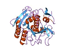
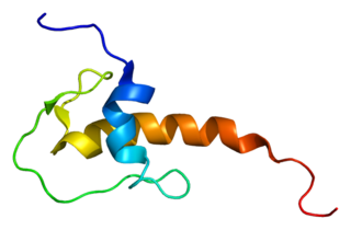
Trypsin is an enzyme in the first section of the small intestine that starts the digestion of protein molecules by cutting long chains of amino acids into smaller pieces. It is a serine protease from the PA clan superfamily, found in the digestive system of many vertebrates, where it hydrolyzes proteins. Trypsin is formed in the small intestine when its proenzyme form, the trypsinogen produced by the pancreas, is activated. Trypsin cuts peptide chains mainly at the carboxyl side of the amino acids lysine or arginine. It is used for numerous biotechnological processes. The process is commonly referred to as trypsinogen proteolysis or trypsinization, and proteins that have been digested/treated with trypsin are said to have been trypsinized. Trypsin was discovered in 1876 by Wilhelm Kühne and was named from the Ancient Greek word for rubbing since it was first isolated by rubbing the pancreas with glycerin.

<span class="mw-page-title-main">Protease</span> Enzyme that cleaves other proteins into smaller peptides

A protease is an enzyme that catalyzes proteolysis, breaking down proteins into smaller polypeptides or single amino acids, and spurring the formation of new protein products. They do this by cleaving the peptide bonds within proteins by hydrolysis, a reaction where water breaks bonds. Proteases are involved in numerous biological pathways, including digestion of ingested proteins, protein catabolism, and cell signaling.

In biology and biochemistry, protease inhibitors, or antiproteases, are molecules that inhibit the function of proteases. Many naturally occurring protease inhibitors are proteins.

<span class="mw-page-title-main">Serine protease</span> Class of enzymes

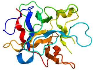
Serine proteases are enzymes that cleave peptide bonds in proteins. Serine serves as the nucleophilic amino acid at the (enzyme's) active site. They are found ubiquitously in both eukaryotes and prokaryotes. Serine proteases fall into two broad categories based on their structure: chymotrypsin-like (trypsin-like) or subtilisin-like.

<span class="mw-page-title-main">Tosyl phenylalanyl chloromethyl ketone</span> Chemical compound

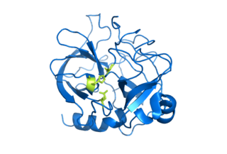
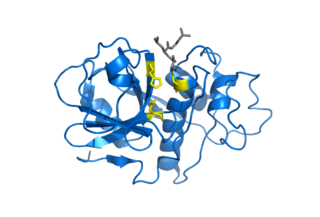
Tosyl phenylalanyl chloromethyl ketone (TPCK) is a protease inhibitor. Its structural formula is 1-chloro-3-tosylamido-4-phenyl-2-butanone.

<span class="mw-page-title-main">Papain</span> Widely used enzyme extracted from papayas

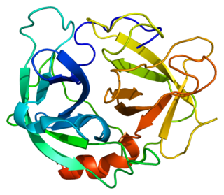
Papain, also known as papaya proteinase I, is a cysteine protease enzyme present in papaya and mountain papaya. It is the namesake member of the papain-like protease family.

<span class="mw-page-title-main">Cysteine protease</span> Class of enzymes

Cysteine proteases, also known as thiol proteases, are hydrolase enzymes that degrade proteins. These proteases share a common catalytic mechanism that involves a nucleophilic cysteine thiol in a catalytic triad or dyad.

Protein metabolism denotes the various biochemical processes responsible for the synthesis of proteins and amino acids (anabolism), and the breakdown of proteins by catabolism.

<span class="mw-page-title-main">Thermolysin</span>

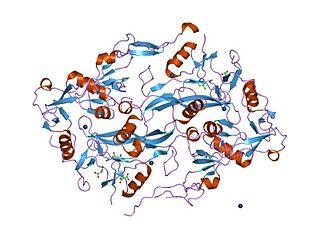
Thermolysin is a thermostable neutral metalloproteinase enzyme produced by the Gram-positive bacteria Bacillus thermoproteolyticus. It requires one zinc ion for enzyme activity and four calcium ions for structural stability. Thermolysin specifically catalyzes the hydrolysis of peptide bonds containing hydrophobic amino acids. However thermolysin is also widely used for peptide bond formation through the reverse reaction of hydrolysis. Thermolysin is the most stable member of a family of metalloproteinases produced by various Bacillus species. These enzymes are also termed 'neutral' proteinases or thermolysin -like proteinases (TLPs).

<span class="mw-page-title-main">Cathepsin L1</span> Protein-coding gene in the species Homo sapiens

Cathepsin L1 is a protein that in humans is encoded by the CTSL1 gene. The protein is a cysteine cathepsin, a lysosomal cysteine protease that plays a major role in intracellular protein catabolism.

<span class="mw-page-title-main">LEKTI</span> Protein-coding gene in the species Homo sapiens

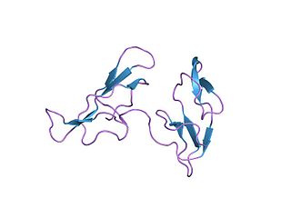
Lympho-epithelial Kazal-type-related inhibitor (LEKTI) also known as serine protease inhibitor Kazal-type 5 (SPINK5) is a protein that in humans is encoded by the SPINK5 gene.

<span class="mw-page-title-main">Kunitz STI protease inhibitor</span>

Kunitz soybean trypsin inhibitor is a type of protein contained in legume seeds which functions as a protease inhibitor. Kunitz-type Soybean Trypsin Inhibitors are usually specific for either trypsin or chymotrypsin. They are thought to protect seeds against consumption by animal predators.

Nepenthesin is an aspartic protease of plant origin that has so far been identified in the pitcher secretions of Nepenthes and in the leaves of Drosera peltata. It is similar to pepsin, but differs in that it also cleaves on either side of Asp residues and at Lys┼Arg. While more pH and temperature stable than porcine pepsin A, it is considerably less stable in urea or guanidine hydrochloride. It is the only known protein with such a stability profile.

<span class="mw-page-title-main">Bowman–Birk protease inhibitor</span>

In molecular biology, the Bowman–Birk protease inhibitor family of proteins consists of eukaryotic proteinase inhibitors, belonging to MEROPS inhibitor family I12, clan IF. They mainly inhibit serine peptidases of the S1 family, but also inhibit S3 peptidases.

<span class="mw-page-title-main">Kazal domain</span>

The Kazal domain is an evolutionary conserved protein domain usually indicative of serine protease inhibitors. However, kazal-like domains are also seen in the extracellular part of agrins, which are not known to be protease inhibitors.

Serratia marcescens nuclease is an enzyme. This enzyme catalyses the following chemical reaction

<span class="mw-page-title-main">Zingibain</span>

Zingibain, zingipain, or ginger protease is a cysteine protease enzyme found in ginger rhizomes. It catalyses the preferential cleavage of peptides with a proline residue at the P2 position. It has two distinct forms, ginger protease I (GP-I) and ginger protease II (GP-II).

<span class="mw-page-title-main">Scytalidopepsin B</span>

Scytalidocarboxyl peptidase B, also known as Scytalidoglutamic peptidase and Scytalidopepsin B is a proteolytic enzyme. It was previously thought to be an aspartic protease, but determination of its molecular structure showed it to belong a novel group of proteases, glutamic protease.

References

  1. Betzel C, Singh TP, Visanji M, Peters K, Fittkau S, Saenger W, Wilson KS (July 1993). "Structure of the complex of proteinase K with a substrate analogue hexapeptide inhibitor at 2.2-A resolution". J. Biol. Chem. 268 (21): 15854–8. doi: 10.1016/S0021-9258(18)82332-8 . PMID   8340410.
  2. Morihara K, Tsuzuki H (1975). "Specificity of proteinase K from Tritirachium album Limber for synthetic peptides". Agric. Biol. Chem. 39 (7): 1489–1492. doi: 10.1271/bbb1961.39.1489 .
  3. Kraus E, Kiltz HH, Femfert UF (February 1976). "The specificity of proteinase K against oxidized insulin B chain". Hoppe-Seyler's Z. Physiol. Chem. 357 (2): 233–7. PMID   943367.
  4. Jany KD, Lederer G, Mayer B (1986). "Amino acid sequence of proteinase K from the mold Tritirachium album Limber". FEBS Lett. 199 (2): 139–144. doi: 10.1016/0014-5793(86)80467-7 . S2CID   85577597.
  5. 1 2 Ebeling W, Hennrich N, Klockow M, Metz H, Orth HD, Lang H (August 1974). "Proteinase K from Tritirachium album Limber". Eur. J. Biochem. 47 (1): 91–7. doi: 10.1111/j.1432-1033.1974.tb03671.x . PMID   4373242.
  6. Müller A, Hinrichs W, Wolf WM, Saenger W (September 1994). "Crystal structure of calcium-free proteinase K at 1.5-A resolution". J. Biol. Chem. 269 (37): 23108–11. doi:10.2210/pdb2pkc/pdb. PMID   8083213.
  7. Ag-Scientific. "Frequently asked questions about Proteinase K".
  8. Hilz H, Wiegers U, Adamietz P (1975). "Stimulation of Proteinase K action by denaturing agents: application to the isolation of nucleic acids and the degradation of 'masked' proteins". European Journal of Biochemistry. 56 (1): 103–108. doi: 10.1111/j.1432-1033.1975.tb02211.x . PMID   1236799.
  9. "PROK - Proteinase K precursor - Parengyodontium album". Uniprot. 2019-12-11. Retrieved 2020-02-07.
  10. 1 2 "Novagen Proteinase K protocol" (PDF). Sigma Aldrich. 2019-12-11. Retrieved 2020-02-07.