Direct fluorescent antibody

Last updated
Direct FA stained mouse brain impression smear reveals the presence of the bacterium Chlamydia psittaci. 400X. Chlamydophila psittaci FA stain.jpg
Direct FA stained mouse brain impression smear reveals the presence of the bacterium Chlamydia psittaci . 400X.

A direct fluorescent antibody (DFA or dFA), also known as "direct immunofluorescence", [1] is an antibody that has been tagged in a direct fluorescent antibody test. Its name derives from the fact that it directly tests the presence of an antigen with the tagged antibody, unlike western blotting, which uses an indirect method of detection, where the primary antibody binds the target antigen, with a secondary antibody directed against the primary, and a tag attached to the secondary antibody.

Contents

Commercial DFA testing kits are available, which contain fluorescently labelled antibodies, designed to specifically target unique antigens present in the bacteria or virus, but not present in mammals (Eukaryotes). This technique can be used to quickly determine if a subject has a specific viral or bacterial infection.

In the case of respiratory viruses, many of which have similar broad symptoms, detection can be carried out using nasal wash samples from the subject with the suspected infection. Although shedding cells in the respiratory tract can be obtained, it is often in low numbers, and so an alternative method can be adopted where compatible cell culture can be exposed to infected nasal wash samples, so if the virus is present it can be grown up to a larger quantity, which can then give a clearer positive or negative reading.

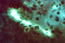
As with all types of fluorescence microscopy, the correct absorption wavelength needs to be determined in order to excite the fluorophore tag attached to the antibody, and detect the fluorescence given off, which indicates which cells are positive for the presence of the virus or bacteria being detected.

Direct immunofluorescence can be used to detect deposits of immunoglobulins and complement proteins in biopsies of skin, kidney and other organs. Their presence is indicative of an autoimmune disease. When skin not exposed to the sun is tested, a positive direct IF (the so-called Lupus band test) is an evidence of systemic lupus erythematosus. [2] Direct fluorescent antibody can also be used to detect parasitic infections, as was pioneered by Sadun, et al. (1960).

See also

Related Research Articles

ELISA Method to detect an antigen using an antibody and enzyme

The enzyme-linked immunosorbent assay (ELISA) is a commonly used analytical biochemistry assay, first described by Engvall and Perlmann in 1971. The assay uses a solid-phase type of enzyme immunoassay (EIA) to detect the presence of a ligand in a liquid sample using antibodies directed against the protein to be measured. ELISA has been used as a diagnostic tool in medicine, plant pathology, and biotechnology, as well as a quality control check in various industries.

Anti-nuclear antibody Autoantibody that binds to contents of the cell nucleus

Antinuclear antibodies are autoantibodies that bind to contents of the cell nucleus. In normal individuals, the immune system produces antibodies to foreign proteins (antigens) but not to human proteins (autoantigens). In some individuals, antibodies to human antigens are produced.

Diagnosis of HIV/AIDS

HIV tests are used to detect the presence of the human immunodeficiency virus (HIV), the virus that causes acquired immunodeficiency syndrome (AIDS), in serum, saliva, or urine. Such tests may detect antibodies, antigens, or RNA.

Venereal Disease Research Laboratory test

The Venereal Disease Research Laboratory test (VDRL) is a blood test for syphilis and related non-venereal treponematoses that was developed by the eponymous lab. The VDRL test is used to screen for syphilis, whereas other, more specific tests are used to diagnose the disease.

Respiratory syncytial virus

Respiratory syncytial virus (RSV), also called human respiratory syncytial virus (hRSV) and human orthopneumovirus, is a very common, contagious virus that causes infections of the respiratory tract. It is a negative-sense, single-stranded RNA virus, and its name is derived from the large syncytium that form when infected cells fuse together.

Immunostaining

In biochemistry, immunostaining is any use of an antibody-based method to detect a specific protein in a sample. The term "immunostaining" was originally used to refer to the immunohistochemical staining of tissue sections, as first described by Albert Coons in 1941. However, immunostaining now encompasses a broad range of techniques used in histology, cell biology, and molecular biology that use antibody-based staining methods.

Immunofluorescence

Immunofluorescence is a technique used for light microscopy with a fluorescence microscope and is used primarily on microbiological samples. This technique uses the specificity of antibodies to their antigen to target fluorescent dyes to specific biomolecule targets within a cell, and therefore allows visualization of the distribution of the target molecule through the sample. The specific region an antibody recognizes on an antigen is called an epitope. There have been efforts in epitope mapping since many antibodies can bind the same epitope and levels of binding between antibodies that recognize the same epitope can vary. Additionally, the binding of the fluorophore to the antibody itself cannot interfere with the immunological specificity of the antibody or the binding capacity of its antigen. Immunofluorescence is a widely used example of immunostaining and is a specific example of immunohistochemistry. This technique primarily makes use of fluorophores to visualise the location of the antibodies.

In immunology, seroconversion is the time period during which a specific antibody develops and becomes detectable in the blood. After seroconversion has occurred, the antibodies can be detected in blood tests for the disease. During an infection or immunization, antigens enter the blood, and the immune system begins to produce antibodies in response. Before seroconversion, the antigen itself may or may not be detectable, but the antibody is, by definition, absent. During seroconversion, the antibody is present but not yet detectable.

A Coombs test, also known as antiglobulin test (AGT) is either of two blood tests used in immunohematology. They are the direct and indirect Coombs tests. The direct Coombs test detects antibodies that are stuck to the surface of the red blood cells. Since these antibodies sometimes destroy red blood cells, a person can be anemic and this test can help clarify the condition. The indirect Coombs detects antibodies that are floating freely in the blood. These antibodies could act against certain red blood cells and the test can be done to diagnose reactions to a blood transfusion.

Heterophile antibodies are antibodies induced by external antigens.

Feline viral rhinotracheitis

Feline viral rhinotracheitis (FVR) is an upper respiratory or pulmonary infection of cats caused by Felid alphaherpesvirus 1 (FeHV-1), of the family Herpesviridae. It is also commonly referred to as feline influenza, feline coryza, and feline pneumonia but, as these terms describe other very distinct collections of respiratory symptoms, they are misnomers for the condition. Viral respiratory diseases in cats can be serious, especially in catteries and kennels. Causing one-half of the respiratory diseases in cats, FVR is the most important of these diseases and is found worldwide. The other important cause of feline respiratory disease is feline calicivirus.

Extractable Nuclear Antigens (ENAs) are over 100 different soluble cytoplasmic and nuclear antigens. They are known as “extractable” because they can be removed from cell nuclei using saline and represent six main proteins: Ro, La, Sm, RNP, Scl-70, Jo1. Most ENAs are part of spliceosomes or nucleosomes complexes and are a type of small nuclear ribonucleoprotein (snRNPS). The location in the nucleus and association with spliceosomes or nucleosomes results in these ENAs being associated with additional RNA and proteins such as polymerases. This quality of ENAs often makes it difficult to purify and quantify their presence for clinical use.

Immunocytochemistry

Immunocytochemistry (ICC) is a common laboratory technique that is used to anatomically visualize the localization of a specific protein or antigen in cells by use of a specific primary antibody that binds to it. The primary antibody allows visualization of the protein under a fluorescence microscope when it is bound by a secondary antibody that has a conjugated fluorophore. ICC allows researchers to evaluate whether or not cells in a particular sample express the antigen in question. In cases where an immunopositive signal is found, ICC also allows researchers to determine which sub-cellular compartments are expressing the antigen.

In the diagnostic laboratory virus infections can be confirmed by a multitude of methods. Diagnostic virology has changed rapidly due to the advent of molecular techniques and increased clinical sensitivity of serological assays.

Heterophile antibody test

The mononuclear spot test or monospot test, a form of the heterophile antibody test, is a rapid test for infectious mononucleosis due to Epstein–Barr virus (EBV). It is an improvement on the Paul–Bunnell test. The test is specific for heterophile antibodies produced by the human immune system in response to EBV infection. Commercially available test kits are 70–92% sensitive and 96–100% specific, with a lower sensitivity in the first two weeks after clinical symptoms begin.

Anti-dsDNA antibodies Group of anti-nuclear antibodies

Anti-double stranded DNA (Anti-dsDNA) antibodies are a group of anti-nuclear antibodies (ANA) the target antigen of which is double stranded DNA. Blood tests such as enzyme-linked immunosorbent assay (ELISA) and immunofluorescence are routinely performed to detect anti-dsDNA antibodies in diagnostic laboratories. They are highly diagnostic of systemic lupus erythematosus (SLE) and are implicated in the pathogenesis of lupus nephritis.

Lupus Human autoimmune disease

Lupus, technically known as systemic lupus erythematosus (SLE), is an autoimmune disease in which the body's immune system mistakenly attacks healthy tissue in many parts of the body. Symptoms vary between people and may be mild to severe. Common symptoms include painful and swollen joints, fever, chest pain, hair loss, mouth ulcers, swollen lymph nodes, feeling tired, and a red rash which is most commonly on the face. Often there are periods of illness, called flares, and periods of remission during which there are few symptoms.

Anti-histone antibodies are autoantibodies that are a subset of the anti-nuclear antibody family, which specifically target histone protein subunits or histone complexes. They were first reported by Henry Kunkel, H.R. Holman, and H.R.G. Dreicher in their studies of cellular causes of lupus erythematosus in 1959–60. Today, anti-histone antibodies are still used as a marker for systemic lupus erythematosus, but are also implicated in other autoimmune diseases like Sjogren's Syndrome, dermatomyositis, or rheumatoid arthritis. Anti-histone antibodies can be used as a marker for drug-induced lupus.

Anti-SSA/Ro autoantibodies

Anti-SSA autoantibodies are a type of anti-nuclear autoantibodies that are associated with many autoimmune diseases, such as systemic lupus erythematosus (SLE), SS/SLE overlap syndrome, subacute cutaneous lupus erythematosus (SCLE), neonatal lupus and primary biliary cirrhosis. They are often present in Sjögren's syndrome (SS). Additionally, Anti-Ro/SSA can be found in other autoimmune diseases such as systemic sclerosis (SSc), polymyositis/dermatomyositis (PM/DM), rheumatoid arthritis (RA), and mixed connective tissue disease (MCTD), and are also associated with heart arrhythmia.

<i>Crithidia luciliae</i> Species of parasitic flagellate protist in the Kinetoplastea class

Crithidia luciliae is a flagellate parasite that uses the housefly, Musca domestica, as a host. As part of the family of Trypanosomatidae, it is characterised by the presence of a kinetoplast, a complex network of interlocking circular double-stranded DNA (dsDNA) molecules. The presence of the kinetoplast makes this organism important in the diagnosis of systemic lupus erythamatosus (SLE). By using C. luciliae as a substrate for immunofluorescence, the organelle can be used to detect anti-dsDNA antibodies, a common feature of the disease.

References

  1. Pober, Jordan S.; Abbas, Abul K.; Lichtman, Andrew H. (2000). Cellular and molecular immunology. Philadelphia: Saunders. ISBN   0-7216-8233-2.
  2. Ther Clin Risk Manag. 2011; 7: 27–32. The lupus band test in systemic lupus erythematosus patients. Adam Reich, Katarzyna Marcinow, and Rafal Bialynicki-Birula