In vitro toxicology

Last updated

In vitro toxicity testing is the scientific analysis of the toxic effects of chemical substances on cultured bacteria or mammalian cells. [1] In vitro (literally 'in glass') testing methods are employed primarily to identify potentially hazardous chemicals and/or to confirm the lack of certain toxic properties in the early stages of the development of potentially useful new substances such as therapeutic drugs, agricultural chemicals and food additives.

Contents

In vitro assays for xenobiotic toxicity are recently carefully considered by key government agencies (e.g., EPA; NIEHS/NTP; FDA), to better assess human risks. There are substantial activities in using in vitro systems to advance mechanistic understanding of toxicant activities, and the use of human cells and tissue to define human-specific toxic effects. [2]

Improvement over animal testing

Most toxicologists believe that in vitro toxicity testing methods can be more useful, more time and cost-effective than toxicology studies in living animals [3] (which are termed in vivo or "in life" methods). However, the extrapolation from in vitro to in vivo requires some careful consideration and is an active research area.

Due to regulatory constraints and ethical considerations, the quest for alternatives to animal testing has gained a new momentum. In many cases the in vitro tests are better than animal tests because they can be used to develop safer products. [4]

The United States Environmental Protection Agency studied 1,065 chemical and drug substances in their ToxCast program (part of the CompTox Chemicals Dashboard) using in silica modelling and a human pluripotent stem cell-based assay to predict in vivo developmental intoxicants based on changes in cellular metabolism following chemical exposure. Major findings from the analysis of this ToxCast_STM dataset published in 2020 include: (1) 19% of 1065 chemicals yielded a prediction of developmental toxicity, (2) assay performance reached 79%–82% accuracy with high specificity (> 84%) but modest sensitivity (< 67%) when compared with in vivo animal models of human prenatal developmental toxicity, (3) sensitivity improved as more stringent weights of evidence requirements were applied to the animal studies, and (4) statistical analysis of the most potent chemical hits on specific biochemical targets in ToxCast revealed positive and negative associations with the STM response, providing insights into the mechanistic underpinnings of the targeted endpoint and its biological domain. [5]

A 96-well microtiter plate being used for ELISA. Microtiter plate.JPG
A 96-well microtiter plate being used for ELISA.

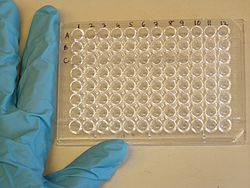
Examples of cell viability and other cytotoxicity assays used for in-vitro toxicology

Many methods of analysis exist for assaying test substances for cytotoxicity and other cellular responses.

Hemolysis assay

The hemolysis assay examines the propensity of chemicals, drugs or medication, or any blood-contacting medical device or material to lyse red blood cells (erythrocytes). The lysis is easily detected due to the release of hemoglobin. [6]

MTT and MTS

MTT assay is used often in determining cell viability and has been validated for use by international organisations. MTT assay involves two steps of introducing the assay to the chemicals and then a solubilisation step.

The colorimetric MTS (3-(4,5-dimethylthiazol-2-yl)-5-(3-carboxymethoxyphenyl)-2-(4-sulfophenyl)-2Htetrazolium) in vitro assay is an updated version of the validated MTT method, MTS assay has the advantage of being soluble. Hence, no solubilisation step is required.

ATP

ATP assay has the main advantage of providing results quickly (within 15 minutes) and only requires fewer sample cells. The assay performs lysis on the cells and the following chemical reaction between the assay and ATP content of cells produces luminescence. The amount of luminescence is then measured by a photometer and can be translated into number cells alive since

Neutral red

Another cell viability endpoint can be neutral red (NR) uptake. Neutral red, a weak cationic dye penetrates cellular membranes by non-diffusion and accumulates intercellularly in lysosomes. Viable cells take up the NR dye, damaged or dead cells do not.

Cytokine quantification via ELISA

ELISA kits can be used to examine up and down regulation of proinflammatory mediators such as cytokines (IL-1, TNF alpha, PGE2)....

Measurement of these types of cellular responses can be windows into the interaction of the test article on the test models (monolayer cell cultures, 3D tissue models, tissue explants).

Types of in vitro studies

Broadly speaking, there are two different types of in vitro studies depending on the type system developed to perform the experiment. The two types of systems generally used are : a) Static well plate system and b) the multi-compartmental perfused systems.

Static well plate system

The static well plate or layer systems are the most traditional and simplest form of assays widely used for in vitro study. These assays are quite beneficial as they are quite simple and provide a very accessible testing environment for monitoring chemicals in the culture medium as well as in the cell. However the disadvantage of using these simple static well plate assays is that, they cannot represent the cellular interactions and physiologic fluid flow conditions taking place inside the body.

Multi-compartmental perfused systems

New testing platforms are now developed to solve problems related to cellular interactions. These new platforms are much more complex based on multi-compartmental perfused systems. [7] The main objective of these systems is to reproduce in vivo mechanisms more reliably by providing cell culture environment close to the in vivo situation. Each compartment in the system represent a specific organ of the living organism and thus each compartment has a specific characteristics and criteria. Each compartment in these systems are connected by tubes and pumps through which the fluid flows thus mimicking the blood flow in the in vivo situation. The draw back behind the use of these perfused systems is that, the adverse effects ( influence of both the biological and non-biological components of the system on the fate of the chemical under study) are more compared to the static systems. In order to reduce the effect of non-biological components of the system, all the compartments are made of glass and the connecting tubes are made up of teflon. A number of kinetic models have been proposed to take care of these non-specific bindings taking place in these in vitro systems. [8]

To improve the biological difficulties arising from the use of different culture in vitro conditions, the traditional models used in flasks or micro-well plates has to be modified. With parallel development in micro-technologies and tissue engineering, these problems are solved using new pertinent tools called "micro-fluidic biochips". [9]

Related Research Articles

<i>In vitro</i> Latin term meaning outside a natural biological environment

In vitro studies are performed with microorganisms, cells, or biological molecules outside their normal biological context. Colloquially called "test-tube experiments", these studies in biology and its subdisciplines are traditionally done in labware such as test tubes, flasks, Petri dishes, and microtiter plates. Studies conducted using components of an organism that have been isolated from their usual biological surroundings permit a more detailed or more convenient analysis than can be done with whole organisms; however, results obtained from in vitro experiments may not fully or accurately predict the effects on a whole organism. In contrast to in vitro experiments, in vivo studies are those conducted in living organisms, including humans, known as clinical trials, and whole plants.

<span class="mw-page-title-main">Toxin</span> Naturally occurring organic poison

A toxin is a naturally occurring organic poison produced by metabolic activities of living cells or organisms. They occur especially as proteins, often conjugated. The term was first used by organic chemist Ludwig Brieger (1849–1919) and is derived from the word "toxic".

<span class="mw-page-title-main">Toxicology</span> Study of substances harmful to living organisms

Toxicology is a scientific discipline, overlapping with biology, chemistry, pharmacology, and medicine, that involves the study of the adverse effects of chemical substances on living organisms and the practice of diagnosing and treating exposures to toxins and toxicants. The relationship between dose and its effects on the exposed organism is of high significance in toxicology. Factors that influence chemical toxicity include the dosage, duration of exposure, route of exposure, species, age, sex, and environment. Toxicologists are experts on poisons and poisoning. There is a movement for evidence-based toxicology as part of the larger movement towards evidence-based practices. Toxicology is currently contributing to the field of cancer research, since some toxins can be used as drugs for killing tumor cells. One prime example of this is ribosome-inactivating proteins, tested in the treatment of leukemia.

The Draize test is an acute toxicity test devised in 1944 by Food and Drug Administration (FDA) toxicologists John H. Draize and Jacob M. Spines. Initially used for testing cosmetics, the procedure involves applying 0.5 mL or 0.5 g of a test substance to the eye or skin of a restrained, conscious animal, and then leaving it for set amount of time before rinsing it out and recording its effects. The animals are observed for up to 14 days for signs of erythema and edema in the skin test, and redness, swelling, discharge, ulceration, hemorrhaging, cloudiness, or blindness in the tested eye. The test subject is commonly an albino rabbit, though other species are used too, including dogs. The animals are euthanized after testing if the test renders irreversible damage to the eye or skin. Animals may be re-used for testing purposes if the product tested causes no permanent damage. Animals are typically reused after a "wash out" period during which all traces of the tested product are allowed to disperse from the test site.

Cytotoxicity is the quality of being toxic to cells. Examples of toxic agents are toxic metals, toxic chemicals, microbe neurotoxins, radiation particles and even specific neurotransmitters when the system is out of balance. Also some types of venom, e.g. from the puff adder or brown recluse spider are toxic to cells.

Genotoxicity is the property of chemical agents that damage the genetic information within a cell causing mutations, which may lead to cancer. While genotoxicity is often confused with mutagenicity, all mutagens are genotoxic, but some genotoxic substances are not mutagenic. The alteration can have direct or indirect effects on the DNA: the induction of mutations, mistimed event activation, and direct DNA damage leading to mutations. The permanent, heritable changes can affect either somatic cells of the organism or germ cells to be passed on to future generations. Cells prevent expression of the genotoxic mutation by either DNA repair or apoptosis; however, the damage may not always be fixed leading to mutagenesis.

Plate readers, also known as microplate readers or microplate photometers, are instruments which are used to detect biological, chemical or physical events of samples in microtiter plates. They are widely used in research, drug discovery, bioassay validation, quality control and manufacturing processes in the pharmaceutical and biotechnological industry and academic organizations. Sample reactions can be assayed in 1-1536 well format microtiter plates. The most common microplate format used in academic research laboratories or clinical diagnostic laboratories is 96-well with a typical reaction volume between 100 and 200 µL per well. Higher density microplates are typically used for screening applications, when throughput and assay cost per sample become critical parameters, with a typical assay volume between 5 and 50 µL per well. Common detection modes for microplate assays are absorbance, fluorescence intensity, luminescence, time-resolved fluorescence, and fluorescence polarization.

<span class="mw-page-title-main">MTT assay</span> Colorimetric analysis for measuring activity of cellular enzymes that reduce a tetrazolium dye

The MTT assay is a colorimetric assay for assessing cell metabolic activity. NAD(P)H-dependent cellular oxidoreductase enzymes may, under defined conditions, reflect the number of viable cells present. These enzymes are capable of reducing the tetrazolium dye MTT, which is chemically 3-(4,5-dimethylthiazol-2-yl)-2,5-diphenyltetrazolium bromide, to its insoluble formazan, which has a purple color. Other closely related tetrazolium dyes including XTT, MTS and the WSTs, are used in conjunction with the intermediate electron acceptor, 1-methoxy phenazine methosulfate (PMS). With WST-1, which is cell-impermeable, reduction occurs outside the cell via plasma membrane electron transport. However, this traditionally assumed explanation is currently contended as proof has also been found of MTT reduction to formazan in lipidic cellular structures without apparent involvement of oxidoreductases.

<span class="mw-page-title-main">Phototoxicity</span> Chemically-induced skin irritation following exposure to light

Phototoxicity, also called photoirritation, is a chemically induced skin irritation, requiring light, that does not involve the immune system. It is a type of photosensitivity.

<span class="mw-page-title-main">Neutral red</span> Chemical compound

Neutral red is a eurhodin dye used for staining in histology. It stains lysosomes red. It is used as a general stain in histology, as a counterstain in combination with other dyes, and for many staining methods. Together with Janus Green B, it is used to stain embryonal tissues and supravital staining of blood. Can be used for staining Golgi apparatus in cells and Nissl granules in neurons.

<span class="mw-page-title-main">Alternatives to animal testing</span> Test methods that avoid the use of animals

Alternatives to animal testing are the development and implementation of test methods that avoid the use of live animals.

<span class="mw-page-title-main">Toxicology testing</span> Biochemical process

Toxicology testing, also known as safety assessment, or toxicity testing, is the process of determining the degree to which a substance of interest negatively impacts the normal biological functions of an organism, given a certain exposure duration, route of exposure, and substance concentration.

An organ-on-a-chip (OOC) is a multi-channel 3-D microfluidic cell culture, integrated circuit (chip) that simulates the activities, mechanics and physiological response of an entire organ or an organ system. It constitutes the subject matter of significant biomedical engineering research, more precisely in bio-MEMS. The convergence of labs-on-chips (LOCs) and cell biology has permitted the study of human physiology in an organ-specific context. By acting as a more sophisticated in vitro approximation of complex tissues than standard cell culture, they provide the potential as an alternative to animal models for drug development and toxin testing.

A 3D cell culture is an artificially created environment in which biological cells are permitted to grow or interact with their surroundings in all three dimensions. Unlike 2D environments, a 3D cell culture allows cells in vitro to grow in all directions, similar to how they would in vivo. These three-dimensional cultures are usually grown in bioreactors, small capsules in which the cells can grow into spheroids, or 3D cell colonies. Approximately 300 spheroids are usually cultured per bioreactor.

Poly(amidoamine), or PAMAM, is a class of dendrimer which is made of repetitively branched subunits of amide and amine functionality. PAMAM dendrimers, sometimes referred to by the trade name Starburst, have been extensively studied since their synthesis in 1985, and represent the most well-characterized dendrimer family as well as the first to be commercialized. Like other dendrimers, PAMAMs have a sphere-like shape overall, and are typified by an internal molecular architecture consisting of tree-like branching, with each outward 'layer', or generation, containing exponentially more branching points. This branched architecture distinguishes PAMAMs and other dendrimers from traditional polymers, as it allows for low polydispersity and a high level of structural control during synthesis, and gives rise to a large number of surface sites relative to the total molecular volume. Moreover, PAMAM dendrimers exhibit greater biocompatibility than other dendrimer families, perhaps due to the combination of surface amines and interior amide bonds; these bonding motifs are highly reminiscent of innate biological chemistry and endow PAMAM dendrimers with properties similar to that of globular proteins. The relative ease/low cost of synthesis of PAMAM dendrimers (especially relative to similarly-sized biological molecules such as proteins and antibodies), along with their biocompatibility, structural control, and functionalizability, have made PAMAMs viable candidates for application in drug development, biochemistry, and nanotechnology.

In vitro to in vivo extrapolation (IVIVE) refers to the qualitative or quantitative transposition of experimental results or observations made in vitro to predict phenomena in vivo, biological organisms.

Albert P. Li is president and CEO of In Vitro ADMET Laboratories (IVAL), Columbia, Maryland, and Malden, Massachusetts. For the past three decades, Li has devoted his scientific career to the advancement of scientific concepts and technologies to accurately predict human drug properties. His research is focused on the development and application of human-based in vitro experimental systems in drug discovery and development. He is a pioneer in the isolation, cryopreservation, and culturing of human hepatocytes and their application in the evaluation of drug metabolism, drug-drug interactions, and drug toxicity.

<span class="mw-page-title-main">Bisphenol F</span> Chemical compound

Bisphenol F is an organic compound with the chemical formula (HOC
6
H
4
)
2
CH
2
. It is structurally related to bisphenol A (BPA), a popular precursor for forming plastics, as both belong to the category of molecules known as bisphenols, which feature two phenol groups connected via a linking group. In BPF, the two aromatic rings are linked by a methylene connecting group. In response to concern about the health effects of BPA, BPF is increasingly used as a substitute for BPA.

<span class="mw-page-title-main">Bioassay</span> Analytical method to determine potency and effect of a substance

A bioassay is an analytical method to determine the potency or effect of a substance by its effect on living animals or plants, or on living cells or tissues. A bioassay can be either quantal or quantitative, direct or indirect. If the measured response is binary, the assay is quantal; if not, it is quantitative.

<span class="mw-page-title-main">CompTox Chemicals Dashboard</span> Chemical database

The CompTox Chemicals Dashboard is a freely accessible online database created and maintained by the U.S. Environmental Protection Agency (EPA). The database provides access to multiple types of data including physicochemical properties, environmental fate and transport, exposure, usage, in vivo toxicity, and in vitro bioassay. EPA and other scientists use the data and models contained within the dashboard to help identify chemicals that require further testing and reduce the use of animals in chemical testing. The Dashboard is also used to provide public access to information from EPA Action Plans, e.g. around perfluorinated alkylated substances.

References

  1. Gavanji S, Bakhtari A, Famurewa AC, Othman EM (January 2023). "Cytotoxic Activity of Herbal Medicines as Assessed in Vitro: A Review". Chemistry & Biodiversity. 20 (2): 3–27. doi: 10.1002/cbdv.202201098 . PMID   36595710. S2CID   255473013.
  2. "Tox21". Source: U.S. Environmental Protection Agency . Retrieved 2011-10-29.
  3. Mahony, Catherine; Ashton, Randolph S.; Birk, Barbara; Boobis, Alan R.; Cull, Tom; Daston, George P.; Ewart, Lorna; Knudsen, Thomas B.; Manou, Irene; Maurer-Stroh, Sebastian; Margiotta-Casaluci, Luigi (July 2020). "New ideas for non-animal approaches to predict repeated-dose systemic toxicity: Report from an EPAA Blue Sky Workshop". Regulatory Toxicology and Pharmacology. 114: 104668. doi: 10.1016/j.yrtph.2020.104668 . PMID   32335207.
  4. "Vision and Roadmap for the 21st Century". Source: National Toxicology Program . Retrieved 2011-10-29.
  5. Zurlinden, TJ; Saili, KS; Rush, N; Kothiya, P; Judson, RS; Houck, KA; Hunter, ES; Baker, NC; Palmer, JA; Thomas, RS; Knudson, TB (2020). "Profiling the ToxCast Library With a Pluripotent Human (H9) Stem Cell Line-Based Biomarker Assay for Developmental Toxicity". Toxicological Sciences. 174 (2): 189–209. doi:10.1093/toxsci/kfaa014. PMC   8527599 . PMID   32073639.
  6. Sæbø PI, Bjørås M, Franzyk H, Helgensen E, Booth J (February 2, 2023). "Optimization of the Hemolysis Assay for the Assessment of Cytotoxicity". Int. J. Mol. Sci. 24 (3): 2914. doi: 10.3390/ijms24032914 . PMC   9917735 . PMID   36769243.
  7. Leclerc E.; Sakai Y.; Fujii T. (2004). "Perfusion culture of fetal human hepatocytes in microfluidic environments". Biochemical Engineering Journal. 20 (2–3): 143–148. doi:10.1016/j.bej.2003.09.010.
  8. Ouattara D.A.; Choi S.-H.; Sakai Y.; Pery A.R.R.; Brochot C. (2011). "Kinetic modelling of in vitro cell-based assays to characterize non-specific bindings and ADME processes in a static and a perfused fluidic system". Toxicology Letters. 205 (3): 310–319. doi:10.1016/j.toxlet.2011.06.021. PMID   21723928.
  9. Baudoin R.; Corlu A.; Griscom L.; Legallais C.; Leclerc E. (2007). "Trends in the development of microfluidic cell biochips for in vitro hepatotoxicity". Toxicology in Vitro. 21 (4): 535–544. doi:10.1016/j.tiv.2006.11.004. PMID   17188836.