McGillivray syndrome

Last updated
McGillivray syndrome
Other namesFamilial scaphocephaly syndrome, McGillivray type, Scaphocephaly-macrocephaly-maxillary retrusion-intellectual disability syndrome
Autosomal dominant - en.svg
This condition is inherited via an autosomal dominant manner
Specialty Cardiology

McGillivray syndrome is a rare syndrome characterized mainly by heart defects, skull and facial abnormalities and ambiguous genitalia. The symptoms of this syndrome are ventricular septal defect, patent ductus arteriosus, small jaw, undescended testes, and webbed fingers. Beside to these symptoms there are more symptoms which is related with bone structure and misshape.

Contents

McGillivray syndrome is a birth defect in which one or more of the joints between the bones of the baby's skull close prematurely, before the baby's brain is fully formed. When the baby has craniosynostosis, the brain cannot grow in its natural shape and the head is misshapen. It can affect one or more of the joints in the baby's skull. In some cases, craniosynostosis is associated with an underlying brain abnormality that prevents the brain from growing properly. Treating McGillivray usually involves surgery to separate the fused bones. If there is no underlying brain abnormality, the surgery allows the baby's brain to grow and develop in adequate space.

Symptoms and signs

The baby's skull has seven bones. Normally, these bones don't fuse until around age 2, giving the baby's brain time to grow. Joints called cranial sutures, made of strong, fibrous tissue, hold these bones together. In the front of the baby's skull, the sutures intersect in the large soft spot (fontanel) on the top of the baby's head. Normally, the sutures remain flexible until the bones fuse. The signs of craniosynostosis may not be noticeable at birth, but they become apparent during the first few months of the baby's life. The symptoms differs from types of synostosis. First of all there is Sagittal synostosis (scaphocephaly). Premature fusion of the suture at the top of the head (sagittal suture) forces the head to grow long and narrow, rather than wide. Scaphocephaly is the most common type of craniosynostosis. The other one is called Coronal synostosis (anterior plagiocephaly). Premature fusion of a coronal suture — one of the structures that run from each ear to the sagittal suture on top of the head — may force the baby's forehead to flatten on the affected side. It may also raise the eye socket and cause a deviated nose and slanted skull. The Bicoronal synostosis (brachycephaly). When both of the coronal sutures fuse prematurely, the baby may have a flat, elevated forehead and brow.[ citation needed ]

Cause

Diagnosis

First of all there is physical exam. Doctors examine the baby's head for abnormalities such as suture ridges and look the facial deformities. Also, they utilizes Computerized Tomography which scan of the baby's skull. Fused sutures are identifiable by their absences. X-rays also may be used to measure precise dimensions of the baby's skull, using a technique called cephalometry.[ citation needed ]

Genetic testing. If the doctor suspects the baby's misshapen skull is caused by an underlying hereditary syndrome, genetic testing may help identify the syndrome. Genetic tests usually require a blood sample. Depending on what type of abnormality is suspected, the doctor may take a sample of the baby's hair, skin or other tissue, such as cells from the inside of the cheek. The sample is sent to a lab for analysis.[ citation needed ]

Rare types

There are two less common types of McGillivray syndromes are: Metopic synostosis (trigonocephaly). The metopic suture runs from the baby's nose to the sagittal suture. Premature fusion gives the scalp a triangular appearance. Another one is Lambdoid synostosis (posterior plagiocephaly). This rare form of craniosynostosis involves the lambdoid suture, which runs across the skull near the back of the head. It may cause flattening of the baby's head on the affected side. A misshapen head doesn't always indicate craniosynostosis. For example, if the back of the baby's head appears flattened, it could be the result of birth trauma or the baby's spending too much time on his or her back. This condition is sometimes treated with a custom-fit helmet that helps mold the baby's head back into a normal position.[ citation needed ]

Treatments

The major treatment is surgery for most babies. The type of surgery which they would undergo differs from age and strength they have. The main reason of doing the surgery is to alleviate pressure on the brain, and create a space for brain developing and growing. It would improve infant's appearance.[ citation needed ]

The first one is Traditional surgery. During surgery, they make an incision in the baby's scalp and cranial bones, and reshape the portion of the skull. Sometimes plates and screws, often made of material that is absorbed over time, are used to hold the bones in place. Surgery, which is performed during general anesthesia, usually takes hours.[ citation needed ]

After surgery, the baby remains in the hospital for at least three days. Some children may require a second surgery later because, the craniosynostosis reoccurs. Also, children with facial deformities often require future surgeries to reshape their faces.[ citation needed ]

Another one is Endoscopic surgery. This less invasive form of surgery isn't an option for everyone. But in certain cases, the surgeon may use a lighted tube (endoscope) inserted through one or two small scalp incisions over the affected suture. The surgeon then opens the suture to enable the baby's brain to grow normally. Endoscopic surgery usually takes about an hour, causes less swelling and blood loss, and shortens the hospital stay, often to one day after surgery.[ citation needed ]

History

McGillivray Syndrome is referenced from the 1851 writings of Virchow. His understanding and descriptions of irregular calvarial growth patterns were the basis of the law of Virchow. According to his observations, the abnormal cranial growth observed in persons with craniosynostosis occurs perpendicular to the involved calvarial sutures. Therefore, if a suture line is prematurely ossified, no growth is present in the direction perpendicular to that suture. Surgical treatment for craniosynostosis was initially advocated by Odilon Lannelongue in 1890.[ citation needed ]

His patients had microcephaly from craniosynostosis and were thought to be imbeciles. These patients accordingly underwent craniectomy to remove the involved suture line and to "release the brain". Soon after, in 1891, linear craniectomy was introduced. As with any new procedure, this one met with much resistance. However, the resistance to a surgical intervention was slowly put to rest with mounting evidence. Several studies indicated that craniosynostectomy was the treatment of choice for the release of fused suture lines in the skull. Studies showed that, over time, cranial suture areas excised during strip craniectomy still became fused and led to an abnormal cranial contour.[ citation needed ]

Strip craniectomy was easier and involved less blood loss compared with the newer cranial vault reconstruction. Strip craniectomy also did not address the frontal bossing and associated abnormalities in calvarial shape and relied on the rapid growth of the brain to correct it. Strip craniectomy was optimal only in the first few months of infancy, while surgeons could use cranial vault reconstruction throughout infancy. Consequently, strip craniectomy lost favor, and the surgical treatment has been modified to include cranial vault remodeling. Recently, with the advent of endoscopy, attention has returned to endoscopic strip craniectomy. The endoscopic technique has only been tried over the last several years, but it offers the advantages of a shorter and safer operation, less cost, less in-hospital time, and less blood loss. The operation was shown to be a success in a study of 12 patients, all younger than 8 months. Critical to this success and a departure from the standard strip craniosynostectomy was the extensive use of a postoperative remodeling helmet. Although first introduced by Persing et al. in 1986, helmet therapy has not been used as extensively as a postoperative therapeutic intervention.[ citation needed ]

Related Research Articles

<span class="mw-page-title-main">Say–Meyer syndrome</span> X-linked recessive disorder characterised by developmental delay

Say–Meyer syndrome is a rare X-linked genetic disorder that is mostly characterized as developmental delay. It is one of the rare causes of short stature. It is closely related with trigonocephaly. People with Say–Meyer syndrome have impaired growth, deficits in motor skills development and mental state.

<span class="mw-page-title-main">Skull</span> Bony structure that forms the head in vertebrates

The skull is a bone protective cavity for the brain. The skull is composed of three types of bone: cranial bones, facial bones, and ear ossicles. Two parts are more prominent: the cranium and the mandible. In humans, these two parts are the neurocranium (braincase) and the viscerocranium that includes the mandible as its largest bone. The skull forms the anterior-most portion of the skeleton and is a product of cephalisation—housing the brain, and several sensory structures such as the eyes, ears, nose, and mouth. In humans, these sensory structures are part of the facial skeleton.

<span class="mw-page-title-main">Fontanelle</span> Anatomical feature of the infant human skull

A fontanelle is an anatomical feature of the infant human skull comprising soft membranous gaps (sutures) between the cranial bones that make up the calvaria of a fetus or an infant. Fontanelles allow for stretching and deformation of the neurocranium both during birth and later as the brain expands faster than the surrounding bone can grow. Premature complete ossification of the sutures is called craniosynostosis.

<span class="mw-page-title-main">Plagiocephaly</span> Medical condition

Plagiocephaly, also known as flat head syndrome, is a condition characterized by an asymmetrical distortion of the skull. A mild and widespread form is characterized by a flat spot on the back or one side of the head caused by remaining in a supine position for prolonged periods.

<span class="mw-page-title-main">Scaphocephaly</span> Cephalic disorder involving premature fusion of the sagittal suture

Scaphocephaly, or sagittal craniosynostosis, is a type of cephalic disorder which occurs when there is a premature fusion of the sagittal suture. Premature closure results in limited lateral expansion of the skull resulting in a characteristic long, narrow head. The skull base is typically spared.

<span class="mw-page-title-main">Trigonocephaly</span> Congenital condition of premature fusion of the metopic suture

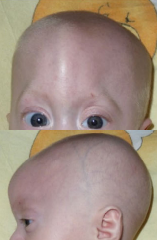
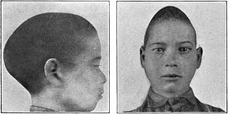
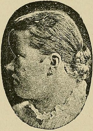
Trigonocephaly is a congenital condition due to premature fusion of the metopic suture, leading to a triangular forehead. The premature merging of the two frontal bones leads to transverse growth restriction and parallel growth expansion. It may occur as one component of a syndrome together with other abnormalities, or in isolated form. The term is from the Greek trigonon, "triangle", and kephale, "head".

<span class="mw-page-title-main">Crouzon syndrome</span> Genetic disorder of the skull and face

Crouzon syndrome is an autosomal dominant genetic disorder known as a branchial arch syndrome. Specifically, this syndrome affects the first branchial arch, which is the precursor of the maxilla and mandible. Because the branchial arches are important developmental features in a growing embryo, disturbances in their development create lasting and widespread effects. The syndrome is caused by a mutation in a gene on chromosome 10 that controls the body's production of fibroblast growth factor receptor 2 (FGFR2).

<span class="mw-page-title-main">Apert syndrome</span> Congenital disorder of the skull and digits

Apert syndrome is a form of acrocephalosyndactyly, a congenital disorder characterized by malformations of the skull, face, hands and feet. It is classified as a branchial arch syndrome, affecting the first branchial arch, the precursor of the maxilla and mandible. Disturbances in the development of the branchial arches in fetal development create lasting and widespread effects.

<span class="mw-page-title-main">Craniosynostosis</span> Premature fusion of bones in the skull

Craniosynostosis is a condition in which one or more of the fibrous sutures in a young infant's skull prematurely fuses by turning into bone (ossification), thereby changing the growth pattern of the skull. Because the skull cannot expand perpendicular to the fused suture, it compensates by growing more in the direction parallel to the closed sutures. Sometimes the resulting growth pattern provides the necessary space for the growing brain, but results in an abnormal head shape and abnormal facial features. In cases in which the compensation does not effectively provide enough space for the growing brain, craniosynostosis results in increased intracranial pressure leading possibly to visual impairment, sleeping impairment, eating difficulties, or an impairment of mental development combined with a significant reduction in IQ.

<span class="mw-page-title-main">Saethre–Chotzen syndrome</span> Medical condition

Saethre–Chotzen syndrome (SCS), also known as acrocephalosyndactyly type III, is a rare congenital disorder associated with craniosynostosis. This affects the shape of the head and face, resulting in a cone-shaped head and an asymmetrical face. Individuals with SCS also have droopy eyelids (ptosis), widely spaced eyes (hypertelorism), and minor abnormalities of the hands and feet (syndactyly). Individuals with more severe cases of SCS may have mild to moderate intellectual or learning disabilities. Depending on the level of severity, some individuals with SCS may require some form of medical or surgical intervention. Most individuals with SCS live fairly normal lives, regardless of whether medical treatment is needed or not.

Craniofacial surgery is a surgical subspecialty that deals with congenital and acquired deformities of the head, skull, face, neck, jaws and associated structures. Although craniofacial treatment often involves manipulation of bone, craniofacial surgery is not tissue-specific; craniofacial surgeons deal with bone, skin, nerve, muscle, teeth, and other related anatomy.

<span class="mw-page-title-main">Carpenter syndrome</span> Medical condition

Carpenter syndrome, also called acrocephalopolysyndactyly type II, is an extremely rare autosomal recessive congenital disorder characterized by craniofacial malformations, obesity, syndactyly, and polydactyly. Acrocephalopolysyndactyly is a variation of acrocephalosyndactyly that presents with polydactyly.

<span class="mw-page-title-main">Sagittal suture</span> Midline joint between the parietal bones of the skull

The sagittal suture, also known as the interparietal suture and the sutura interparietalis, is a dense, fibrous connective tissue joint between the two parietal bones of the skull. The term is derived from the Latin word sagitta, meaning arrow.

<span class="mw-page-title-main">Jackson–Weiss syndrome</span> Medical condition

Jackson–Weiss syndrome (JWS) is a genetic disorder characterized by foot abnormalities and the premature fusion of certain bones of the skull (craniosynostosis), which prevents further growth of the skull and affects the shape of the head and face. This genetic disorder can also sometimes cause intellectual disability and crossed eyes. It was characterized in 1976.

<span class="mw-page-title-main">Synostosis</span>

Synostosis is fusion of two or more bones. It can be normal in puberty, fusion of the epiphyseal plate to become the epiphyseal line, or abnormal. When synostosis is abnormal it is a type of dysostosis. Examples of synostoses include:

<span class="mw-page-title-main">Acrocephalosyndactyly</span> Group of diseases

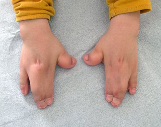
Acrocephalosyndactyly is a group of congenital conditions characterized by irregular features of the face and skull (craniosynostosis) and hands and feet (syndactyly). Craniosynostosis occurs when the cranial sutures, the fibrous tissue connecting the skull bones, fuse the cranial bones early in development. Cranial sutures allow the skull bones to continue growing until they fuse at age 24. Premature fusing of the cranial sutures can result in alterations to the skull shape and interfere with brain growth. Syndactyly occurs when digits of the hands or feet are fused together. When polydactyly is also present, the classification is acrocephalopolysyndactyly. Polydactyly occurs when the hands or feet possess additional digits. Acrocephalosyndactyly is usually diagnosed after birth, although prenatal diagnosis is sometimes possible if the genetic variation is present in family members, as the conditions are typically inherited in an autosomal dominant pattern Treatment often involves surgery in early childhood to correct for craniosynostosis and syndactyly.

<span class="mw-page-title-main">Muenke syndrome</span> Medical condition

Muenke syndrome, also known as FGFR3-related craniosynostosis, is a human specific condition characterized by the premature closure of certain bones of the skull during development, which affects the shape of the head and face. First described by Maximilian Muenke, the syndrome occurs in about 1 in 30,000 newborns. This condition accounts for an estimated 8 percent of all cases of craniosynostosis.

<span class="mw-page-title-main">Calvaria (skull)</span> Top part of the skull

The calvaria is the top part of the skull. It is the superior part of the neurocranium and covers the cranial cavity containing the brain. It forms the main component of the skull roof.

<span class="mw-page-title-main">Baller–Gerold syndrome</span> Medical condition

Baller–Gerold syndrome (BGS) is a rare genetic syndrome that involves premature fusion of the skull bones and malformations of facial, forearm and hand bones. The symptoms of Baller–Gerold syndrome overlap with features of a few other genetics disorders: Rothmund–Thomson syndrome and RAPADILINO syndrome. The prevalence of BGS is unknown, as there have only been a few reported cases, but it is estimated to be less than 1 in a million. The name of the syndrome comes from the researchers Baller and Gerold who discovered the first three cases.

<span class="mw-page-title-main">Kleeblattschaedel</span> Medical condition

Kleeblattschaedel is a rare malformation of the head where there is a protrusion of the skull and broadening of the face. This condition is a severe type of craniosynostosis.

References