Mesangial proliferative glomerulonephritis

Last updated
Mesangial proliferative glomerulonephritis
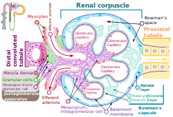
Renal corpuscle-en.svg
Renal corpuscle showing intraglomerular mesangium, where mesangial proliferation occurs.
Specialty Nephrology   OOjs UI icon edit-ltr-progressive.svg

Mesangial proliferative glomerulonephritis (MesPGN) is a morphological pattern characterized by a numerical increase in mesangial cells and expansion of the extracellular matrix within the mesangium of the glomerulus. [1] The increase in the number of mesangial cells can be diffuse or local and immunoglobulin and/or complement deposition can also occur. MesPGN is associated with a variety of disease processes affecting the glomerulus, though can be idiopathic. The clinical presentation of MesPGN usually consists of hematuria or nephrotic syndrome. [2] Treatment is often consistent with the histologic pattern of and/or disease process contributing to mesangial proliferative glomerulonephritis, and usually involves some form of immunosuppressant.

Contents

Mechanism

MesPGN often occurs as a result of glomerular injury, though can be idiopathic. MesPGN has been associated with disease processes such as: IgA nephropathy, IgM nephropathy, systemic lupus erythematous, Alport's syndrome, resolving post-infectious glomerulonephritis, and complement nephropathy, such as C1Q nephropathy. [1]

IgA nephropathy is the most common cause of MesPGN. [3] It is thought abnormally glycosylated IgA form polymers and deposit in the mesangium. [3] Subsequently, IgA immune complexes bind to IgA receptors on mesangial cells and induce injury to mesangial cells through release of cytokines and growth factors that promote infiltration of leukocytes, mesangial cell proliferation, and mesangial matrix expansion. [3]

In the context of resolving post-infectious glomerulonephritis, MesPGN can be seen after an infection with a nephritogenic strain of group A streptococci. [4] Pathogenesis of post-streptococcal glomerulonephritis includes injury to the glomerulus by immune complexes (IgG) passively trapped in the glomerulus, which leads to an inflammatory response from recruited immune cells, cytokines, chemical mediators, and complement and coagulation cascade activation. [5] The inflammatory response includes endothelial and mesangial cell proliferation. [3]

Mesangial proliferative glomerulonephritis of Lupus nephritis, Class II is also noted by mesangial hypercellularity and matrix expansion. Microscopic haematuria with or without proteinuria may be seen in Class II Lupus nephritis. Hypertension, nephrotic syndrome, and acute kidney injury are very rare at this stage. [6]

Idiopathic mesangial proliferative glomerulonephritis is less established in the literature. Idiopathic mesangial proliferative glomerulonephritis does not involve the deposition of either IgA or IgG immune complexes, though there can be focal or diffuse IgM deposits in the mesangium. [7] The relationship of IgM and mesangial proliferative glomerulonephritis is hypothesized to involve either formed or deposited IgM complexes in the mesangium leading to T-cell mediated inflammatory response, mesangial proliferation, and glomerular injury or, as a result of mesangial proliferation, decreased clearance of monocytic IgM complexes. [7]

Diagnosis

Most glomerulonephritis' classification and prognosis are aided by histological evaluation by renal biopsy. [3] The renal biopsy is classically evaluated with light microscopy, electron microscopy, and immunohistology to diagnose a histological pattern, which is then compared to clinical evaluation through history, physical, and laboratory evaluation. [3] The laboratory evaluation usually follows that of a standard nephrology work up and will likely be targeted to a differential diagnosis. Studies focusing on mesangial proliferative glomerulonephritis often use defined clinical criteria and histological criteria to select patients for research. For example, one study used the following histological criteria: "Glomeruli with mesangial hypercellularity (four or more cells/mesangium with or without mesangial matrix expansion and immune complex deposits)". [1] The histologic pattern of injury can also provide insight into the prognosis of the glomerular disorder. Mesangial proliferation indicates a mild, though active, lesion. [8] Overall, a kidney biopsy should address the following: [8]

The kidney biopsy is foundational to informing the diagnosis of mesangial proliferative glomerulonephritis, as it is a morphological pattern.

Clinical Presentation

Mesangial proliferative glomerulonephritis often presents with hematuria (gross or microscopic) or nephrotic syndrome. [2] Presentation can also include asymptomatic proteinuria. [2] These presenting symptoms are relatively non-specific and are often seen in other glomerular disorders. Preceding upper respiratory tract infection or post-streptococcal glomerulonephritis may contribute to hematuria, as both have been identified in patients presenting with hematuria in the context of mesangial proliferative glomerulonephritis. [2] Preceding infection was not as readily identified in patients presenting with either asymptomatic proteinuria or nephrotic syndrome. However, it has been shown patients presenting with nephrotic syndrome have some histo- and clinic-pathologic similarities to minimal change disease. [9]

Treatment

Treatment for glomerular disorders is often established for specific histological patterns. [3] Presentations of hematuria in the context of mesangial proliferative glomerulonephritis often resolve spontaneously, with a relatively benign course. [2] Presentation of nephrotic syndrome in the context of mesangial proliferative glomerulonephritis have been treated with immunosuppressants, such as steroids and cyclophosphamide. [2] Presentation with nephrotic syndrome can resolve with treatment, but can also progress. [10] Patients can become resistant to steroids or specific immunosuppressive agents, in which case it may be necessary to use different immunosuppressive agents. [2]

Related Research Articles

<span class="mw-page-title-main">Proteinuria</span> Presence of an excess of serum proteins in the urine

Proteinuria is the presence of excess proteins in the urine. In healthy persons, urine contains very little protein; an excess is suggestive of illness. Excess protein in the urine often causes the urine to become foamy. Severe proteinuria can cause nephrotic syndrome in which there is worsening swelling of the body.

<span class="mw-page-title-main">Nephrotic syndrome</span> Collection of symptoms due to kidney damage

Nephrotic syndrome is a collection of symptoms due to kidney damage. This includes protein in the urine, low blood albumin levels, high blood lipids, and significant swelling. Other symptoms may include weight gain, feeling tired, and foamy urine. Complications may include blood clots, infections, and high blood pressure.

<span class="mw-page-title-main">Nephritis</span> Inflammation of the kidneys

Nephritis is inflammation of the kidneys and may involve the glomeruli, tubules, or interstitial tissue surrounding the glomeruli and tubules. It is one of several different types of nephropathy.

<span class="mw-page-title-main">Hematuria</span> Medical condition

Hematuria or haematuria is defined as the presence of blood or red blood cells in the urine. "Gross hematuria" occurs when urine appears red, brown, or tea-colored due to the presence of blood. Hematuria may also be subtle and only detectable with a microscope or laboratory test. Blood that enters and mixes with the urine can come from any location within the urinary system, including the kidney, ureter, urinary bladder, urethra, and in men, the prostate. Common causes of hematuria include urinary tract infection (UTI), kidney stones, viral illness, trauma, bladder cancer, and exercise. These causes are grouped into glomerular and non-glomerular causes, depending on the involvement of the glomerulus of the kidney. But not all red urine is hematuria. Other substances such as certain medications and foods can cause urine to appear red. Menstruation in women may also cause the appearance of hematuria and may result in a positive urine dipstick test for hematuria. A urine dipstick test may also give an incorrect positive result for hematuria if there are other substances in the urine such as myoglobin, a protein excreted into urine during rhabdomyolysis. A positive urine dipstick test should be confirmed with microscopy, where hematuria is defined by three of more red blood cells per high power field. When hematuria is detected, a thorough history and physical examination with appropriate further evaluation can help determine the underlying cause.

<span class="mw-page-title-main">Kidney disease</span> Damage to or disease of a kidney

Kidney disease, or renal disease, technically referred to as nephropathy, is damage to or disease of a kidney. Nephritis is an inflammatory kidney disease and has several types according to the location of the inflammation. Inflammation can be diagnosed by blood tests. Nephrosis is non-inflammatory kidney disease. Nephritis and nephrosis can give rise to nephritic syndrome and nephrotic syndrome respectively. Kidney disease usually causes a loss of kidney function to some degree and can result in kidney failure, the complete loss of kidney function. Kidney failure is known as the end-stage of kidney disease, where dialysis or a kidney transplant is the only treatment option.

<span class="mw-page-title-main">Glomerulus (kidney)</span> Functional unit of nephron

The glomerulus is a network of small blood vessels (capillaries) known as a tuft, located at the beginning of a nephron in the kidney. Each of the two kidneys contains about one million nephrons. The tuft is structurally supported by the mesangium, composed of intraglomerular mesangial cells. The blood is filtered across the capillary walls of this tuft through the glomerular filtration barrier, which yields its filtrate of water and soluble substances to a cup-like sac known as Bowman's capsule. The filtrate then enters the renal tubule of the nephron.

<span class="mw-page-title-main">IgA nephropathy</span> Disease of the kidney

IgA nephropathy (IgAN), also known as Berger's disease, or synpharyngitic glomerulonephritis, is a disease of the kidney and the immune system; specifically it is a form of glomerulonephritis or an inflammation of the glomeruli of the kidney. Aggressive Berger's disease can attack other major organs, such as the liver, skin and heart.

Mesangial cells are specialised cells in the kidney that make up the mesangium of the glomerulus. Together with the mesangial matrix, they form the vascular pole of the renal corpuscle. The mesangial cell population accounts for approximately 30-40% of the total cells in the glomerulus. Mesangial cells can be categorized as either extraglomerular mesangial cells or intraglomerular mesangial cells, based on their relative location to the glomerulus. The extraglomerular mesangial cells are found between the afferent and efferent arterioles towards the vascular pole of the glomerulus. The extraglomerular mesangial cells are adjacent to the intraglomerular mesangial cells that are located inside the glomerulus and in between the capillaries. The primary function of mesangial cells is to remove trapped residues and aggregated protein from the basement membrane thus keeping the filter free of debris. The contractile properties of mesangial cells have been shown to be insignificant in changing the filtration pressure of the glomerulus.

<span class="mw-page-title-main">Henoch–Schönlein purpura</span> Medical condition

Henoch–Schönlein purpura (HSP), also known as IgA vasculitis, is a disease of the skin, mucous membranes, and sometimes other organs that most commonly affects children. In the skin, the disease causes palpable purpura, often with joint pain and abdominal pain. With kidney involvement, there may be a loss of small amounts of blood and protein in the urine, but this usually goes unnoticed; in a small proportion of cases, the kidney involvement proceeds to chronic kidney disease. HSP is often preceded by an infection, such as a throat infection.

<span class="mw-page-title-main">Glomerulonephritis</span> Term for several kidney diseases

Glomerulonephritis (GN) is a term used to refer to several kidney diseases. Many of the diseases are characterised by inflammation either of the glomeruli or of the small blood vessels in the kidneys, hence the name, but not all diseases necessarily have an inflammatory component.

<span class="mw-page-title-main">Membranous glomerulonephritis</span> Medical condition

Membranous glomerulonephritis (MGN) is a slowly progressive disease of the kidney affecting mostly people between ages of 30 and 50 years, usually white people.

<span class="mw-page-title-main">Nephritic syndrome</span> Medical condition

Nephritic syndrome is a syndrome comprising signs of nephritis, which is kidney disease involving inflammation. It often occurs in the glomerulus, where it is called glomerulonephritis. Glomerulonephritis is characterized by inflammation and thinning of the glomerular basement membrane and the occurrence of small pores in the podocytes of the glomerulus. These pores become large enough to permit both proteins and red blood cells to pass into the urine. By contrast, nephrotic syndrome is characterized by proteinuria and a constellation of other symptoms that specifically do not include hematuria. Nephritic syndrome, like nephrotic syndrome, may involve low level of albumin in the blood due to the protein albumin moving from the blood to the urine.

<span class="mw-page-title-main">Focal segmental glomerulosclerosis</span> Kidney disease

Focal segmental glomerulosclerosis (FSGS) is a histopathologic finding of scarring (sclerosis) of glomeruli and damage to renal podocytes. This process damages the filtration function of the kidney, resulting in protein loss in the urine. FSGS is a leading cause of excess protein loss—nephrotic syndrome—in children and adults. Signs and symptoms include proteinuria, water retention, and edema. Kidney failure is a common long-term complication of disease. FSGS can be classified as primary versus secondary depending on whether a particular toxic or pathologic stressor can be identified as the cause. Diagnosis is established by renal biopsy, and treatment consists of glucocorticoids and other immune-modulatory drugs. Response to therapy is variable, with a significant portion of patients progressing to end-stage kidney failure. FSGS is estimated to occur in 2-3 persons per million, with males and African peoples at higher risk.

Thin basement membrane disease is, along with IgA nephropathy, the most common cause of hematuria without other symptoms. The only abnormal finding in this disease is a thinning of the basement membrane of the glomeruli in the kidneys. Its importance lies in the fact that it has a benign prognosis, with patients maintaining a normal kidney function throughout their lives.

<span class="mw-page-title-main">Glomerular basement membrane</span>

The glomerular basement membrane of the kidney is the basal lamina layer of the glomerulus. The glomerular endothelial cells, the glomerular basement membrane, and the filtration slits between the podocytes perform the filtration function of the glomerulus, separating the blood in the capillaries from the filtrate that forms in Bowman's capsule. The glomerular basement membrane is a fusion of the endothelial cell and podocyte basal laminas, and is the main site of restriction of water flow. Glomerular basement membrane is secreted and maintained by podocyte cells.

<span class="mw-page-title-main">Rapidly progressive glomerulonephritis</span> Medical condition

Rapidly progressive glomerulonephritis (RPGN) is a syndrome of the kidney that is characterized by a rapid loss of kidney function, with glomerular crescent formation seen in at least 50% or 75% of glomeruli seen on kidney biopsies. If left untreated, it rapidly progresses into acute kidney failure and death within months. In 50% of cases, RPGN is associated with an underlying disease such as Goodpasture syndrome, systemic lupus erythematosus or granulomatosis with polyangiitis; the remaining cases are idiopathic. Regardless of the underlying cause, RPGN involves severe injury to the kidneys' glomeruli, with many of the glomeruli containing characteristic glomerular crescents.

Glomerulonephrosis is a non-inflammatory disease of the kidney (nephrosis) presenting primarily in the glomerulus as Nephrotic Syndrome. The nephron is the functional unit of the kidney and it contains the glomerulus, which acts as a filter for blood to retain proteins and blood lipids. Damage to these filtration units results in important blood contents being released as waste in urine. This disease can be characterized by symptoms such as fatigue, swelling, and foamy urine, and can lead to chronic kidney disease and ultimately end-stage renal disease, as well as cardiovascular diseases. Glomerulonephrosis can present as either primary glomerulonephrosis or secondary glomerulonephrosis.

Focal proliferative nephritis is a type of glomerulonephritis seen in 20% to 35% of cases of lupus nephritis, classified as type III. As the name suggests, lesions are seen in less than half of the glomeruli. Typically, one or two foci within an otherwise normal glomerulus show swelling and proliferation of endothelial and mesangial cells, infiltration by neutrophils, and/or fibrinoid deposits with capillary thrombi. Focal glomerulonephritis is usually associated with only mild microscopic hematuria and proteinuria; a transition to a more diffuse form of renal involvement is associated with more severe disease.

Diffuse proliferative glomerulonephritis (DPGN) is a type of glomerulonephritis that is the most serious form of renal lesions in SLE and is also the most common, occurring in 35% to 60% of patients. In absence of SLE, DPGN pathology looks more like Membranoproliferative glomerulonephritis

Monoclonal gammopathy of renal significance (MGRS) are a group of kidney disorders that present with kidney damage due to nephrotoxic monoclonal immunoglobulins secreted by clonal plasma cells or B cells. By definition, people with MGRS do not meet criteria for multiple myeloma or other hematologic malignancies. The term MGRS was introduced in 2012 by the International Kidney and Monoclonal Gammopathy Research Group (IKMG). MGRS is associated with monoclonal gammopathy of undetermined significance (MGUS). People with MGUS have a monoclonal gammopathy but does not meet the criteria for the clonal burden nor the presence of end organ damage seen in hematologic malignancies. In a population based study based on the NHANES III health survey; 6% of patients with MGUS were subsequently classified as having MGRS. The prevalence and incidence of MGRS in the general population or in specific populations is not known but it is more prevalent in those over the age of 50 as there is a monoclonal protein (M-protein) present in 3% of those 50 and years older and 5% of those 70 years and older, placing those 50 and older at increased risk of MGRS.

References

  1. 1 2 3 Mokhtar, Ghadeer A.; Jalalah, Sawsan; Sultana, Shabnum (2014). "Pathological patterns of mesangioproliferative glomerulonephritis seen at a tertiary care center". Journal of Nephropharmacology. 3 (2): 33–37. ISSN   2345-4202. PMC   5297525 . PMID   28197459.
  2. 1 2 3 4 5 6 7 Brown, E. A.; Upadhyaya, K.; Hayslett, J. P.; Kashgarian, M.; Siegel, N. J. (July 1979). "The clinical course of mesangial proliferative glomerulonephritis". Medicine. 58 (4): 295–303. doi:10.1097/00005792-197907000-00002. ISSN   0025-7974. PMID   449664. S2CID   19581682.
  3. 1 2 3 4 5 6 7 Johnson, Richard J.; Floege, Jürgen; Feehally, John (2010), "Introduction to Glomerular Disease", Comprehensive Clinical Nephrology, Elsevier, pp. 208–217, doi:10.1016/b978-0-323-05876-6.00016-2, ISBN   9780323058766 , retrieved 2021-11-11
  4. Sagel, Inge (1973-10-01). "Occurrence and Nature of Glomerular Lesions after Group A Streptococci Infections in Children". Annals of Internal Medicine. 79 (4): 492–499. doi:10.7326/0003-4819-79-4-492. ISSN   0003-4819. PMID   4795879.
  5. Balow, James E.; Cho, Monique E.; Waldman, Meryl; Austin, Howard A. (2013), "Immunologic renal diseases", Clinical Immunology, Elsevier, pp. 812–827, doi:10.1016/b978-0-7234-3691-1.00081-7, ISBN   978-0-7234-3691-1 , retrieved 2021-11-13
  6. Lewis, Edmund J.; Schwartz, Melvin M. (2010-11-04). Lupus Nephritis. OUP Oxford. pp. 174–177. ISBN   9780199568055.
  7. 1 2 Little, Mark A.; Dorman, Anthony; Gill, Denis; Walshe, J. Joseph; Walshe, John J. (January 2000). "MESANGIOPROLIFERATIVE GLOMERULONEPHRITIS WITH IgM DEPOSITION: CLINICAL CHARACTERISTICS AND OUTCOME". Renal Failure. 22 (4): 445–457. doi:10.1081/JDI-100100886. ISSN   0886-022X. PMID   10901182. S2CID   8527496.
  8. 1 2 Sethi, Sanjeev; Fervenza, Fernando C (2019-02-01). "Standardized classification and reporting of glomerulonephritis". Nephrology Dialysis Transplantation. 34 (2): 193–199. doi: 10.1093/ndt/gfy220 . ISSN   0931-0509. PMID   30124958.
  9. Hogg, R J (July 1983). "Childhood nephrotic syndrome associated with diffuse mesangial hypercellularity. A report of the Southwest Pediatric Nephrology Study Group". Kidney International. 24 (1): 87–94. doi: 10.1038/ki.1983.129 . PMID   6620853.
  10. Waldherr, R.; Gubler, M. C.; Levy, M.; Broyer, M.; Habib, R. (November 1978). "The significance of pure diffuse mesangial proliferation in idiopathic nephrotic syndrome". Clinical Nephrology. 10 (5): 171–179. ISSN   0301-0430. PMID   365403.