Neural tissue engineering

Last updated

Neural tissue engineering is a specific sub-field of tissue engineering. Neural tissue engineering is primarily a search for strategies to eliminate inflammation and fibrosis upon implantation of foreign substances. Often foreign substances in the form of grafts and scaffolds are implanted to promote nerve regeneration and to repair damage caused to nerves of both the central nervous system (CNS) and peripheral nervous system (PNS) by an injury.

Contents

Introduction

There are two parts of the nervous system: the central nervous system (CNS) and the peripheral nervous system (PNS). General body functions are supervised by the central nervous system (CNS), which includes the brain and spinal cord. The PNS delivers motor signals to control body activities and receives sensory data from the CNS. The PNS It is made up of nerve fibers arranged into nerves. The PNS's autonomic nervous system (ANS), whose sympathetic and parasympathetic branches preserve homeostasis, regulates involuntary physiological functions. [1]

The "fight-or-flight" reaction is triggered by the sympathetic nervous system (SNS), which is derived from the thoracic and upper lumbar spinal cord. It readies the body for quick reactions under pressure. The parasympathetic nervous system (PSNS), on the other hand, is derived from the brainstem and sacral spinal cord and facilitates normal physiological processes by encouraging rest and energy conservation. One of the main nerves in the PSNS, the vagus nerve, has its start in the brainstem and travels throughout the body, affecting different organs. It has sensory and motor fibers. Sensory messages tell the brain what the body is doing, allowing it to maintain homeostasis and control activities. Additionally, the vagus nerve influences emotions and memory through connections to several brain regions.

Neuroimmune Interactions The immune system's role is to identify and protect the body against external chemicals and infections. It is separated into innate and adaptive immunity and consists of immune organs, cells, and active ingredients. Remarkably, under certain circumstances, a variety of non-immune cells can display immunological properties. The immune system and the neurological system, which control body processes, are interdependent. [2] By controlling humoral chemicals on a systemic level, the central nervous system CNS affects the immune system. Sleep and other psychosocial variables can affect immunological responses. [3] Obesity and sleep deprivation, for example, can impair immunity, and long-term stress can erode immunological responses, making people more vulnerable to infections like COVID-19. [4] In diseases like asthma that are made worse by psychological stress or depression, neuroimmune interactions are clearly seen. The immune response can impact brain activity, and neuroendocrine hormones control the release of cytokines. [5] Fever symptoms like drowsiness and decreased appetite are caused by proinflammatory mediators. Immune system organs get autonomic innervation from the peripheral nervous system (PNS), which facilitates specialized communication between the two systems. Comprehensive information on bidirectional crosstalk pathways is frequently lacking, despite evidence of functional links between the neurological and immune systems already in place. [1] lymph nodes are essential components of the immune system because they serve as both collecting places for various immune cells and act as filters for dangerous chemicals. Their well-structured composition promotes efficient immune responses, protecting the body against external chemicals, infections, and malignancies. [6] Regional innervation of lymph nodes involves complex participation from the sympathetic and parasympathetic branches of the autonomic nervous system (ANS). [7] Furthermore, there is afferent innervation, which is in charge of immune responses in particular areas. Through the use of neuropeptides, nociceptors—specialized nerve endings that feel pain—control the immune system. Distinct nerve fibers inside lymph nodes are identified by several markers, such as TH, anti-β2-AR, ChAT, and VAChT. Studies have shown that nerve fibers originate from the hilum, travel along blood vessels, cross medullary areas, and form subscapular plexuses. [7] Some limitations do, however, remain. These include the sparse identification of neurons and nerve fibers, the lack of a thorough examination of fine nerve fibers, the incomplete knowledge of innervation in particular regions, and the inadequate documentation in certain studies of close interactions between immune and non-immune cells and nerve fibers. [8]

Neuroimmune interplays have possible therapeutical approaches [9] Novel approaches focusing on neuroimmune interactions may alter the course of the disease or reduce symptoms. Targeting neuroimmune pathways is a holistic approach that seeks to affect both immune responses and brain functioning. The term "acupuncture" refers to the ancient Chinese medical technique of gently stimulating nociceptors and receptors with tiny needles inserted into certain body sites in order to treat various ailments, including pain and inflammation. [10] The FDA-approved therapy for depression and epilepsy, vagus nerve stimulation (VNS), may also be beneficial for non-neurological conditions such rheumatoid arthritis and inflammatory bowel disease. Chemical therapies, such as peripheral nervous system (PNS) modulation, are being investigated for the treatment of infectious and inflammatory disorders, such as rheumatoid arthritis and issues associated with diabetes. [11] Targeting tumor innervation is being explored as a potential new treatment approach. Intratumoral innervation, which involves nerves inside or around tumors, influences the biology of cancer. [12] Peripheral neuropathy is one of the PNS-associated disorders that can be treated with immunotherapy manipulation. [13] According to many experimental researchers, extensive clinical studies are necessary to confirm the safety, effectiveness, and regulatory approval of these experimental techniques prior to their establishment as established therapies. [14] [11]

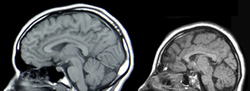
Tissue Engineering The need for neural tissue engineering arises from the difficulty of the nerve cells and neural tissues to regenerate on their own after neural damage has occurred. The PNS has some, but limited, regeneration of neural cells. Adult stem cell neurogenesis in the CNS has been found to occur in the hippocampus, the subventricular zone (SVZ), and spinal cord. [15] CNS injuries can be caused by stroke, neurodegenerative disorders, trauma, or encephalopathy. A few methods currently being investigated to treat CNS injuries are: implanting stem cells directly into the injury site, delivering morphogens to the injury site, or growing neural tissue in vitro with neural stem or progenitor cells in a 3D scaffold. [16] Proposed use of electrospun polymeric fibrous scaffolds for neural repair substrates dates back to at least 1986 in a NIH SBIR application from Simon. [17] For the PNS, a severed nerve can be reconnected and reinnervated using grafts or guidance of the existing nerve through a channel. [18]

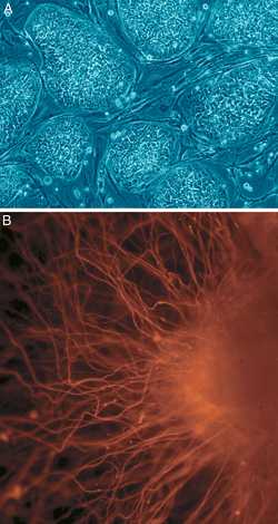
Recent research into creating miniature cortexes, known as corticopoiesis, and brain models, known as cerebral organoids, are techniques that could further the field of neural tissue regeneration. The native cortical progenitors in corticopoiesis are neural tissues that could be effectively embedded into the brain. [19] Cerebral organoids are 3D human pluripotent stem cells developed into sections of the brain cortex, showing that there is a potential to isolate and develop certain neural tissues using neural progenitors. [20]

Another situation that calls for implanting of foreign tissue is use of recording electrodes. Chronic Electrode Implants are a tool being used in research applications to record signals from regions of the cerebral cortex. Research into the stimulation of PNS neurons in patients with paralysis and prosthetics could further the knowledge of reinnervation of neural tissue in both the PNS and the CNS. [21] This research is capable of making one difficult aspect of neural tissue engineering, functional innervation of neural tissue, more manageable. [21]

CNS

Causes of CNS injury

There are three main causes of CNS injury: stroke, traumatic brain injury (TBI), or developmental complications. Strokes are classified as either hemorrhagic (when a vessel is damaged to the point of bleeding into the brain) or ischemic (when a clot blocks the blood flow through the vessel in the brain). When a hemorrhage occurs, blood seeps into the surrounding tissue, resulting in tissue death, while ischemic hemorrhages result in a lack of blood flow to certain tissues. Traumatic brain injury is caused by external forces impacting the cranium or the spinal cord. Problems with CNS development results in abnormal tissue growth during development, thus decreasing the function of the CNS. [16]

Normal Brain Development (left), Microcephaly, a type of encephalopathy (right) Microcephaly.png
Normal Brain Development (left), Microcephaly, a type of encephalopathy (right)

CNS treatments and research

Implantation of stem cells to the injury site

One method to treat CNS injury involves culturing stem cells in vitro and implanting the non-directed stem cells into the brain injury site. Implanting stem cells directly into the injury site prevents glial scar formation and promotes neurogenesis originating from the patient, but also runs the risk of tumor development, inflammation, and migration of the stem cells out of the injury location. Tumorigenesis can occur due to the uncontrolled nature of the stem cell differentiation, inflammation can occur due to rejection of the implanted cells by the host cells, and the highly migratory nature of stem cells results in the cells moving away from the injury site, thus not having the desired effect on the injury site. Other concerns of neural tissue engineering include establishing safe sources of stem cells and getting reproducible results from treatment to treatment. [16]

Alternatively, these stem cells can act as carriers for other therapies, though the positive effects of using stem cells as a delivery mechanism has not been confirmed. Direct stem cell delivery has an increased beneficial effect if they are directed to be neuronal cells in vitro. This way, the risks associated with undirected stem cells are decreased; additionally, injuries that do not have a specific boundary could be treated efficiently. [16]

Human embryonic stem colonies (A), axonal outgrowths (B) Human embryonic stem cells.png
Human embryonic stem colonies (A), axonal outgrowths (B)

Delivery of molecules to the injury site

Molecules that promote the regeneration of neural tissue, including pharmaceutical drugs, growth factors known as morphogens, and miRNA can also be directly introduced to the injury site of the damaged CNS tissue. Neurogenesis has been seen in animals that are treated with psychotropic drugs through the inhibition of serotonin reuptake and induction of neurogenesis in the brain. When stem cells are differentiating, the cells secrete morphogens such as growth factors to promote healthy development. These morphogens help maintain homeostasis and neural signaling pathways, and they can be delivered into the injury site to promote the growth of the injured tissues. Currently, morphogen delivery has minimal benefits because of the interactions the morphogens have with the injured tissue. Morphogens that are not innate in the body have a limited effect on the injured tissue due to the physical size and their limited mobility within CNS tissue. To be an effective treatment, the morphogens must be present at the injury site at a specific and constant concentration. miRNA has also been shown to affect neurogenesis by directing the differentiation of undifferentiated neural cells. [16]

Implantation of neural tissue developed in vitro

A third method for treating CNS injuries is to artificially create tissue outside of the body to implant into the injury site. This method could treat injuries that consist of large cavities, where larger amounts of neural tissue needs to be replaced and regenerated. Neural tissue is grown in vitro with neural stem or progenitor cells in a 3D scaffold, forming embryoid bodies (EBs). These EBs consist of a sphere of stem cells, where the inner cells are undifferentiated neural cells, and the surrounding cells are increasingly more differentiated. 3D scaffolds are used to transplant tissue to the injury site and to make the appropriate interface between the artificial and the brain tissue. The scaffolds must be: biocompatible, biodegradable, fit injury site, similar to existing tissue in elasticity and stiffness, and support growing cells and tissues. The combination of using directed stem cells and scaffolds to support the neural cells and tissues increase the survival of the stem cells in the injury site, increasing the efficacy of the treatment. [16]

Mouse Embryonic Stem Cells (mESCs) embryoid bodies (EBs) MESC EBs.jpg
Mouse Embryonic Stem Cells (mESCs) embryoid bodies (EBs)

There are 6 different types of scaffolds that are being researched to use in this method for treating neural tissue injury:

  • Liquid hydrogels are cross-linked hydrophobic polymer chains, and the neural stem cells are either grown on the surface of the gel or integrated into the gel during cross-linking of the polymer chains. The major drawback of liquid hydrogels is there is limited protection of the cells that are transplanted.
  • Supportive scaffolds are made from solid bead-shaped or microporous structures, and can act as carriers for the transplanted cells or for the growth factors that the stem cells secrete when they are differentiating. The cells adhere to the surface of the matrix in 2D layers. The supportive scaffolds are easily transplanted into the brain injury site because of the scaffold size. They provide a matrix promoting cell adhesion and aggregation, thus increasing increased healthy cell culture.
  • Aligning scaffolds can be silk-based, polysaccharide-based, or based on other materials such as a collagen-rich hydrogel. These gels are now enhanced with micro-patterns on the surface for the promotion of neuronal outgrowths. These scaffolds are primarily used for regeneration that needs to occur in a specific orientation, such as in spinal cord injuries.
  • Integrative scaffolds are mainly used to protect the transplanted cells from mechanical forces that they are exposed to in the process of implantation into the site of the injury. These scaffolds also decrease the likelihood of having the inflammatory cells located at the site of the injury migrate into the scaffold with the stem cells. Blood vessels have been observed to grow through the scaffold, thus the scaffold and cells are being integrated into the host tissue.
  • A combination of engineered scaffolds presents an option for a 3D scaffold that can have both the necessary patterns for cell adhesion and the flexibility to adapt to the ever changing environment at the injury site. Decellularized ECM scaffolds is an option for scaffolds because they more closely mimc the native tissue, but these scaffolds can only currently be harvested from amputations and cadavers. [16]

These 3D scaffolds can be fabricated using particulate leaching, gas foaming, fiber bonding, solvent casting, or electrospinning techniques; each technique creates a scaffold with different properties than the other techniques. [22]

Incorporation success of 3D scaffolds into the CNS has been shown to depend on the stage at which the cells have differentiated. Later stages provide a more efficient implantation, while earlier staged cells need to be exposed to factors that coerce the cells to differentiate and thus respond appropriately to the signals the cells will receive at the CNS injury site. [23] Brain-derived neurotrophic factor is a potential co-factor to promote functional activation of ES cell-derived neurons into the CNS injury sites. [24]

PNS

Causes of PNS injury

Trauma to the PNS can cause damage as severe as a severance of the nerve, splitting the nerve into a proximal and distal section. The distal nerve degenerates over time due to inactivity, while the proximal end swells over time. The distal end does not degenerate right away, and the swelling of the proximal end does not render it nonfunctional, so methods to reestablish the connection between the two ends of the nerve are being investigated. [18]

PNS treatments and research

Surgical reconnection

One method to treat PNS injury is surgical reconnection of the severed nerve by taking the two ends of the nerve and suturing them together. When suturing the nerves together, the fascicles of the nerve are each reconnected, bridging the nerve back together. Though this method works for severances that create a small gap between the proximal and distal nerve ends, this method does not work over gaps of greater distances due to the tension that must be put on the nerve endings. This tension results in the nerve degeneration, and therefore the nerve cannot regenerate and form a functional neural connection. [18]

Tissue grafts

Tissue grafts utilize nerves or other materials to bridge the two ends of the severed nerve. There are three categories of tissue grafts: autologous tissue grafts, nonautologous tissue grafts, and acellular grafts.

Autologous tissue grafts transplant nerves from a different part of the body of the patient to fill the gap between either end of the injured nerve. These nerves are typically cutaneous nerves, but other nerves have been researched as well with encouraging results. These autologous nerve grafts are the current gold standard for PNS nerve grafting because of the highly biocompatible nature of the autologous nerve graft, but there are issues concerning harvesting the nerve from the patients themselves and being able to store a large amount of autologous grafts for future use.

Nonautologous and acellular grafts (including ECM-based materials) are tissues that do not come from the patient, but instead can be harvested from cadavers (known as allogenic tissue) or animals (known as xenogeneic tissue). While these tissues have an advantage over autologous tissue grafts because the tissue does not need to be taken from the patient, difficulty arises with the potential of disease transmission and thus immunogenic problems. Methods of eliminating the immunogenic cells, thus leaving behind only the ECM-components of the tissue, are currently being investigated to increase the efficacy of nonautologous tissue grafts. [18]

Guidance

Regeneration with guidance in zebrafish peripheral axons

Guidance methods of PNS regeneration use nerve guide channels to help axons regrow along the correct path, and may direct growth factors secreted by both ends of the nerve to promote growth and reconnection. Guidance methods reduce scarring of the nerves, increasing the functionality of the nerves to transmit action potentials after reconnection. Two types of materials are used in guidance methods of PNS regeneration: natural-based materials and synthetic materials.

Natural-based materials are modified scaffolds stemming from ECM components and glycosaminoglycans. Laminin, collagen, and fibronectin, which are all ECM components, guide axonal development and promote neural stimulation and activity. Other molecules that have the potential to promote nerve repair are: hyaluronic acid, fibrinogen, fibrin gels, self-assembling peptide scaffolds, alginate, agarose, and chitosan.

Synthetic materials also provide another method for tissue regeneration in which the graft's chemical and physical properties can be controlled. Since the properties of a material may be specified for the situation in which it is being used, synthetic materials are an attractive option for PNS regeneration. The use of synthetic materials come with certain concerns, such as: easy formation of the graft material into the necessary dimensions, biodegradable, sterilizable, tear resistant, easy to operate with, low risk of infection, and low inflammation response due to the material. The material must also maintain the channel during the nerve regeneration. Currently, the materials most commonly researched mainly focus on polyesters, but biodegradable polyurethane, other polymers, and biodegradable glass are also being investigated. Other possibilities for synthetic materials are conducting polymers and polymers biologically modified to promote cell axon growth and maintain the axon channel. [18]

Neuroimmune Enhancement Through EVs

Extracellular vesicles (EVs) are bilayer-bound lipid particles that participate in intercellular communication by releasing a variety of substances, including nucleic acids, lipids, and proteins. [25] Exosomes, macrovesicles, and apoptotic bodies are the three primary forms; each has unique properties. EVs have the potential to be used as therapeutic delivery vehicles [26] and diagnostic biomarkers [27] and play roles in immunological responses, cancer, tissue regeneration, and neurological diseases. Damaged neurons generate neuron-derived exosomes (NDEs), which can influence target cells by transferring a variety of cargos, including the Zika virus. [28] [29] Neurodegenerative illnesses are linked to NDEs. Immune cell exosomes (IEEs) have the potential to be used in immunotherapy and vaccine development since they influence immune responses and interact with other cells. Immune cells such as DCs, macrophages, B cells, and T cells produce IEEs. EVs have been shown to promote neuroimmune crosstalk, allowing for both local and distant tissue and cell communication. [27]

Difficulty of research

Because there are so many factors that contribute to the success or failure of neural tissue engineering, there are many difficulties that arise in using neural tissue engineering to treat CNS and PNS injuries. First, the therapy needs to be delivered to the site of the injury. This means that the injury site needs to be accessed by surgery or drug delivery. Both of these methods have inherent risks and difficulties in themselves, compounding the problems associated with the treatments. A second concern is keeping the therapy at the site of the injury. Stem cells have a tendency to migrate out of the injury site to other sections of the brain, thus the therapy is not as effective as it could be as when the cells stay at the injury site. Additionally, the delivery of stem cells and other morphogens to the site of injury can cause more harm than good if they induce tumorigenesis, inflammation, or other unforeseen effects. Finally, the findings in laboratories may not translate to practical clinical treatments. Treatments are successful in a lab, or even an animal model of the injury, may not be effective in a human patient. [30]

Modeling brain tissue development in vitro

Two models for brain tissue development are cerebral organoids and corticopoiesis. These models provide an "in vitro" model for normal brain development, [20] but they can be manipulated to represent neural defects. Therefore, the mechanisms behind healthy and malfunctioning development can be studied by researchers using these models. [20] These tissues can be made with either mouse embryonic stem cells (ESC)s or human ESCs. Mouse ESCs are cultured in a protein called Sonic Hedgehog inhibitor to promote the development of dorsal forebrain and study cortical fate. [19] This method has been shown to produce axonal layers that mimic a broad range of cortical layers. [31] Human ESC-derived tissues use pluripotent stem cells to form tissues on scaffold, forming human EBs. These human ESC-derived tissues are formed by culturing human pluripotent EBs in a spinning bioreactor. [20]

Targeted reinnervation

Targeted reinnervation is a method to reinnervate the neural connections in the CNS and PNS, specifically in paralyzed patients and amputees using prosthetic limbs. Currently, devices are being investigated that take in and record the electrical signals that are propagated through neurons in response to a person's intent to move. This research could shed light on how to reinnervate the neural connections between severed PNS nerves and the connections between the transplanted 3D scaffolds into the CNS. [21]

Related Research Articles

<span class="mw-page-title-main">Central nervous system</span> Brain and spinal cord

The central nervous system (CNS) is the part of the nervous system consisting of the brain and spinal cord, the retina and optic nerve, and the olfactory nerve and epithelia. The CNS is so named because the brain integrates the received information and coordinates and influences the activity of all parts of the bodies of bilaterally symmetric and triploblastic animals—that is, all multicellular animals except sponges and diploblasts. It is a structure composed of nervous tissue positioned along the rostral to caudal axis of the body and may have an enlarged section at the rostral end which is a brain. Only arthropods, cephalopods and vertebrates have a true brain, though precursor structures exist in onychophorans, gastropods and lancelets.

<span class="mw-page-title-main">Nerve</span> Enclosed, cable-like bundle of axons in the peripheral nervous system

A nerve is an enclosed, cable-like bundle of nerve fibers in the peripheral nervous system.

<span class="mw-page-title-main">Nervous system</span> Part of an animal that coordinates actions and senses

In biology, the nervous system is the highly complex part of an animal that coordinates its actions and sensory information by transmitting signals to and from different parts of its body. The nervous system detects environmental changes that impact the body, then works in tandem with the endocrine system to respond to such events. Nervous tissue first arose in wormlike organisms about 550 to 600 million years ago. In vertebrates, it consists of two main parts, the central nervous system (CNS) and the peripheral nervous system (PNS). The CNS consists of the brain and spinal cord. The PNS consists mainly of nerves, which are enclosed bundles of the long fibers, or axons, that connect the CNS to every other part of the body. Nerves that transmit signals from the brain are called motor nerves or efferent nerves, while those nerves that transmit information from the body to the CNS are called sensory nerves or afferent. Spinal nerves are mixed nerves that serve both functions. The PNS is divided into three separate subsystems, the somatic, autonomic, and enteric nervous systems. Somatic nerves mediate voluntary movement. The autonomic nervous system is further subdivided into the sympathetic and the parasympathetic nervous systems. The sympathetic nervous system is activated in cases of emergencies to mobilize energy, while the parasympathetic nervous system is activated when organisms are in a relaxed state. The enteric nervous system functions to control the gastrointestinal system. Both autonomic and enteric nervous systems function involuntarily. Nerves that exit from the cranium are called cranial nerves while those exiting from the spinal cord are called spinal nerves.

<span class="mw-page-title-main">Schwann cell</span> Glial cell type

Schwann cells or neurolemmocytes are the principal glia of the peripheral nervous system (PNS). Glial cells function to support neurons and in the PNS, also include satellite cells, olfactory ensheathing cells, enteric glia and glia that reside at sensory nerve endings, such as the Pacinian corpuscle. The two types of Schwann cells are myelinating and nonmyelinating. Myelinating Schwann cells wrap around axons of motor and sensory neurons to form the myelin sheath. The Schwann cell promoter is present in the downstream region of the human dystrophin gene that gives shortened transcript that are again synthesized in a tissue-specific manner.

<span class="mw-page-title-main">Nervous tissue</span> Main component of the nervous system

Nervous tissue, also called neural tissue, is the main tissue component of the nervous system. The nervous system regulates and controls body functions and activity. It consists of two parts: the central nervous system (CNS) comprising the brain and spinal cord, and the peripheral nervous system (PNS) comprising the branching peripheral nerves. It is composed of neurons, also known as nerve cells, which receive and transmit impulses, and neuroglia, also known as glial cells or glia, which assist the propagation of the nerve impulse as well as provide nutrients to the neurons.

<span class="mw-page-title-main">Motor nerve</span> Nerve located in the central nervous system

A motor nerve is a nerve that transmits motor signals from the central nervous system (CNS) to the muscles of the body. This is different from the motor neuron, which includes a cell body and branching of dendrites, while the nerve is made up of a bundle of axons. Motor nerves act as efferent nerves which carry information out from the CNS to muscles, as opposed to afferent nerves, which transfer signals from sensory receptors in the periphery to the CNS. Efferent nerves can also connect to glands or other organs/issues instead of muscles. In addition, there are nerves that serve as both sensory and motor nerves called mixed nerves.

<span class="mw-page-title-main">Glia</span> Support cells in the nervous system

Glia, also called glial cells(gliocytes) or neuroglia, are non-neuronal cells in the central nervous system (brain and spinal cord) and the peripheral nervous system that do not produce electrical impulses. The neuroglia make up more than one half the volume of neural tissue in our body. They maintain homeostasis, form myelin in the peripheral nervous system, and provide support and protection for neurons. In the central nervous system, glial cells include oligodendrocytes, astrocytes, ependymal cells and microglia, and in the peripheral nervous system they include Schwann cells and satellite cells.

<span class="mw-page-title-main">Wallerian degeneration</span> Biological process of axonal degeneration

Wallerian degeneration is an active process of degeneration that results when a nerve fiber is cut or crushed and the part of the axon distal to the injury degenerates. A related process of dying back or retrograde degeneration known as 'Wallerian-like degeneration' occurs in many neurodegenerative diseases, especially those where axonal transport is impaired such as ALS and Alzheimer's disease. Primary culture studies suggest that a failure to deliver sufficient quantities of the essential axonal protein NMNAT2 is a key initiating event.

Neural engineering is a discipline within biomedical engineering that uses engineering techniques to understand, repair, replace, or enhance neural systems. Neural engineers are uniquely qualified to solve design problems at the interface of living neural tissue and non-living constructs.

Stem-cell therapy uses stem cells to treat or prevent a disease or condition. As of 2016, the only established therapy using stem cells is hematopoietic stem cell transplantation. This usually takes the form of a bone marrow transplantation, but the cells can also be derived from umbilical cord blood. Research is underway to develop various sources for stem cells as well as to apply stem-cell treatments for neurodegenerative diseases and conditions such as diabetes and heart disease.

<span class="mw-page-title-main">Neuroimmune system</span>

The neuroimmune system is a system of structures and processes involving the biochemical and electrophysiological interactions between the nervous system and immune system which protect neurons from pathogens. It serves to protect neurons against disease by maintaining selectively permeable barriers, mediating neuroinflammation and wound healing in damaged neurons, and mobilizing host defenses against pathogens.

Neural stem cells (NSCs) are self-renewing, multipotent cells that firstly generate the radial glial progenitor cells that generate the neurons and glia of the nervous system of all animals during embryonic development. Some neural progenitor stem cells persist in highly restricted regions in the adult vertebrate brain and continue to produce neurons throughout life. Differences in the size of the central nervous system are among the most important distinctions between the species and thus mutations in the genes that regulate the size of the neural stem cell compartment are among the most important drivers of vertebrate evolution.

<span class="mw-page-title-main">Nerve injury</span> Damage to nervous tissue

Nerve injury is an injury to a nerve. There is no single classification system that can describe all the many variations of nerve injuries. In 1941, Seddon introduced a classification of nerve injuries based on three main types of nerve fiber injury and whether there is continuity of the nerve. Usually, however, nerve injuries are classified in five stages, based on the extent of damage to both the nerve and the surrounding connective tissue, since supporting glial cells may be involved.

Neuroregeneration involves the regrowth or repair of nervous tissues, cells or cell products. Neuroregenerative mechanisms may include generation of new neurons, glia, axons, myelin, or synapses. Neuroregeneration differs between the peripheral nervous system (PNS) and the central nervous system (CNS) by the functional mechanisms involved, especially in the extent and speed of repair. When an axon is damaged, the distal segment undergoes Wallerian degeneration, losing its myelin sheath. The proximal segment can either die by apoptosis or undergo the chromatolytic reaction, which is an attempt at repair. In the CNS, synaptic stripping occurs as glial foot processes invade the dead synapse.

A nerve guidance conduit is an artificial means of guiding axonal regrowth to facilitate nerve regeneration and is one of several clinical treatments for nerve injuries. When direct suturing of the two stumps of a severed nerve cannot be accomplished without tension, the standard clinical treatment for peripheral nerve injuries is autologous nerve grafting. Due to the limited availability of donor tissue and functional recovery in autologous nerve grafting, neural tissue engineering research has focused on the development of bioartificial nerve guidance conduits as an alternative treatment, especially for large defects. Similar techniques are also being explored for nerve repair in the spinal cord but nerve regeneration in the central nervous system poses a greater challenge because its axons do not regenerate appreciably in their native environment.

A fibrin scaffold is a network of protein that holds together and supports a variety of living tissues. It is produced naturally by the body after injury, but also can be engineered as a tissue substitute to speed healing. The scaffold consists of naturally occurring biomaterials composed of a cross-linked fibrin network and has a broad use in biomedical applications.

Preferential motor reinnervation (PMR) refers to the tendency of a regenerating axon in the peripheral nervous system (PNS) to reinnervate a motor pathway as opposed to a somatosensory pathway. PMR affects how nerves regenerate and reinnervate within the PNS after surgical procedures or traumatic injuries. It is important to understand in order to further develop axonal regrowth surgical techniques. Further research of preferential motor reinnervation will lead to a better understanding of peripheral nervous system function in the human body regarding cell roles and abilities.

Spinal cord injury research seeks new ways to cure or treat spinal cord injury in order to lessen the debilitating effects of the injury in the short or long term. There is no cure for SCI, and current treatments are mostly focused on spinal cord injury rehabilitation and management of the secondary effects of the condition. Two major areas of research include neuroprotection, ways to prevent damage to cells caused by biological processes that take place in the body after the injury, and neuroregeneration, regrowing or replacing damaged neural circuits.

Nano neuro knitting is an emerging technology for repairing nervous system tissues via nano scaffolding techniques. Currently being explored in numerous research endeavors, nano neuro knitting has been shown to allow partial reinnervation in damaged areas of the nervous system through the interactions between potentially regenerative axons and peptide scaffolds. This interaction has been shown to lead to sufficient axon density renewal to the point that functionality is restored. While nano neuro knitting shows promise, the uncertainty of the effects in human subjects warrants further investigation before clinical trials initiate.

Craniofacial regeneration refers to the biological process by which the skull and face regrow to heal an injury. This page covers birth defects and injuries related to the craniofacial region, the mechanisms behind the regeneration, the medical application of these processes, and the scientific research conducted on this specific regeneration. This regeneration is not to be confused with tooth regeneration. Craniofacial regrowth is broadly related to the mechanisms of general bone healing.

References

  1. 1 2 Boahen, A.; Hu, D.; Adams, M. J.; Nicholls, P. K.; Greene, W. K.; Ma, B. (2023). "Bidirectional crosstalk between the peripheral nervous system and lymphoid tissues/Organs". Frontiers in Immunology. 14. doi: 10.3389/fimmu.2023.1254054 . PMC   10520967 . PMID   37767094.
  2. Marshall, J. S.; Warrington, R.; Watson, W.; Kim, H. L. (2018). "An introduction to immunology and immunopathology". Allergy, Asthma, and Clinical Immunology. 14 (Suppl 2): 49. doi: 10.1186/s13223-018-0278-1 . PMC   6156898 . PMID   30263032.
  3. Lang, G. P.; Ndongson-Dongmo, B.; Lajqi, T.; Brodhun, M.; Han, Y.; Wetzker, R.; Frasch, M. G.; Bauer, R. (2020). "Impact of ambient temperature on inflammation-induced encephalopathy in endotoxemic mice-role of phosphoinositide 3-kinase gamma". Journal of Neuroinflammation. 17 (1): 292. doi: 10.1186/s12974-020-01954-7 . PMC   7541275 . PMID   33028343.
  4. Peters EMJ; Schedlowski, M.; Watzl, C.; Gimsa, U. (2021). "To stress or not to stress: Brain-behavior-immune interaction may weaken or promote the immune response to SARS-CoV-2". Neurobiology of Stress. 14. doi:10.1016/j.ynstr.2021.100296. PMC   7839386 . PMID   33527083.
  5. Quatrini, L.; Vivier, E.; Ugolini, S. (2018). "Neuroendocrine regulation of innate lymphoid cells". Immunological Reviews. 286 (1): 120–136. doi:10.1111/imr.12707. PMC   6221181 . PMID   30294960.
  6. Grant, Spencer M.; Lou, Meng; Yao, Li; Germain, Ronald N.; Radtke, Andrea J. (March 6, 2020). "The lymph node at a glance - how spatial organization optimizes the immune response". Journal of Cell Science. 133 (5): jcs241828. doi:10.1242/jcs.241828. PMC   7063836 . PMID   32144196.
  7. 1 2 De Virgiliis, Francesco; Oliva, Valeria Maria; Kizil, Burak; Scheiermann, Christoph (September 26, 2022). "Control of lymph node activity by direct local innervation". Trends in Neurosciences. 45 (9): 704–712. doi:10.1016/j.tins.2022.06.006. PMID   35820971. S2CID   250361701 via PubMed.
  8. Cleypool, Cindy G. J.; Mackaaij, Claire; Lotgerink Bruinenberg, Dyonne; Schurink, Bernadette; Bleys, Ronald L. A. W. (August 26, 2021). "Sympathetic nerve distribution in human lymph nodes". Journal of Anatomy. 239 (2): 282–289. doi:10.1111/joa.13422. PMC   8273593 . PMID   33677834.
  9. Chang, E. H.; Carnevale, D.; Chavan, S. S. (2023). "Editorial: Understanding and targeting neuro-immune interactions within disease and inflammation". Frontiers in Immunology. 14. doi: 10.3389/fimmu.2023.1201669 . PMC   10154784 . PMID   37153559.
  10. Musial, F. (2019). "Acupuncture for the Treatment of Pain - A Mega-Placebo?". Frontiers in Neuroscience. 13: 1110. doi: 10.3389/fnins.2019.01110 . PMC   6811493 . PMID   31680841.
  11. 1 2 Johnson, R. L.; Wilson, C. G. (2018). "A review of vagus nerve stimulation as a therapeutic intervention". Journal of Inflammation Research. 11: 203–213. doi: 10.2147/JIR.S163248 . PMC   5961632 . PMID   29844694.
  12. Baraldi, J. H.; Martyn, G. V.; Shurin, G. V.; Shurin, M. R. (2022). "Tumor Innervation: History, Methodologies, and Significance". Cancers. 14 (8): 1979. doi: 10.3390/cancers14081979 . PMC   9029781 . PMID   35454883.
  13. Oaklander, A. L.; Mills, A. J.; Kelley, M.; Toran, L. S.; Smith, B.; Dalakas, M. C.; Nath, A. (2022). "Peripheral Neuropathy Evaluations of Patients with Prolonged Long COVID". Neurology(R) Neuroimmunology & Neuroinflammation. 9 (3). doi:10.1212/NXI.0000000000001146. PMC   8889896 . PMID   35232750.
  14. Austelle, C. W.; O'Leary, G. H.; Thompson, S.; Gruber, E.; Kahn, A.; Manett, A. J.; Short, B.; Badran, B. W. (2022). "A Comprehensive Review of Vagus Nerve Stimulation for Depression". Neuromodulation. 25 (3): 309–315. doi:10.1111/ner.13528. PMC   8898319 . PMID   35396067.
  15. Temple, Sally (November 2001). "The development of neural stem cells". Nature. 414 (6859): 112–117. Bibcode:2001Natur.414..112T. doi:10.1038/35102174. PMID   11689956. S2CID   4302192.
  16. 1 2 3 4 5 6 7 Forraz, N.; Wright, K. E.; Jurga, M.; McGuckin, C. P. (2013). "Experimental therapies for repair of the central nervous system: stem cells and tissue engineering". Journal of Tissue Engineering and Regenerative Medicine. 7 (7): 523–536. doi: 10.1002/term.552 . PMID   22467493. S2CID   42424867.
  17. Simon, Eric M. (1986). "NIH SBIR PHASE I GRANT APPLICATION: FIBROUS SUBSTRATES FOR CELL CULTURE (PDF Download Available)". doi:10.13140/RG.2.2.22440.16647 . Retrieved 2017-05-22 via ResearchGate.
  18. 1 2 3 4 5 Schmidt, Christine; Jennie Leach (June 2003). "Neural Tissue Engineering: Strategies for Repair and Regeneration". Annual Review of Biomedical Engineering. 5: 293–347. doi:10.1146/annurev.bioeng.5.011303.120731. PMID   14527315.
  19. 1 2 Gaspard, N.; Gaillard, A.; Vanderhaegen, P. (August 2009). "Making cortex in a dish: In vitro corticopoiesis from embryonic stem cells". Cell Cycle. 8 (16): 2491–6. doi: 10.4161/cc.8.16.9276 . PMID   19597331.
  20. 1 2 3 4 Lancaster, M.A.; et al. (August 2013). "Cerebral organoids model human brain development and microcephaly". Nature. 501 (7467): 373–379. Bibcode:2013Natur.501..373L. doi:10.1038/nature12517. PMC   3817409 . PMID   23995685.
  21. 1 2 3 Tenore, Francesco; Vogelstein, Jacob (2011). "Revolutionizing Prothetics: Devices for Neural Integration". Johns Hopkins APL Technical Digest. 30 (3): 230–39.
  22. Ko, J; Mohtaram NK; Ahmed F; et al. (September 2013). "Fabrication of poly (ϵ-caprolactone) microfiber scaffolds with varying topography and mechanical properties for stem cell-based tissue engineering applications". Journal of Biomaterials Science, Polymer Edition. 25 (1): 1–17. doi:10.1080/09205063.2013.830913. hdl: 1828/7315 . PMID   23998440. S2CID   205620361.
  23. Sheen, V. L.; Arnold, M. W.; Wang, Y.; Macklis, J. D. (July 1999). "Neural precursor differentiation following transplantation into neocortex is dependent on intrinsic developmental state and receptor competence". Experimental Neurology. 158 (1): 47–62. doi:10.1006/exnr.1999.7104. PMID   10448417. S2CID   12291422.
  24. Copi, A; Jungling, K.; Gottmann, K. (November 2005). "Activity- and BDNF-induced plasticity of miniature synaptic currents in ES cell-derived neurons integrated in a neocortical network". Journal of Neurophysiology. 94 (6): 4538–43. doi:10.1152/jn.00155.2005. PMID   16293594.
  25. Doyle, L. M.; Wang, M. Z. (2019). "Overview of Extracellular Vesicles, Their Origin, Composition, Purpose, and Methods for Exosome Isolation and Analysis". Cells. 8 (7): 727. doi: 10.3390/cells8070727 . PMC   6678302 . PMID   31311206.
  26. Butreddy, A.; Kommineni, N.; Dudhipala, N. (2021). "Exosomes as Naturally Occurring Vehicles for Delivery of Biopharmaceuticals: Insights from Drug Delivery to Clinical Perspectives". Nanomaterials (Basel, Switzerland). 11 (6): 1481. doi: 10.3390/nano11061481 . PMC   8229362 . PMID   34204903.
  27. 1 2 Hazrati, A.; Soudi, S.; Malekpour, K.; Mahmoudi, M.; Rahimi, A.; Hashemi, S. M.; Varma, R. S. (2022). "Immune cells-derived exosomes function as a double-edged sword: Role in disease progression and their therapeutic applications". Biomarker Research. 10 (1): 30. doi: 10.1186/s40364-022-00374-4 . PMC   9102350 . PMID   35550636.
  28. Pineles, B.; Mani, A.; Sura, L.; Rossignol, C.; Albayram, M.; Weiss, M. D.; Goetzl, L. (2022). "Neuronal exosome proteins: Novel biomarkers for predicting neonatal response to therapeutic hypothermia". Archives of Disease in Childhood. Fetal and Neonatal Edition. 107 (1): 60–64. doi:10.1136/archdischild-2020-321096. PMID   34021027. S2CID   235076563.
  29. Zhou, W.; Woodson, M.; Sherman, M. B.; Neelakanta, G.; Sultana, H. (2019). "Exosomes mediate Zika virus transmission through SMPD3 neutral Sphingomyelinase in cortical neurons". Emerging Microbes & Infections. 8 (1): 307–326. doi:10.1080/22221751.2019.1578188. PMC   6455149 . PMID   30866785.
  30. LaPlaca, Michele (3 October 2013). "Personal Interview".
  31. Gaspard, N; et al. (September 2008). "An intrinsic mechanism of corticogenesis from embryonic stem cells" (PDF). Nature. 455 (7211): 351–357. Bibcode:2008Natur.455..351G. doi:10.1038/nature07287. hdl:2268/239873. PMID   18716623. S2CID   2611446.