
Acremonium is a genus of fungi in the family Hypocreaceae. It used to be known as Cephalosporium.
Acremonium strictum is an environmentally widespread saprotroph species found in soil, plant debris, and rotting mushrooms. Isolates have been collected in North and Central America, Asia, Europe and Egypt. A. strictum is an agent of hyalohyphomycosis and has been identified as an increasingly frequent human pathogen in immunosuppressed individuals, causing localized, disseminated and invasive infections. Although extremely rare, A. strictum can infect immunocompetent individuals, as well as neonates. Due to the growing number of infections caused by A. strictum in the past few years, the need for new medical techniques in the identification of the fungus as well as for the treatment of human infections has risen considerably.

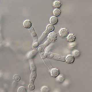
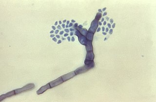
Acrophialophora fusispora is a poorly studied ascomycete fungus found in soil, air and various plants. A. fusispora is morphologically similar to the genera Paecilomyces and Masonia, but differ in the presence of pigmented conidiophores, verticillate phialides, and frequent sympodial proliferation. Moreover, A. fusispora is distinguished by its pigmented spindle-shaped conidia, covered with spiral bands. The fungus is naturally found in soils of tropical to temperate regions. The fungus has been identified as a plant and animal pathogen, and has recently been recognized as an emerging opportunistic human pathogen. A. fusispora infection in human is rare and has few documented clinical cases, but due to the rarity of the fungus and potential misidentification, the infections may be underdiagnosed. Clinical cases of A. fusispora include cases of keratitis, pulmonary colonization and infection, and cerebral infections. The fungus also has two documented cases of infection in dogs.
Exophiala jeanselmei is a saprotrophic fungus in the family Herpotrichiellaceae. Four varieties have been discovered: Exophiala jeanselmei var. heteromorpha, E. jeanselmei var. lecanii-corni, E. jeanselmei var. jeanselmei, and E. jeanselmei var. castellanii. Other species in the genus Exophiala such as E. dermatitidis and E. spinifera have been reported to have similar annellidic conidiogenesis and may therefore be difficult to differentiate.

Fonsecaea pedrosoi is a fungal species in the family Herpotrichiellaceae, and the major causative agent of chromoblastomycosis. This species is commonly found in tropical and sub-tropical regions, especially in South America, where it grows as a soil saprotroph. Farming activities in the endemic zone are a risk factor for the development of chromoblastomycosis.

Exophiala dermatitidis is a thermophilic black yeast, and a member of the Herpotrichiellaceae. While the species is only found at low abundance in nature, metabolically active strains are commonly isolated in saunas, steam baths, and dish washers. Exophiala dermatitidis only rarely causes infection in humans, however cases have been reported around the world. In East Asia, the species has caused lethal brain infections in young and otherwise healthy individuals. The fungus has been known to cause cutaneous and subcutaneous phaeohyphomycosis, and as a lung colonist in people with cystic fibrosis in Europe. In 2002, an outbreak of systemic E. dermatitidis infection occurred in women who had received contaminated steroid injections at North Carolina hospitals.
Coniochaeta hoffmannii, also known as Lecythophora hoffmannii, is an ascomycete fungus that grows commonly in soil. It has also been categorized as a soft-rot fungus capable of bringing the surface layer of timber into a state of decay, even when safeguarded with preservatives. Additionally, it has pathogenic properties, although it causes serious infection only in rare cases. A plant pathogen lacking a known sexual state, C. hoffmannii has been classified as a "dematiaceous fungus" despite its contradictory lack of pigmentation; both in vivo and in vitro, there is no correlation between its appearance and its classification.
Microascus brevicaulis is a microfungus in the Ascomycota. It is the teleomorph form of Scopulariopsis brevicaulis.Microascus brevicaulis occurs world-wide as a saprotroph in soil, a common agent of biodeterioration, an irregular plant pathogen, and an occasional agent of human nail infection.
Phialemonium curvatum is a pathogenic fungus in the phylum Ascomycota. The genus was created to accommodate taxa intermediate to Acremonium and Phialophora. This genus is characterized by its abundance of adelophialides and few discrete phialides with no signs of collarettes. Specifically, P. curvatum is characterized by its grayish white colonies and its allantoid conidia. Phialemonium curvatum is typically found in a variety of environments including air, soil, industrial water and sewage. Furthermore, P. curvatum affects mainly immunocompromised and is rarely seen in immunocompetent people. The species has been known to cause peritonitis, endocarditis, endovascular infections, osteomyelitis as well as cutaneous infections of wounds and burns.
Phialemonium is a genus of fungi in the family Cephalothecaceae of the Ascomycota. The genus was circumscribed by Walter Gams and Michael McGinnis in 1983. The genus is intermediate between Acremonium and Phialophora. It is classified as a dematiaceous (dark-walled) fungus.
Aspergillus unguis is a species of fungus in the genus Aspergillus, and the asexual state (anamorph) of Emericella unguis. Aspergillus unguis is a filamentous soil-borne fungus found on decomposing plant matter and other moist substrates including with building materials and household dust. Aspergillus unguis occurs mainly in tropical and subtropical soils but has also been isolated from various marine and aquatic habitats. The species was first isolated in 1935 by Weill and L. Gaudin. Historically, A. unguis was assigned to the A. nidulans group, a common group of soil-borne fungi due to the resemblance of its ascospores and cleistothecia to those of Emericella nidulans. Aspergillus unguis is distinctive, however, in possessing spicular hyphae. A number of synonyms have been collapsed into this species, including Sterigmatocystis unguis, Aspergillus laokiashanensis and Aspergillus mellinus.
Fonsecaea compacta is a saprophytic fungal species found in the family Herpotrichiellaceae. It is a rare etiological agent of chromoblastomycosis, with low rates of correspondence observed from reports. The main active components of F. compacta are glycolipids, yet very little is known about its composition. F. compacta is widely regarded as a dysplastic variety of Fonsecaea pedrosoi, its morphological precursor. The genus Fonsecaea presently contains two species, F. pedrosoi and F. compacta. Over 100 strains of F. pedrosoi have been isolated but only two of F. compacta.

Phialophora verrucosa is a pathogenic, dematiaceous fungus that is a common cause of chromoblastomycosis. It has also been reported to cause subcutaneous phaeohyphomycosis and mycetoma in very rare cases. In the natural environment, it can be found in rotting wood, soil, wasp nests, and plant debris. P. verrucosa is sometimes referred to as Phialophora americana, a closely related environmental species which, along with P. verrucosa, is also categorized in the P. carrionii clade.
Exophiala pisciphila is a mesophilic black yeast and member of the dark septate endophytes. This saprotrophic fungus is found commonly in marine and soil environments. It is abundant in harsh environments like soil contaminated with heavy metals. E. pisciphila forms symbiotic relationships with various plants by colonizing on roots, conferring resistance to drought and heavy metal stress. It is an opportunistic pathogen that commonly causes infections in captive fish and amphibians, while rarely causing disease in humans. Secondary metabolites produced by this species have potential clinical antibiotic and antiretroviral applications.
Sarocladium kiliense is a saprobic fungus that is occasionally encountered as a opportunistic pathogen of humans, particularly immunocompromised and individuals. The fungus is frequently found in soil and has been linked with skin and systemic infections. This species is also known to cause disease in the green alga, Cladophora glomerata as well as various fruit and vegetable crops grown in warmer climates.

Phialophora fastigiata is a mitosporic, saprophytic fungus commonly found in soil, and on wood, and wood-pulp. This species was initially placed in the genus Cadophora but was later transferred to the genus Phialophora based on morphological and growth characteristics. In culture, P. fastigiata produces olive-brown, velvety colonies. The fungus is recognizable microscopically due to the presence of distinctive, funnel-shaped cuffs (collarettes) encircling the tips of phialides that bear slimy conidia. The fungus is often implicated in soft-rot wood decay due to its ability to degrade lignin, cellulose and pectin. It has also been reported to cause blue staining of wood and wood pulp.
Cladophialophora arxii is a black yeast shaped dematiaceous fungus that is able to cause serious phaeohyphomycotic infections. C. arxii was first discovered in 1995 in Germany from a 22-year-old female patient suffering multiple granulomatous tracheal tumours. It is a clinical strain that is typically found in humans and is also capable of acting as an opportunistic fungus of other vertebrates Human cases caused by C. arxii have been reported from all parts of the world such as Germany and Australia.
Gliomastix murorum is one of four species of fungus in the genus Gliomastix. G. murorum is a type of saprophyte. One of the techniques that is used to isolate this fungus is through dilution plate.
Cephalotheca foveolata is a species of fungus. It is rarely opportunistic and generally manifests as a minor subcutaneous infection.
Rhopalophora is a genus of lichen-like fungus in the family Dactylosporaceae. It contains the sole species R. clavispora, previously belonging to the genus Phialophora but redescribed in 2016 to compose this monotypic genus.

