Timeline of human prenatal development

Last updated

Changes by weeks of gestation

Gestational age vs. embryonic age

Gestational age is the time that has passed since the onset of the last menstruation, which generally or as standard occurs 2 weeks before the actual fertilization. Embryonic age , in contrast measures the actual age of the embryo or fetus from the time of fertilization. Nevertheless, menstruation has historically been the only means of estimating embryonal/fetal age, and is still the presumed measure if not else specified. However, the actual duration between last menstruation and fertilization may in fact differ from the standard 2 weeks by several days.

Contents

Thus, the first week of embryonic age is already week three counting with gestational age.

Furthermore, the number of the week is one more than the actual age of the embryo/fetus. For example, the embryo is 0 whole weeks old during the 1st week after fertilization.

The following table summarizes the various expression systems during week number x of gestation.

Week
number
Initial age
(whole weeks)
Gestationalxx-1
Embryonicx-2x-3

Week 3

Gestational age: 2 weeks and 0 days until 2 weeks and 6 days old. 15–21 days from last menstruation.

Embryonic age: Week nr 1. 0 (whole) weeks old. 1–7 days from fertilization.

Week 4

Gestational age: 3 weeks and 0 days until 3 weeks and 6 days old. 22–28 days from last menstruation.

Embryonic age: Week nr 2. 1 week old. 8–14 days from fertilization.

Week 5

Gestational age: 4 weeks and 0 days until 4 weeks and 6 days old. 29–35 days from last menstruation.

Embryonic age: Week nr 3. 2 weeks old. 15–21 days from fertilization.

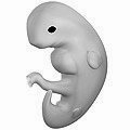
Embryo at 4 weeks after fertilization. 6 weeks pregnant.png
Embryo at 4 weeks after fertilization.
A 10mm embryo from an ectopic pregnancy, still in the oviduct. This embryo is about five weeks old (or from the seventh week of menstrual age). Tubal Pregnancy with embryo.jpg
A 10mm embryo from an ectopic pregnancy, still in the oviduct. This embryo is about five weeks old (or from the seventh week of menstrual age).

Week 6

Gestational age: 5 weeks and 0 days until 5 weeks and 6 days old. 36–42 days from last menstruation.

Embryonic age: Week nr 4. 3 weeks old. 22–28 days from fertilization.

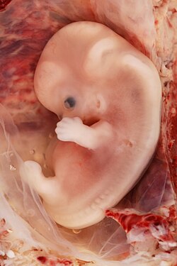
A six-week embryonic age or eight-week gestational age intact human embryo. Human Embryo.JPG
A six-week embryonic age or eight-week gestational age intact human embryo.

Week 7

Gestational age: 6 weeks and 0 days until 6 weeks and 6 days old. 43–49 days from last menstruation.

Embryonic age: Week nr 5. 4 weeks old. 29–35 days from fertilization.

[5]

This embryo is also from an ectopic pregnancy, this one in the cornu (the part of the uterus to which the Fallopian tube is attached). The features are consistent with a developmental age of seven weeks (reckoned as the ninth week of pregnancy). 9-Week Human Embryo from Ectopic Pregnancy.jpg
This embryo is also from an ectopic pregnancy, this one in the cornu (the part of the uterus to which the Fallopian tube is attached). The features are consistent with a developmental age of seven weeks (reckoned as the ninth week of pregnancy).

Week 8

Gestational age: 7 weeks and 0 days until 7 weeks and 6 days old. 50–56 days from last menstruation.

Embryonic age: Week nr 6. 5 weeks old. 36–42 days from fertilization.

Week 9

Gestational age: 8 weeks and 0 days until 8 weeks and 6 days old. 57–63 days from last menstruation.

Embryonic age: Week nr 7. 6 weeks old. 43–49 days from fertilization.

Changes by weeks of gestation

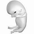
Fetus at 8 weeks after fertilization. 10 weeks pregnant.png
Fetus at 8 weeks after fertilization.

Weeks 10 to 12

Gestational age: 9 weeks and 0 days until 11 weeks and 6 days old.

Embryonic age: 7 weeks and 0 days until 9 weeks and 6 days old.

Fetus at 10 weeks Human fetus 10 weeks with amniotic sac - therapeutic abortion.jpg
Fetus at 10 weeks
  • Embryo measures 30–80 mm (1.2–3.1 in) in length.
  • Ventral and dorsal pancreatic buds fuse during the 8th week
  • Intestines rotate.
  • Facial features continue to develop.
  • The eyelids are more developed.
  • The external features of the ear begin to take their final shape.
  • The head comprises nearly half of the fetus' size.
  • The face is well formed.
  • The eyelids close and will not reopen until about the 27th week.
  • Tooth buds, which will form the baby teeth, appear.
  • The limbs are long and thin.
  • The fetus can make a fist with its fingers.
  • Genitals appear well differentiated.
  • Red blood cells are produced in the liver.
  • Heartbeat can be detected by ultrasound. [7]

Weeks 13 to 16

Gestational age: 12 weeks and 0 days until 15 weeks and 6 days old.

Embryonic age: 10 weeks and 0 days until 13 weeks and 6 days old.

  • The fetus reaches a length of about 15 cm (6 in).
  • A fine hair called lanugo develops on the head.
  • Fetal skin is almost transparent.
  • More muscle tissue and bones have developed, and the bones become harder.
  • The fetus makes active movements.
  • Sucking motions are made with the mouth.
  • Meconium is made in the intestinal tract.
  • The liver and pancreas produce fluid secretions.
  • From week 13, sex prediction by obstetric ultrasonography is almost 100% accurate. [8]
  • At week 15, main development of external genitalia is finished.
Fetus at 18 weeks after fertilization. 20 weeks pregnant.png
Fetus at 18 weeks after fertilization.

Week 21

Gestational age: 20 weeks old.

Embryonic age: 18 weeks old.

  • The fetus reaches a length of 20 cm (8 in).
  • Lanugo covers the entire body.
  • Eyebrows and eyelashes appear.
  • Nails appear on fingers and toes.
  • The fetus is more active with increased muscle development.
  • "Quickening" usually occurs (the mother and others can feel the fetus moving).
  • The fetal heartbeat can be heard with a stethoscope.

Week 23

Gestational age: 22 weeks old.

Embryonic age: 20 weeks old.

  • The fetus reaches a length of 28 cm (11 in).
  • The fetus weighs about 500g.
  • Eyebrows and eyelashes are well formed.
  • All of the eye components are developed.
  • The fetus has a hand and startle reflex.
  • Footprints and fingerprints continue forming.
  • Alveoli (air sacs) are forming in lungs.

Week 26

Gestational age: 24 weeks old.

Embryonic age: Week nr 25. 24 weeks old.

  • The fetus reaches a length of 32.5 cm (13 in). [10]
  • The fetus weighs about 650 g (1 lb 7 oz). [11]
  • The brain develops rapidly.
  • The nervous system develops enough to control some body functions.
  • The eyelids open and close.
  • The cochleae are now developed, though the myelin sheaths in neural portion of the auditory system will continue to develop until 18 months after birth.
  • The respiratory system, while immature, has developed to the point where gas exchange is possible.

Week 31

Gestational age: 30 weeks old.

Embryonic age: Week nr 29. 28 weeks old.

  • The fetus reaches a length of about 38–43 cm (15–17 in).
  • The fetus weighs about 1.5 kg (3 lb 5 oz).
  • The amount of body fat rapidly increases.
  • Rhythmic breathing movements occur, but lungs are not fully mature.
  • Thalamic brain connections, which mediate sensory input, form.
  • Bones are fully developed, but are still soft and pliable.
  • The fetus begins storing a lot of iron, calcium and phosphorus.

Week 35

Gestational age: 34 weeks old.

Embryonic age: Week nr 33. 32 weeks old.

  • The fetus reaches a length of about 40–48 cm (16–19 in).
  • The fetus weighs about 2.5 to 3 kg (6 lb 10 oz)to 6 lb 12 oz).
  • Lanugo begins to disappear.
  • Body fat increases.
  • Fingernails reach the end of the fingertips.
  • A baby born at 36 weeks has a high chance of survival, but may require medical interventions.
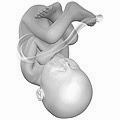
Fetus at 38 weeks after fertilization. 40 weeks pregnant.png
Fetus at 38 weeks after fertilization.

Weeks 36 to 40

Gestational age: 35 and 0 days until 39 weeks and 6 days old.

Embryonic age: Weeks nr 34–38. 33–37 weeks old.

  • The fetus is considered full-term at the end of the 39th week of gestational age.
  • It may be 48 to 53 cm (19 to 21 in) in length.
  • The lanugo is gone except on the upper arms and shoulders.
  • Fingernails extend beyond fingertips.
  • Small breast buds are present on both sexes.
  • Head hair is now coarse and thickest.

Related Research Articles

<span class="mw-page-title-main">Pregnancy (mammals)</span> Period of reproduction

In mammals, pregnancy is the period of reproduction during which a female carries one or more live offspring from implantation in the uterus through gestation. It begins when a fertilized zygote implants in the female's uterus, and ends once it leaves the uterus.

Development of the human body is the process of growth to maturity. The process begins with fertilization, where an egg released from the ovary of a female is penetrated by a sperm cell from a male. The resulting zygote develops through mitosis and cell differentiation, and the resulting embryo then implants in the uterus, where the embryo continues development through a fetal stage until birth. Further growth and development continues after birth, and includes both physical and psychological development that is influenced by genetic, hormonal, environmental and other factors. This continues throughout life: through childhood and adolescence into adulthood.

<span class="mw-page-title-main">Amniocentesis</span> Sampling of amniotic fluid done mainly to detect fetal chromosomal abnormalities

Amniocentesis is a medical procedure used primarily in the prenatal diagnosis of genetic conditions. It has other uses such as in the assessment of infection and fetal lung maturity. Prenatal diagnostic testing, which includes amniocentesis, is necessary to conclusively diagnose the majority of genetic disorders, with amniocentesis being the gold-standard procedure after 15 weeks' gestation.

<span class="mw-page-title-main">Blastocyst</span> Structure formed around day 5 of mammalian embryonic development

The blastocyst is a structure formed in the early embryonic development of mammals. It possesses an inner cell mass (ICM) also known as the embryoblast which subsequently forms the embryo, and an outer layer of trophoblast cells called the trophectoderm. This layer surrounds the inner cell mass and a fluid-filled cavity known as the blastocoel. In the late blastocyst, the trophectoderm is known as the trophoblast. The trophoblast gives rise to the chorion and amnion, the two fetal membranes that surround the embryo. The placenta derives from the embryonic chorion and the underlying uterine tissue of the mother.

<span class="mw-page-title-main">Obstetric ultrasonography</span> Use of medical ultrasonography in pregnancy

Obstetric ultrasonography, or prenatal ultrasound, is the use of medical ultrasonography in pregnancy, in which sound waves are used to create real-time visual images of the developing embryo or fetus in the uterus (womb). The procedure is a standard part of prenatal care in many countries, as it can provide a variety of information about the health of the mother, the timing and progress of the pregnancy, and the health and development of the embryo or fetus.

<span class="mw-page-title-main">Trophoblast</span> Early embryonic structure that gives rise to the placenta

The trophoblast is the outer layer of cells of the blastocyst. Trophoblasts are present four days after fertilization in humans. They provide nutrients to the embryo and develop into a large part of the placenta. They form during the first stage of pregnancy and are the first cells to differentiate from the fertilized egg to become extraembryonic structures that do not directly contribute to the embryo. After blastulation, the trophoblast is contiguous with the ectoderm of the embryo and is referred to as the trophectoderm. After the first differentiation, the cells in the human embryo lose their totipotency because they can no longer form a trophoblast. They become pluripotent stem cells.

<span class="mw-page-title-main">Gestational sac</span> Cavity of fluid surrounding an embryo

The gestational sac is the large cavity of fluid surrounding the embryo. During early embryogenesis, it consists of the extraembryonic coelom, also called the chorionic cavity. The gestational sac is normally contained within the uterus. It is the only available structure that can be used to determine if an intrauterine pregnancy exists until the embryo can be identified.

<span class="mw-page-title-main">Lanugo</span> Type of soft baby hair

Lanugo is very thin, soft, usually unpigmented, downy hair that is sometimes found on the body of a fetus or newborn. It is the first hair to be produced by the fetal hair follicles, and it usually appears around sixteen weeks of gestation and is abundant by week twenty. It is normally shed before birth, around seven or eight months of gestation, but is sometimes present at birth. It disappears on its own within a few weeks.

In obstetrics, gestational age is a measure of the age of a pregnancy taken from the beginning of the woman's last menstrual period (LMP), or the corresponding age of the gestation as estimated by a more accurate method, if available. Such methods include adding 14 days to a known duration since fertilization, or by obstetric ultrasonography. The popularity of using this measure of pregnancy is largely due to convenience: menstruation is usually noticed, while there is generally no convenient way to discern when fertilization or implantation occurred.

<span class="mw-page-title-main">Pregnancy</span> Time of offspring development in mothers body

Pregnancy is the time during which one or more offspring develops (gestates) inside a woman's uterus (womb). A multiple pregnancy involves more than one offspring, such as with twins.

Prenatal development involves the development of the embryo and of the fetus during a viviparous animal's gestation. Prenatal development starts with fertilization, in the germinal stage of embryonic development, and continues in fetal development until birth.

<span class="mw-page-title-main">Human fertilization</span> Union of a human egg and sperm

Human fertilization is the union of an egg and sperm, occurring primarily in the ampulla of the fallopian tube. The result of this union leads to the production of a fertilized egg called a zygote, initiating embryonic development. Scientists discovered the dynamics of human fertilization in the 19th century.

<span class="mw-page-title-main">Human reproduction</span> Procreative biological processes of humanity

Human reproduction is sexual reproduction that results in human fertilization to produce a human offspring. It typically involves sexual intercourse between a sexually mature human male and female. During sexual intercourse, the interaction between the male and female reproductive systems results in fertilization of the ovum by the sperm to form a zygote. While normal cells contain 46 chromosomes, gamete cells only contain 23 single chromosomes, and it is when these two cells merge into one zygote cell that genetic recombination occurs and the new zygote contains 23 chromosomes from each parent, giving it 46 chromosomes. The zygote then undergoes a defined development process that is known as human embryogenesis, and this starts the typical 9-month gestation period that is followed by childbirth. The fertilization of the ovum may be achieved by artificial insemination methods, which do not involve sexual intercourse. Assisted reproductive technology also exists.

<span class="mw-page-title-main">Crown-rump length</span> Ultrasound measurement of a foetus

Crown-rump length (CRL) is the measurement of the length of human embryos and fetuses from the top of the head (crown) to the bottom of the buttocks (rump). It is typically determined from ultrasound imagery and can be used to estimate gestational age.

<span class="mw-page-title-main">Fetal movement</span>

Fetal movement refers to motion of a fetus caused by its own muscle activity. Locomotor activity begins during the late embryological stage and changes in nature throughout development. Muscles begin to move as soon as they are innervated. These first movements are not reflexive, but arise from self-generated nerve impulses originating in the spinal cord. As the nervous system matures, muscles can move in response to stimuli.

A fetus or foetus is the unborn offspring that develops from a mammal embryo. Following embryonic development, the fetal stage of development takes place. In human prenatal development, fetal development begins from the ninth week after fertilization and continues until the birth of a newborn. Prenatal development is a continuum, with no clear defining feature distinguishing an embryo from a fetus. However, a fetus is characterized by the presence of all the major body organs, though they will not yet be fully developed and functional and some not yet situated in their final anatomical location.

The estimated date of delivery (EDD), also known as expected date of confinement, and estimated due date or simply due date, is a term describing the estimated delivery date for a pregnant woman. Normal pregnancies last between 38 and 42 weeks. Children are delivered on their expected due date about 4% of the time.

<span class="mw-page-title-main">Prenatal nutrition</span>

Prenatal nutrition addresses nutrient recommendations before and during pregnancy. Nutrition and weight management before and during pregnancy has a profound effect on the development of infants. This is a rather critical time for healthy development since infants rely heavily on maternal stores and nutrient for optimal growth and health outcome later in life.

<span class="mw-page-title-main">Heart development</span> Prenatal development of the heart

Heart development, also known as cardiogenesis, refers to the prenatal development of the heart. This begins with the formation of two endocardial tubes which merge to form the tubular heart, also called the primitive heart tube. The heart is the first functional organ in vertebrate embryos.

The anomaly scan, also sometimes called the anatomy scan, 20-week ultrasound, or level 2 ultrasound, evaluates anatomic structures of the fetus, placenta, and maternal pelvic organs. This scan is an important and common component of routine prenatal care. The function of the ultrasound is to measure the fetus so that growth abnormalities can be recognized quickly later in pregnancy, to assess for congenital malformations and multiple pregnancies, and to plan method of delivery.

References

  1. 1 2 3 4 5 6 7 8 9 10 11 12 13 14 15 16 17 18 19 20 William J. Larsen (2001). Human embryology. Edinburgh: Churchill Livingstone. ISBN   978-0-443-06583-5.[ page needed ]
  2. Scott F. Gilbert; with a chapter on plant development by Susan R. Singer (2000). Developmental biology . Sunderland, Mass: Sinauer Associates. ISBN   978-0-87893-243-6.{{cite book}}: CS1 maint: multiple names: authors list (link)
  3. 3D Pregnancy Archived 27 September 2007 at the Wayback Machine (large image of fetus at 4 weeks after fertilization). Retrieved 2007-08-28. A rotatable 3D version of this photo is available here Archived 14 September 2007 at the Wayback Machine , and a sketch is available here Archived 27 September 2007 at the Wayback Machine .
  4. "7 Weeks Pregnant – Symptoms, Fetal Development, Tips" . Retrieved 2016-07-18.
  5. Wagner F, Erdösová B, Kylarová D (December 2004). "Degradation phase of apoptosis during the early stages of human metanephros development". Biomed Pap Med Fac Univ Palacky Olomouc Czech Repub. 148 (2): 255–6. doi: 10.5507/bp.2004.054 . PMID   15744391.
  6. 3D Pregnancy Archived 27 September 2007 at the Wayback Machine (large image of fetus at 10 weeks after fertilization). Retrieved 2007-08-28. A rotatable 3D version of this photo is available here Archived 16 September 2007 at the Wayback Machine , and a sketch is available here Archived 27 September 2007 at the Wayback Machine .
  7. Jeve, Y (13 October 2011). "Accuracy of first-trimester ultrasound in the diagnosis of early embryonic demise: a systematic review". Ultrasound in Obstetrics and Gynecology. 38 (5): 489–496. doi: 10.1002/uog.10108 . PMID   21997875.
  8. Mazza V, Falcinelli C, Paganelli S, et al. (June 2001). "Sonographic early fetal gender assignment: a longitudinal study in pregnancies after in vitro fertilization". Ultrasound Obstet Gynecol. 17 (6): 513–6. doi: 10.1046/j.1469-0705.2001.00421.x . PMID   11422974.
  9. 3D Pregnancy Archived 27 September 2007 at the Wayback Machine (large image of fetus at 18 weeks after fertilization). Retrieved 2007-08-28. A rotatable 3D version of this photo is available here Archived 16 September 2007 at the Wayback Machine , and a sketch is available here Archived 27 September 2007 at the Wayback Machine .
  10. Pregnancy Calendar: 25 Weeks Pregnant
  11. Pregnancy Calendar: 25 Weeks Pregnant
  12. 3D Pregnancy Archived 27 September 2007 at the Wayback Machine (large image of fetus at 38 weeks after fertilization). Retrieved 2007-08-28. A rotatable 3D version of this photo is available here Archived 16 September 2007 at the Wayback Machine , and a sketch is available here Archived 27 September 2007 at the Wayback Machine .