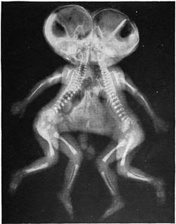
Conjoined twins | |
---|---|
Other names | Siamese twins, monstra duplicia |
X-ray of conjoined twins, Cephalothoracopagus. | |
Specialty | Medical genetics |
Symptoms | Bodies fused |
Complications | Depends on type |
Usual onset | Beginning of pregnancy |
Duration | Lifelong |
Types | see article |
Causes | Incomplete fission |
Treatment | Surgery, symptomatic care |
Prognosis | Depends on type; occasionally may survive |
Conjoined twins, popularly referred to as Siamese twins, [1] [2] are twins joined in utero . [a] It is a very rare phenomenon, estimated to occur in anywhere between one in 50,000 births to one in 200,000 births, with a somewhat higher incidence in southwest Asia and Africa. [5] Approximately half are stillborn, and an additional one-third die within 24 hours. Most live births are female, with a ratio of 3:1. [5] [6]
Two possible explanations of the cause of conjoined twins have been proposed. The one that is generally accepted is fission, in which the fertilized egg splits partially. [7] The other explanation, no longer believed to be accurate, [7] is fusion, in which the fertilized egg completely separates, but stem cells (that search for similar cells) find similar stem cells on the other twin and fuse the twins together. Conjoined twins share a single common chorion, placenta, and amniotic sac in utero , but so do some monozygotic but non-conjoined twins. [8]
Chang and Eng Bunker (1811–1874) were brothers born in Siam (now Thailand) who traveled widely for many years and were known internationally as the Siamese Twins. Chang and Eng were joined at the torso by a band of flesh and cartilage, and by their fused livers. In modern times, they could easily have been separated. [9] Due to the brothers' fame and the rarity of the condition, the term Siamese twins came to be associated with conjoined twins.
There are two hypotheses about the development of conjoined twins.
Partial splitting of the primitive node and streak may result in the formation of conjoined twins. These twins are classified according to the nature and degree of their union. Occasionally, monozygotic twins are connected only by a common skin bridge or by a common liver bridge. The type of twins formed depends on when and to what extent abnormalities of the node and streak occurred. Misexpression of genes, such as goosecoid, may also result in conjoined twins. [11] Goosecoid activates inhibitors of BMP4 and contributes to regulation of head development. Over- or underexpression of this gene in laboratory animals results in severe malformations of the head region, including duplications, similar to some types of conjoined twins. [12]
Conjoined twins are typically classified by the point at which their bodies are joined. The most common types of conjoined twins are:
Other, less common types of conjoined twins include:
Surgery to separate conjoined twins may range from very easy to very difficult depending on the point of attachment and the internal parts that are shared. [19] Most cases of separation are extremely risky and life-threatening. Though there have been a number of successful separations throughout history, in many cases, the surgery results in the death of one or both of the twins, particularly if they are joined at the head or share a vital organ. This makes the ethics of surgical separation, where the twins can survive if not separated, contentious. Alice Dreger of Northwestern University found the quality of life of twins who remain conjoined to be higher than is commonly supposed. [20] Lori and George Schappell and Abby and Brittany Hensel are notable examples.
The first recorded separation of conjoined twins took place in the Byzantine Empire in the 900s. One of the conjoined twins had already died, so the doctors of the town attempted to separate the dead twin from the surviving twin. The result was briefly successful, as the remaining twin lived for three days after separation. The next recorded case of separating conjoined twins was several centuries later, in Germany, in 1689. [21] [22] The first recorded successful separation of conjoined twins was performed in 1689 by Johannes Fatio. [23] Around this same time Dr. Böhm of Gunzenhausen separated his own children, a pair of omphalopagus or xiphopagus twins; the feebler twin died four days later, but the healthier one was still alive and well at age five, when the case was reported. [24] In 1955, neurosurgeon Harold Voris (1902-1980) [25] and his team at Mercy Hospital in Chicago performed the first successful operation to separate craniopagus twins (conjoined at the head), which resulted in long-term survival for both. [26] [27] [28] The larger girl was reported in 1963 as developing normally, but the smaller girl was permanently impaired. [29]
In 1957, Bertram Katz and his surgical team made international medical history performing the world's first successful separation of conjoined twins sharing a vital organ. [30] Omphalopagus twins John Nelson and James Edward Freeman (Johnny and Jimmy) were born in Youngstown, Ohio, on April 27, 1956. The boys shared a liver but had separate hearts and were successfully separated at North Side Hospital in Youngstown, Ohio, by Bertram Katz. The operation was funded by the Ohio Crippled Children's Service Society. [31]
Recent successful separations of conjoined twins include that of the separation of Ganga and Jamuna Shreshta in 2001, who were born in Kathmandu, Nepal, in 2000. The 97-hour surgery on the pair of craniopagus twins was a landmark one which took place in Singapore; the team was led by neurosurgeons Chumpon Chan and Keith Goh. [32] The surgery left Ganga with brain damage and Jamuna unable to walk. Seven years later, Ganga Shrestha died at the Model Hospital in Kathmandu in July 2009, at the age of eight, three days after being admitted for treatment of a severe chest infection. [33]
Infants Rose and Grace Attard, conjoined twins from Malta, were separated in the United Kingdom by court order Re A over the religious objections of their parents, Michaelangelo and Rina Attard. The twins were attached at the lower abdomen and spine. The surgery took place in November 2000, at St Mary's Hospital in Manchester. The operation was controversial because Rose, the weaker twin, would die as a result of the procedure as her heart and lungs were dependent upon Grace's. However, if the operation had not taken place, it was certain that both twins would die. [34] [35] Grace survived to enjoy a normal childhood. [36]
In 2003, two 29-year-old women from Iran, Ladan and Laleh Bijani, who were joined at the head but had separate brains (craniopagus), were surgically separated in Singapore, despite surgeons' warnings that the operation could be fatal to one or both. Their complex case was accepted only because technologically advanced graphical imagery and modeling would allow the medical team to plan the risky surgery. However, an undetected major vein hidden from the scans was discovered during the operation. [37] The separation was completed but both women died while still in surgery.
In 2019 Safa and Marwa Ullah were separated at Great Ormond Street Hospital in London, England. The twins, born January 2017, were joined at the top of the head with separate brains and a cylindrical shared skull with the twins each facing in opposite directions to one another. The surgery was jointly led by neurosurgeon Owase Jeelani and plastic surgeon Professor David Dunaway. The surgery presented particular difficulties due to a number of shared veins and a distortion in the shape of the girls' brains, causing them to overlap. The distortion would need to be corrected in order for the separation to go ahead. The surgery utilized a team of more than 100 including bio engineers, 3D modelers and a virtual reality designer. The separation was completed in February 2019 following a total of 52 hours of surgery over three separate operations. As of July 2019, both girls remained healthy and the family planned to return to their home in Pakistan in 2020. [38] [39]
The Moche culture of ancient Peru depicted conjoined twins in their ceramics dating back to 300 CE. [40] Writing around 415 AD, St. Augustine of Hippo, in his book, City of God , refers to a man "double in his upper, but single in his lower half—having two heads, two chests, four hands, but one body and two feet like an ordinary man." [41]
According to Theophanes the Confessor, a Byzantine historian of the 9th century, around 385/386 AD, he writes that "in the village of Emmaus in Palestine, a child was born perfectly normal below the navel but divided above it, so that it had two chests and two heads, each possessing the senses. One would eat and drink but the other did not eat; one would sleep but the other stayed awake. There were times when they played with each other, when both cried and hit each other. They lived for a little over two years. One died while the other lived for another four days and it, too, died." [42]
In Arabia, the twin brothers Hashim ibn Abd Manaf and 'Abd Shams were born with Hashim's leg attached to his twin brother's head. Legend says that their father, Abd Manaf ibn Qusai, separated his conjoined sons with a sword and that some priests believed that the blood that had flowed between them signified wars between their progeny (confrontations did occur between Banu al'Abbas and Banu Ummaya ibn 'Abd Shams in the year 750 AH). [43] The Muslim polymath Abū al-Rayhān al-Bīrūnī described conjoined twins in his book Kitab-al-Saidana. [44]
The English twin sisters Mary and Eliza Chulkhurst, who were conjoined at the back (pygopagus), lived from 1100 to 1134 (or 1500 to 1534) and were perhaps the best-known early historical example of conjoined twins. Other early conjoined twins to attain notice were the "Scottish brothers", allegedly of the dicephalus type, essentially two heads sharing the same body (1460–1488, although the dates vary); the pygopagus Helen and Judith of Szőny, Hungary (1701–1723), who enjoyed a brief career in music before being sent to live in a convent; and Rita and Cristina of Parodi of Sardinia, born in 1829. Rita and Cristina were dicephalus tetrabrachius (one body with four arms) twins and although they died at only eight months of age, they gained much attention as a curiosity when their parents exhibited them in Paris.
Several sets of conjoined twins lived during the nineteenth century and made careers for themselves in the performing arts, though none achieved quite the same level of fame and fortune as Chang and Eng. Most notably, Millie and Christine McCoy (or McKoy), pygopagus twins, were born into slavery in North Carolina in 1851. They were sold to a showman, J.P. Smith, at birth, but were soon kidnapped by a rival showman. The kidnapper fled to England but was thwarted because England had already banned slavery. Smith traveled to England to collect the girls and brought with him their mother, Monimia, from whom they had been separated. He and his wife provided the twins with an education and taught them to speak five languages, play music, and sing. For the rest of the century, the twins enjoyed a successful career as "The Two-Headed Nightingale" and appeared with the Barnum Circus. In 1912, they died of tuberculosis, 17 hours apart.
Giacomo and Giovanni Tocci, from Locana, Italy, were immortalized in Mark Twain's short story "Those Extraordinary Twins" as fictitious twins Angelo and Luigi. The Toccis, born in 1877, were dicephalus tetrabrachius twins, having one body with two legs, two heads, and four arms. From birth they were forced by their parents to perform and never learned to walk, as each twin controlled one leg (in modern times, physical therapy allows twins like the Toccis to learn to walk on their own). They are said to have disliked show business. In 1886, after touring the United States, the twins returned to Europe with their family. They are believed to have died around this time, though some sources claim they survived until 1940, living in seclusion in Italy.
Conjoined twins have been the focus of several works of fiction, including:
Craniopagus parasiticus is an extremely rare type of parasitic twinning occurring in about 4 to 6 of 10,000,000 births. In craniopagus parasiticus, a parasitic twin head with an undeveloped body is attached to the head of a developed twin. Fewer than a dozen cases of this type of conjoined twin have been documented in literature.
A parasitic twin, also known as an asymmetrical twin or unequal conjoined twin, occurs when a twin embryo begins developing in utero, but the pair does not fully separate, and one embryo maintains dominant development at the expense of the other. It results from the same processes that also produces vanishing twins and conjoined twins, and may represent a continuum between the two. In parasitic twins, one ceases development during gestation and is vestigial to a mostly fully formed, otherwise healthy individual twin. The undeveloped twin is termed as parasitic, because it is incompletely formed or wholly dependent on the body functions of the complete fetus. The independent twin is called the autosite. The autosite, together with the parasite, are collectively referred to as heteropagus twins.
Keith Goh is a neurosurgeon from Singapore. Goh is known for his operations in separating conjoined twins with two known successful cases and a failed attempt in separating Ladan and Laleh Bijani.
Abigail Loraine Hensel and Brittany Lee Hensel are American conjoined twins. They are dicephalic parapagus twins, and are highly symmetric for conjoined twins. Each has a heart, stomach, spine, pair of lungs, and spinal cord. Each twin controls one arm and one leg. When they were infants, learning to crawl, walk, and clap required cooperation. They can eat and write separately and simultaneously. Activities such as running, swimming, hair-brushing, playing piano or volleyball, riding a bicycle, or driving a car require coordination.
Sirenomelia, also called mermaid syndrome, is a rare congenital deformity in which the legs are fused together, giving the appearance of a mermaid's tail, hence the nickname.
Ischiopagi comes from the Greek word ischio- meaning hip (ilium) and -pagus meaning fixed or united. It is the medical term used for conjoined twins who are united at the pelvis. The twins are classically joined with the vertebral axis at 180°. The conjoined twins usually have four arms; two, three or four legs; and typically one external genitalia and anus.
Clarence and Carl Aguirre are former conjoined twins born in Manila. They were conjoined at the top of the head and shared 8 centimetres (3.1 in) of brain. More than 1–2 centimetres (0.4–0.8 in) will affect brain functionality in one or both of twins. Without separation, they were expected to live around 6–8 months.
Kendra Deene Herrin and Maliyah Mae Herrin are former conjoined twins. They were separated in August 2006. They were the first set of conjoined twins to be separated to share a kidney. Kendra retained their shared kidney following the separation surgery, while Maliyah Herrin underwent dialysis until she was transplanted with a kidney donated by her mother in April 2007.
Krista and Tatiana Hogan are Canadians who are conjoined craniopagus twins. They are joined at the head and share a skull and a brain. They were born in Vancouver, British Columbia, and are the only unseparated conjoined twins of that type currently alive in Canada. They live with their mother, Felicia Simms, in Vernon, British Columbia, have two sisters and a brother and often travel to Vancouver for care at BC Children's Hospital and Sunny Hill Health Centre for Children.
Yarlagadda Nayudamma is a consultant paediatric surgeon from Guntur, India. Previously, he operated at the Guntur General Hospital where he was the head of the paediatric surgery department.
Lakshmi Tatma is an Indian girl born in 2005 in a village in Araria district, Bihar, with four arms and four legs. She was actually one of a pair of ischiopagus conjoined twins, one of which was headless because its head had atrophied and chest had not fully developed in the womb, causing the appearance of one child with four arms and four legs. She has undergone surgery to remove these extra limbs.
Ascher Lawrence Mestel was an American pediatric surgeon and artist who was based in Brooklyn, New York. He was one of the pioneers in the field of pediatric surgery and was widely published. Mestel was especially well known for the groundbreaking first successful separation of Ischiopagus Tripus conjoined twins.
Viet Nguyen and Duc Nguyen were a pair of Vietnamese conjoined twins surgically separated in 1988. Viet died in 2007 of natural causes.
Dicephalic parapagus is a rare form of partial twinning with two heads side by side on one torso. Infants conjoined this way are sometimes called "two-headed babies" in popular media. The condition is also called parapagus dicephalus.
Craniopagus twins are conjoined twins who are fused at the cranium. The union may occur on any portion of the cranium, but does not primarily involve either the face or the foramen magnum; the two brains are usually separate, but they may share some brain tissue. Conjoined twins are genetically identical and always share the same sex. The thorax and abdomen are separate and each twin has their own umbilicus and umbilical cord.
Bothwell Anesu Mbuwayesango is a Zimbabwean pediatric surgeon who successfully led an all Zimbabwean team that separated conjoined twins in 2014 during an eight-hour operation at Harare hospital; it was the country's second successful separation, the first was in 1985. The two-month-old male twins were joined at the chest and abdomen. In 2021 Mbuwayesango led another successful separation during an eighteen-hour surgery at the same hospital. Mr Mbuwayesango was a Council member of the Medical and Dental Practitioners Council of Zimbabwe. He has published articles in the medical literature.
Maria and Teresa Tapia are formerly conjoined twins born in the Dominican Republic. The twins were joined by their lower chest and abdomen and were therefore classified as omphalopagus, sharing a liver, pancreas, and a small portion of their small intestine. On November 7, 2011, the twins underwent successful separation surgery at the Children's Hospital of Richmond in Virginia.
James Tait Goodrich was an American neurosurgeon. He was the director of the Division of Pediatric Neurosurgery at Montefiore Health System and Professor of Clinical Neurological Surgery, Pediatrics, Plastic and Reconstructive Surgery at the Albert Einstein College of Medicine, and gained worldwide recognition for performing multiple successful separations of conjoined twins. He assisted in two craniopagus separations with Dr. Alferayan A in Riyadh, Saudi Arabia, with the first one done May 5, 2014 and the second one done February 14, 2016. Both pairs were successfully separated and are doing well.
Noor ul Owase Jeelani is a Kashmiri-British neurosurgeon and academic. He is a consultant paediatric neurosurgeon at Great Ormond Street Hospital for Children (GOSH) and was the Head of the Department of Neurosurgery from 2012 until 2018. He is an Honorary Associate Professor at the Institute of Child Health, University College London. He leads the FaceValue research group in Craniofacial Morphometrics, device design, and clinical outcomes.
András Csókay, is a Hungarian neurosurgeon with international recognition in the field of neurosurgery for his development of a technique to enhance microsurgical precision in the vascular tunnel and for the separation of a pair of Bangladeshi (Islam) Craniopagus Twins.
one could predict dizygotic conjoined twins occurring less than 1 in every 4.7 billion ovulation cycles