Trichosporon asteroides

Last updated

Trichosporon asteroides
Trichosporon asteroides culture.jpg
Scientific classification
Kingdom:
Division:
Class:
Order:
Genus:
Species:
T. asteroides
Binomial name
Trichosporon asteroides
M. Ota (1926)
Synonyms
  • Parendomyces asteroides Rischin (1921)
  • Geotrichoides asteroides(Rischin) Langeron & Talice (1932)
  • Proteomyces asteroides(Rischin) C.W. Dodge (1935)
  • Prototheca filamentaP. Arnold & Ahearn (1972)
  • Fissuricella filamenta(P. Arnold & Ahearn) Pore, D'Amatao & Ajello (1977)

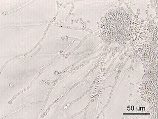
Trichosporon asteroides is an asexual basidiomycetous fungus first described from human skin but now mainly isolated from blood and urine. [1] [2] [3] [4] [5] T. asteroides is a hyphal fungus with a characteristically yeast-like appearance due to the presence of slimy arthroconidia. [3] [6] Infections by this species usually respond to treatment with azoles and amphotericin B. [2] [7] [8]

Contents

History and taxonomy

Trichosporon asteroides was isolated from lesion of male chin skin by Rinchin in Bern in 1926 and named in the genus, Parendomyces. T. asteroides was later reevaluated by Masao Ota and transferred to the genus Trichosporon as T. asteroides. [1] Ota noted that its hyphae were more sparsely branched than other species in Trichosporon, and it lacked the ability to ferment glucose, maltose, sucrose and fructose. [1] Molecular phylogenetic study has since supported the placement of T. asteroides in the genus Trichsporon. [9] T. asteroides was determined to be conspecific with Fissuricella filamenta on the basis of DNA/DNA reassociation. [3] [6] Modern classifications support the placement of this species within the order Trichosporonales. [10]

Growth and morphology

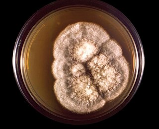
Trichosporon asteroides grows in a range of media including in the presence of the antifungal agent, cycloheximide. [4] Colonies of T. asteroides grown on Sabouraud agar appear white with a brain-like texture, reaching a diameter of 10 mm after 10 days growth at 25 °C (77 °F). [3] [6] Colonies have a radially furrowed outer zone and matt appearance that is not powdery like the related species, T. asahii. [3] Antibodies produced by infected patients can also be used to differentiate T. asahii and T. asteroides in disease. [11] Weak growth is seen at 37 °C (99 °F) but is absent at higher temperatures. [6] [12] T. asteroides can be distinguished from related Trichosporon species by its ability to utilize D-galactose, L-rhamnose, erythritol and L-arabinitol. [3]

Physiology and pathogenesis

Trichosporon asteroides does not ferment glucose, maltose, sucrose or fructose, a feature common to all Trichosporon species. [1] T. asteroides causes trichosporonosis, causing deep-seated, mucosa-associated, superficial, and systematic infections including blood. [2] [4] [6] [13] [14] This species is one of the three most common Trichosporon species isolated in clinical settings. [13] The fungus is sometimes recovered from specimens of blood, urine, and aspiration fluid, vaginal mucosa, male perigenital skin area, and catheters. [4] [15] [5] The first record of this agent in a systemic infection involved a bloodstream infection. [4] Main types of latent infection include fungemia, urinary tract infections, peritonitis, endocarditis, arthritis, esophagitis, meningitis, brain abscess, splenic abscess, and uterine infections. [2] Allergies and systemic infections can also be caused by this fungus particularly in immunosuppressed people. [16] Recently, T. asteroides was implicated in a case of kerion celsi, a rare, highly inflammatory scalp infection seen mainly in children. [17]

This fungus is rarely encountered in zoonotic infections [16] and has never been shown to cause disease in insects. [13] However, T. asteroides was isolated from multifocal, irregularly raised skin lesions on a female Bottle-Nosed Dolphin, Tursiops truncatus , in Japan. [16] In the absence of cultures and molecular biological analysis, the agent was initially suspected to be Paracoccidioides ceti, a worldwide causative agent of Paracoccidioidomycosis in dolphins. [16]

Treatment

Azole antifungals are often used as a front-line therapy in trichosporonosis. [7] Resistance to amphotericin, flucytosine, fluconazole, and itraconazole have been described. [8] [2] Echinocandins, as a group, are ineffective against Trichosporon species. [6] Triazoles show better in vitro and in vivo antifungal activity than amphotericin B, while voriconazole also has excellent in vitro activity against Trichosporon asteroides. [2] [8] [18] Combination therapy regimens such as voriconazole and amphotericin B are also effective in serious infections. [18] Fluconazole has been used successfully in subcutaneous and deep infections. [17]

Related Research Articles

<span class="mw-page-title-main">Candidiasis</span> Fungal infection due to any type of Candida

Candidiasis is a fungal infection due to any species of the genus Candida. When it affects the mouth, in some countries it is commonly called thrush. Signs and symptoms include white patches on the tongue or other areas of the mouth and throat. Other symptoms may include soreness and problems swallowing. When it affects the vagina, it may be referred to as a yeast infection or thrush. Signs and symptoms include genital itching, burning, and sometimes a white "cottage cheese-like" discharge from the vagina. Yeast infections of the penis are less common and typically present with an itchy rash. Very rarely, yeast infections may become invasive, spreading to other parts of the body. This may result in fevers, among other symptoms, depending on the parts involved.

<span class="mw-page-title-main">Antifungal</span> Pharmaceutical fungicide or fungistatic used to treat and prevent mycosis

An antifungal medication, also known as an antimycotic medication, is a pharmaceutical fungicide or fungistatic used to treat and prevent mycosis such as athlete's foot, ringworm, candidiasis (thrush), serious systemic infections such as cryptococcal meningitis, and others. Such drugs are usually obtained by a doctor's prescription, but a few are available over the counter (OTC). The evolution of antifungal resistance is a growing threat to health globally.
<i>Candida</i> (fungus) Genus of ascomycete fungi

Candida is a genus of yeasts. It is the most common cause of fungal infections worldwide and the largest genus of medically important yeast.
<i>Coccidioides immitis</i> Species of fungus

Coccidioides immitis is a pathogenic fungus that resides in the soil in certain parts of the southwestern United States, northern Mexico, and a few other areas in the Western Hemisphere.
<span class="mw-page-title-main">Sporotrichosis</span> Medical condition

Sporotrichosis, also known as rose handler's disease, is a fungal infection that may be localised to skin, lungs, bone and joint, or become systemic. It presents with firm painless nodules that later ulcerate. Following initial exposure to Sporothrix schenckii, the disease typically progresses over a period of a week to several months. Serious complications may develop in people who have a weakened immune system.

Pichia kudriavzevii is a budding yeast involved in chocolate production. P. kudriavzevii is an emerging fungal nosocomial pathogen primarily found in the immunocompromised and those with hematological malignancies. It has natural resistance to fluconazole, a standard antifungal agent. It is most often found in patients who have had prior fluconazole exposure, sparking debate and conflicting evidence as to whether fluconazole should be used prophylactically. Mortality due to P. kudriavzevii fungemia is much higher than the more common C. albicans. Other Candida species that also fit this profile are C. parapsilosis, C. glabrata, C. tropicalis, C. guillermondii and C. rugosa.
<i>Nakaseomyces glabratus</i> Species of fungus

Nakaseomyces glabratus is a species of haploid yeast of the genus Nakaseomyces, previously known as Candida glabrata. Despite the fact that no sexual life cycle has been documented for this species, N. glabratus strains of both mating types are commonly found. C. glabrata is generally a commensal of human mucosal tissues, but in today's era of wider human immunodeficiency from various causes, N. glabratus is often the second or third most common cause of candidiasis as an opportunistic pathogen. Infections caused by N. glabratus can affect the urogenital tract or even cause systemic infections by entrance of the fungal cells in the bloodstream (Candidemia), especially prevalent in immunocompromised patients.
<span class="mw-page-title-main">Echinocandin</span> Group of chemical compounds

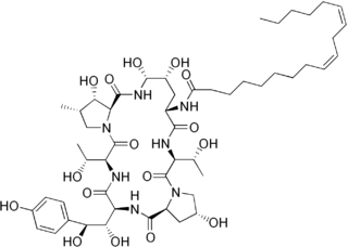
Echinocandins are a class of antifungal drugs that inhibit the synthesis of β-glucan in the fungal cell wall via noncompetitive inhibition of the enzyme 1,3-β glucan synthase. The class has been termed the "penicillin of antifungals," along with the related papulacandins, as their mechanism of action resembles that of penicillin in bacteria. β-glucans are carbohydrate polymers that are cross-linked with other fungal cell wall components, the fungal equivalent to bacterial peptidoglycan. Caspofungin, micafungin, and anidulafungin are semisynthetic echinocandin derivatives with limited clinical use due to their solubility, antifungal spectrum, and pharmacokinetic properties.

<i>Geotrichum</i> Genus of fungi

Geotrichum is a genus of fungi found worldwide in soil, water, air, and sewage, as well as in plants, cereals, and dairy products; it is also commonly found in normal human flora and is isolated from sputum and feces. It was first described in 1809 by Johann Heinrich Friedrich Link.

Exophiala jeanselmei is a saprotrophic fungus in the family Herpotrichiellaceae. Four varieties have been discovered: Exophiala jeanselmei var. heteromorpha, E. jeanselmei var. lecanii-corni, E. jeanselmei var. jeanselmei, and E. jeanselmei var. castellanii. Other species in the genus Exophiala such as E. dermatitidis and E. spinifera have been reported to have similar annellidic conidiogenesis and may therefore be difficult to differentiate.

<i>Trichosporon</i> Genus of fungi


Trichosporon is a genus of anamorphic fungi in the family Trichosporonaceae. All species of Trichosporon are yeasts with no known teleomorphs. Most are typically isolated from soil, but several species occur as a natural part of the skin microbiota of humans and other animals. Proliferation of Trichosporon yeasts in the hair can lead to an unpleasant but non-serious condition known as white piedra. Trichosporon species can also cause severe opportunistic infections (trichosporonosis) in immunocompromised individuals.

<i>Exophiala dermatitidis</i> Species of fungus

Exophiala dermatitidis is a thermophilic black yeast, and a member of the Herpotrichiellaceae. While the species is only found at low abundance in nature, metabolically active strains are commonly isolated in saunas, steam baths, and dish washers. Exophiala dermatitidis only rarely causes infection in humans, however cases have been reported around the world. In East Asia, the species has caused lethal brain infections in young and otherwise healthy individuals. The fungus has been known to cause cutaneous and subcutaneous phaeohyphomycosis, and as a lung colonist in people with cystic fibrosis in Europe. In 2002, an outbreak of systemic E. dermatitidis infection occurred in women who had received contaminated steroid injections at North Carolina hospitals.
<i>Aspergillus ustus</i> Species of fungus

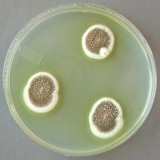
Aspergillus ustus is a microfungus and member of the division Ascomycota. It is commonly found in indoor environments and soil. Isolated cases of human infection resulting from A. ustus have been described; however the majority of these are nail infections.

Scedosporiosis is the general name for any mycosis – i.e., fungal infection – caused by a fungus from the genus Scedosporium. Current population-based studies suggest Scedosporium prolificans and Scedosporium apiospermum to be among the most common infecting agents from the genus, although infections caused by other members thereof are not unheard of. The latter is an asexual form (anamorph) of another fungus, Pseudallescheria boydii. The former is a "black yeast", currently not characterized as well, although both of them have been described as saprophytes.

Histoplasma duboisii is a saprotrophic fungus responsible for the invasive infection known as African histoplasmosis. This species is a close relative of Histoplasma capsulatum, the agent of classical histoplasmosis, and the two occur in similar habitats. Histoplasma duboisii is restricted to continental Africa and Madagascar, although scattered reports have arisen from other places usually in individuals with an African travel history. Like, H. capsulatum, H. duboisii is dimorphic – growing as a filamentous fungus at ambient temperature and a yeast at body temperature. It differs morphologically from H. capsulatum by the typical production of a large-celled yeast form. Both agents cause similar forms of disease, although H. duboisii predominantly causes cutaneous and subcutaneous disease in humans and non-human primates. The agent responds to many antifungal drug therapies used to treat serious fungal diseases.

<i>Candida auris</i> Species of fungus

Candida auris is a species of fungus that grows as yeast. It is one of the few species of the genus Candida which cause candidiasis in humans. Often, candidiasis is acquired in hospitals by patients with weakened immune systems. C. auris can cause invasive candidiasis (fungemia) in which the bloodstream, the central nervous system, and internal organs are infected. It has attracted widespread attention because of its multiple drug resistance. Treatment is also complicated because it is easily misidentified as other Candida species.

Invasive candidiasis is an infection (candidiasis) that can be caused by various species of Candida yeast. Unlike Candida infections of the mouth and throat or vagina, invasive candidiasis is a serious, progressive, and potentially fatal infection that can affect the blood (fungemia), heart, brain, eyes, bones, and other parts of the body.
<i>Candida tropicalis</i> Species of fungus

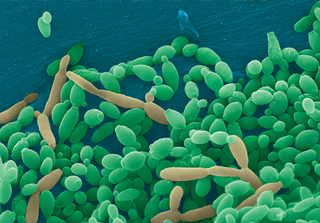
Candida tropicalis is a species of yeast in the genus Candida. It is a common pathogen in neutropenic hosts, in whom it may spread through the bloodstream to peripheral organs. For invasive disease, treatments include amphotericin B, echinocandins, or extended-spectrum triazole antifungals.
<i>Metarhizium granulomatis</i> Species of fungus

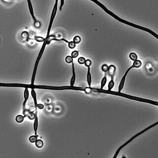
Metarhizium granulomatis is a fungus in the family Clavicipitaceae associated with systemic mycosis in veiled chameleons. The genus Metarhizium is known to infect arthropods, and collectively are referred to green-spored asexual pathogenic fungi. This species grows near the roots of plants and has been reported as an agent of disease in captive veiled chameleons. The etymology of the species epithet, "granulomatis" refers to the ability of the fungus to cause granulomatous disease in susceptible reptiles.

Candida haemulonii is a yeast fungal pathogen that is known to cause infections in humans. C. haemulonii is an emerging opportunistic pathogen that is found in hospitals and healthcare settings. Infections are difficult to treat because the fungus has resistance to antifungal agents. Since its emergence, little research has been conducted on this fungus. However, in recent years, research has been conducted to help identify the various properties of C. haemulonii.

References

  1. 1 2 3 4 Masao, Ota (1926). "Sur quelques champignons pathogènes du type Trichosporum beigeli Vuillemin". Annales de Parasitologie Humaine et Comparée. 4 (1): 1–13. doi: 10.1051/parasite/1926041001 .
  2. 1 2 3 4 5 6 Colombo, A. L.; Padovan, A. C. B.; Chaves, G. M. (2011). "Current Knowledge of Trichosporon spp. and Trichosporonosis". Clinical Microbiology Reviews. 24 (4): 682–700. doi:10.1128/CMR.00003-11. PMC   3194827 . PMID   21976604.
  3. 1 2 3 4 5 6 Guého, E.; Smith, M. T.; De Hoog, G. S.; Billon-Grand, G.; Christen, R.; van der Vegte, W. B. (1992). "Contributions to a revision of the genus Trichosporon". Antonie van Leeuwenhoek. 61 (4): 289–316. doi:10.1007/BF00713938. PMID   1497334. S2CID   24841877.
  4. 1 2 3 4 5 Kustimur, Semra; Kalkanci, Ayse; Caglar, Kayhan; Dizbay, Murat; Aktas, Firdevs; Sugita, Takashi (2002). "Nosocomial fungemia due to Trichosporon asteroides: firstly described bloodstream infection". Diagnostic Microbiology and Infectious Disease. 43 (3): 167–170. doi:10.1016/S0732-8893(02)00385-1. PMID   12088626.
  5. 1 2 Silvestre Junior, Agenor Messias; Miranda, Mauricio Alexandre Bandeira Rampazzo; Camargo, Zoilo Pires de (2010). "Trichosporon species isolated from the perigenital region, urine and catheters of a Brazilian population". Brazilian Journal of Microbiology. 41 (3): 628–34. doi:10.1590/S1517-83822010000300013. PMC   3768649 . PMID   24031538.
  6. 1 2 3 4 5 6 Guého, Eveline; Improvisi, Luce; Dupont, B.; De Hoog, G. S. (1994). "Trichosporon on humans: a practical account: Trichosporon am Menschen: Eine Darstellung für die Praxis". Mycoses. 37 (1–2): 3–10. doi:10.1111/j.1439-0507.1994.tb00277.x. PMID   7935589. S2CID   24703812.
  7. 1 2 Anaissie, E.; Gokaslan, A.; Hachem, R.; Rubin, R.; Griffin, G.; Robinson, R.; Sobel, J.; Bodey, G. (1992). "Azole Therapy for Trichosporonosis: Clinical Evaluation of Eight Patients, Experimental Therapy for Murine Infection, and Review". Clinical Infectious Diseases. 15 (5): 781–787. doi:10.1093/clind/15.5.781. ISSN   1058-4838. PMID   1445976.
  8. 1 2 3 Garg, Vaibhav; Jones, Elizabeth K.; Friedman, Ben J.; Lee, Jason B.; Yang, Sherry (2018). "Invasive trichosporonosis treated with voriconazole". JAAD Case Reports. 4 (4): 362–364. doi:10.1016/j.jdcr.2017.11.003. ISSN   2352-5126. PMC   5911819 . PMID   29693071.
  9. Sugita, Takashi; Nakase, Takashi (1998). "Molecular phylogenetic study of the basidiomycetous anamorphic yeast genus Trichosporon and related taxa based on small subunit ribosomal DNA sequences". Mycoscience. 39 (1): 7–13. doi:10.1007/BF02461572. S2CID   83647795.
  10. Liu XZ, Wang QM, Göker M, Groenewald M, Kachalkin AV, Lumbsch HT, Millanes AM, Wedin M, Yurkov AM, Boekhout T, Bai FY (2015). "Towards an integrated phylogenetic classification of the Tremellomycetes". Studies in Mycology. 81: 85–147. doi: 10.1016/j.simyco.2015.12.001 . PMC   4777781 .
  11. Davies, Genna E.; Thornton, Christopher R. (2014). "Differentiation of the emerging human pathogens Trichosporon asahii and Trichosporon asteroides from other pathogenic yeasts and moulds by using species-specific monoclonal antibodies". PLOS ONE. 9 (1): e84789. doi: 10.1371/journal.pone.0084789 . PMC   3879316 . PMID   24392156.
  12. Howard, Dexter H. (2007). Pathogenic fungi in humans and animals (2nd ed.). New York: Marcel Dekker. ISBN   9780203909102.
  13. 1 2 3 Mariné, Marçal; Bom, Vinicius Leite Pedro; Castro, Patricia Alves de; Winkelstrote, Lizziane Kretli; Ramalh, Leandra Naira; Brown, Neil Andrew; Goldman, Gustavo Henrique (2015). "The development of animal infection models and antifungal efficacy assays against clinical isolates of Trichosporon asahii, T. asteroides and T. inkin". Virulence. 6 (5): 476–86. doi:10.1080/21505594.2015.1020273. PMC   4601256 . PMID   25751127.
  14. Sugita, T; Nishikawa, A; Shinoda, T (1998). "Rapid detection of species of the opportunistic yeast Trichosporon by PCR". Journal of Clinical Microbiology. 36 (5): 1458–60. doi:10.1128/JCM.36.5.1458-1460.1998. PMC   104855 . PMID   9574732.
  15. Paula, Claudete R.; Krebs, Vera L. J.; Auler, Marcos E.; Ruiz, Luciana S.; Matsumoto, Flavia E.; Silva, Elza H.; Diniz, Edna M. A.; Vaz, Flavio A. C. (2006). "Nosocomial infection in newborns by in a Brazilian intensive care unit". Medical Mycology. 44 (5): 479–484. doi: 10.1080/13693780600561809 . PMID   16882616.
  16. 1 2 3 4 Ueda, Keiichi; Nakamura, Ichiro; Itano, Eiko Nakagawa; Takemura, Kazunori; Nakazato, Yasutomo; Sano, Ayako (2017). "Trichosporon asteroides Isolated from Cutaneous Lesions of a Suspected Case of "paracoccidioidomycosis ceti" in a Bottlenose Dolphin (Tursiops truncatus)". Mycopathologia. 182 (9–10): 937–946. doi:10.1007/s11046-017-0147-3. PMID   28547342. S2CID   11893217.
  17. 1 2 Kocabaş, Bilge Aldemİr; Karbuz, Adem; Çİftçİ, Ergin; Beğde, Fırat; Ametoglou, Selver; Fouad, Ali Adil; Kalkancı, Ayşe; Karahan, Zeynep Ceren; Aysev, Derya; İnce, Erdal (2016). "Trichosporon asteroides: A novel etiologic agent of Kerion celsi in a child" (PDF). İzmir Dr. Behçet Uz Çocuk Hastanesi Dergisi. 6 (2): 151–154.
  18. 1 2 Maxfield, Luke; Matthews, Juliana J.; Ambrosetti, David Ryan; Ephtimios, Issa E. (2015). "Trichosporon fungemia in a pediatric patient with acute lymphoblastic leukemia". IDCases. 2 (4): 106–108. doi:10.1016/j.idcr.2015.09.007. ISSN   2214-2509. PMC   4712209 . PMID   26793473.