The abducens nerve or abducent nerve, also known as the sixth cranial nerve, cranial nerve VI, or simply CN VI, is a cranial nerve in humans and various other animals that controls the movement of the lateral rectus muscle, one of the extraocular muscles responsible for outward gaze. It is a somatic efferent nerve.
Articles related to anatomy include:

The pons is part of the brainstem that in humans and other bipeds lies inferior to the midbrain, superior to the medulla oblongata and anterior to the cerebellum.

The brainstem is the stalk-like part of the brain that interconnects the cerebrum and diencephalon with the spinal cord. In the human brain the brainstem is composed of the midbrain, the pons, and the medulla oblongata. The midbrain is continuous with the thalamus of the diencephalon through the tentorial notch.

The oculomotor nerve, also known as the third cranial nerve, cranial nerve III, or simply CN III, is a cranial nerve that enters the orbit through the superior orbital fissure and innervates extraocular muscles that enable most movements of the eye and that raise the eyelid. The nerve also contains fibers that innervate the intrinsic eye muscles that enable pupillary constriction and accommodation. The oculomotor nerve is derived from the basal plate of the embryonic midbrain. Cranial nerves IV and VI also participate in control of eye movement.

The midbrain or mesencephalon is the rostral-most portion of the brainstem connecting the diencephalon and cerebrum with the pons. It consists of the cerebral peduncles, tegmentum, and tectum.

The medial longitudinal fasciculus (MLF) is an area of crossed over tracts, on each side of the brainstem. These bundles of axons are situated near the midline of the brainstem. They are made up of both ascending and descending fibers that arise from a number of sources and terminate in different areas, including the superior colliculus, the vestibular nuclei, and the cerebellum. It contains the interstitial nucleus of Cajal, responsible for oculomotor control, head posture, and vertical eye movement.

The pontine tegmentum, or dorsal pons, is located within the brainstem, and is one of two parts of the pons, the other being the ventral pons or basilar part of the pons. The pontine tegmentum can be defined in contrast to the basilar pons: basilar pons contains the corticospinal tract running craniocaudally and can be considered the rostral extension of the ventral medulla oblongata; however, basilar pons is distinguished from ventral medulla oblongata in that it contains additional transverse pontine fibres that continue laterally to become the middle cerebellar peduncle. The pontine tegmentum is all the material dorsal from the basilar pons to the fourth ventricle. Along with the dorsal surface of the medulla, it forms part of the rhomboid fossa – the floor of the fourth ventricle.

Eye movement includes the voluntary or involuntary movement of the eyes. Eye movements are used by a number of organisms to fixate, inspect and track visual objects of interests. A special type of eye movement, rapid eye movement, occurs during REM sleep.

In humans, the tectospinal tract is a nerve tract that coordinates head and eye movements. This tract is part of the extrapyramidal system and connects the midbrain tectum, and cervical regions of the spinal cord.

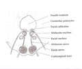
The abducens nucleus is the originating nucleus from which the abducens nerve (VI) emerges—a cranial nerve nucleus. This nucleus is located beneath the fourth ventricle in the caudal portion of the pons near the midline, medial to the sulcus limitans.

Internuclear ophthalmoplegia (INO) is a disorder of conjugate lateral gaze in which the affected eye shows impairment of adduction. When an attempt is made to gaze contralaterally, the affected eye adducts minimally, if at all. The contralateral eye abducts, however with nystagmus. Additionally, the divergence of the eyes leads to horizontal diplopia. That is if the right eye is affected the patient will "see double" when looking to the left, seeing two images side-by-side. Convergence is generally preserved.
The paramedian pontine reticular formation, also known as PPRF or paraabducens nucleus, is part of the pontine reticular formation, a brain region without clearly defined borders in the center of the pons. It is involved in the coordination of eye movements, particularly horizontal gaze and saccades.

The nucleus of the trochlear nerve is a motor nucleus in the medial midbrain giving rise to the trochlear nerve.
The paramedian reticular nucleus sends its connections to the spinal cord in a mostly ipsilateral manner, although there is some decussation.
Conjugate gaze palsies are neurological disorders affecting the ability to move both eyes in the same direction. These palsies can affect gaze in a horizontal, upward, or downward direction. These entities overlap with ophthalmoparesis and ophthalmoplegia.
The term gaze is frequently used in physiology to describe coordinated motion of the eyes and neck. The lateral gaze is controlled by the paramedian pontine reticular formation (PPRF). The vertical gaze is controlled by the rostral interstitial nucleus of medial longitudinal fasciculus and the interstitial nucleus of Cajal.
Conjugate eye movement refers to motor coordination of the eyes that allows for bilateral fixation on a single object. A conjugate eye movement is a movement of both eyes in the same direction to maintain binocular gaze. This is in contrast to vergence eye movement, where binocular gaze is maintained by moving eyes in opposite directions, such as going “cross eyed” to view an object moving towards the face. Conjugate eye movements can be in any direction, and can accompany both saccadic eye movements and smooth pursuit eye movements.
Perihypoglossal nuclei, called also perihypoglossal complex or perihypoglossal nuclear complex or satellite nuclei is a group of neurons in the floor of the fourth ventricle, in close proximity to the nucleus of the hypoglossal nerve in the gray substance of the medulla oblongata, all of which contain cells with characteristics suggestive of reticular connections.