Ovarian vein syndrome

Last updated
Ovarian vein syndrome
Gray1120.png
Relation of arteries and veins to ureters, view from the back of a male (the ovarian veins substitute for the internal spermatic veins in females [1] )

Ovarian vein syndrome is a rare (possibly not uncommon, certainly under-diagnosed) condition in which a dilated ovarian vein compresses the ureter (the tube that brings the urine from the kidney to the bladder). This causes chronic or colicky abdominal pain, back pain and/or pelvic pain. The pain can worsen on lying down or between ovulation and menstruation. [2] [3] There can also be an increased tendency towards urinary tract infection or pyelonephritis (kidney infection). The right ovarian vein is most commonly involved, although the disease can be left-sided or affect both sides. It is currently classified as a form of pelvic congestion syndrome.

Contents

Signs and symptoms

Mechanism

Normally, the ovarian vein crosses over the ureter at the level of the fourth or fifth lumbar vertebra. The ureter itself courses over the external iliac artery and vein. [4] Thus, these vessels can impinge on the ureter causing obstruction. The left ovarian vein ends in the left renal vein whereas the right ovarian vein normally enters into the inferior vena cava. [1] In the case of right ovarian vein syndrome, the vein often ends in the renal vein.[ citation needed ] This is thought to contribute to venous engorgement, in analogy to what is seen in varicoceles, which arise more commonly on the left side. The straight angle between the ovarian vein (or testicular vein in males in the case of varicocoele) and the renal vein has been proposed as a cause of decreased blood return.[ citation needed ]

A related diagnosis is nutcracker syndrome where the left renal vein is described as being compressed between the aorta and the superior mesenteric artery. This is reported to cause collateral flow paths to open up to drain the left kidney i.e. reversed flow (reflux caudally) in the left renal vein. Pelvic Congestion Syndrome, vaginal and vulval varices, lower limb varices are clinical sequelae. Virtually all such patient are female and have been pregnant, often multiply.[ citation needed ]

The ovarian vein often displays incompetent valves. This has been observed more often in women with a higher number of previous pregnancies. Pressure from the baby might hinder the return of blood through the ovarian vein. However, dilation of the urinary tract is a normal observation in pregnancy, due to mechanical compression and the hormonal action of progesterone. Ovarian vein dilatation might also follow venous thrombosis (clotting inside the vein).[ citation needed ]

Another proposed mechanism of obstruction is when the ovarian vein and ureter both run through a sheath of fibrous tissue, following a local inflammation. This could be seen as a localised form of retroperitoneal fibrosis. [4]

Following obstruction, the ureter displays an abnormal peristalsis (contractions) towards the kidney instead of towards the bladder. This is thought to cause the colicky pain (similar to renal colic), and it is relieved after surgical decompression.[ citation needed ]

Diagnosis

Since it is a rare disease, it remains a diagnosis of exclusion of other conditions with similar symptoms. The diagnosis is supported by the results of imaging studies such as computed tomography or magnetic resonance imaging, ultrasound of the abdomen (with or without doppler imaging) or intravenous urography.[ citation needed ]

Specialist vascular ultrasonographers should routinely look for left ovarian vein reflux in patients with lower limb varices especially if not associated with long or short saphenous reflux. The clinical pattern of varices differs between the two types of lower limb varices. CT scanning is used to exclude abdominal or pelvic pathology. CT-Angiography/Venography can often demonstrate left ovarian vein reflux and image an enlarged left ovarian vein but is less sensitive and much more expensive than duplex Doppler ultrasound examination. Ultrasound requires that the ultrasonographer be experienced in venous vascular ultrasound and so is not always readily available. A second specialist ultrasound exam remains preferable to a CT scan.[ citation needed ]

As a wide range of pelvic and abdominal pathology can cause symptoms consistent with those symptoms due to left ovarian vein reflux, prior to embolisation of the left ovarian vein, a careful search for such diagnoses is essential. Consultation with general surgeons, gynaecologists, and possibly CT scanning should always be considered.[ citation needed ]

Treatment

Treatment consists of painkillers and surgical ablation of the dilated vein. This can be accomplished with open abdominal surgery (laparotomy) or keyhole surgery (laparoscopy). [5] Recently, the first robot-assisted surgery was described. [6]

Another approach to treatment involves catheter-based embolisation, [7] often preceded by phlebography to visualise the vein on X-ray fluoroscopy. [3] [8]

Ovarian vein coil embolisation is an effective and safe treatment for pelvic congestion syndrome and lower limb varices of pelvic origin. Many patients with lower limb varices of pelvic origin respond to local treatment i.e. ultrasound guided sclerotherapy. In those cases, ovarian vein coil embolisation should be considered second line treatment to be used if veins recur in a short time period i.e. 1–3 years. This approach allows further pregnancies to proceed if desired. Bilateral coil embolisation is not advised if a future pregnancy is possible. This treatment has largely superseded operative options.[ citation needed ]

Coil embolisation requires exclusion of other pelvic pathology, expertise in endovascular surgery, correct placement of appropriate sized coils in the pelvis and also in the upper left ovarian vein, careful pre- and post-procedure specialist vascular ultrasound imaging, a full discussion of the procedure with the patient i.e. informed consent. Complications, such as coil migration, are rare but reported. Their sequelae are usually minor.[ citation needed ]

If a Nutcracker compression (see below) is discovered, operative relocation of the renal vein should be considered before embolization of the ovarian vein. Stenting is not advised. Reducing outflow obstruction should always be the main objective.[ citation needed ]

History

The entity was first described by Clark in 1964. [9] Following initial scepticism and lively discussion in the medical literature, [10] [11] it is now recognised by many authors. [2]

Related Research Articles

<span class="mw-page-title-main">Varicose veins</span> Medical condition in which superficial veins become large and twisted

Varicose veins, also known as varicoses, are a medical condition in which superficial veins become enlarged and twisted. These veins typically develop in the legs, just under the skin. Varicose veins usually cause few symptoms. However, some individuals may experience fatigue or pain in the area. Complications can include bleeding or superficial thrombophlebitis. Varices in the scrotum are known as a varicocele, while those around the anus are known as hemorrhoids. Due to the various physical, social, and psychological effects of varicose veins, they can negatively affect one's quality of life.

<span class="mw-page-title-main">Ureter</span> Tubes used in the urinary system in most animals

The ureters are tubes made of smooth muscle that propel urine from the kidneys to the urinary bladder. In a human adult, the ureters are usually 20–30 cm (8–12 in) long and around 3–4 mm (0.12–0.16 in) in diameter. The ureter is lined by urothelial cells, a type of transitional epithelium, and has an additional smooth muscle layer that assists with peristalsis in its lowest third.

<span class="mw-page-title-main">Interventional radiology</span> Medical subspecialty

Interventional radiology (IR) is a medical specialty that performs various minimally-invasive procedures using medical imaging guidance, such as x-ray fluoroscopy, computed tomography, magnetic resonance imaging, or ultrasound. IR performs both diagnostic and therapeutic procedures through very small incisions or body orifices. Diagnostic IR procedures are those intended to help make a diagnosis or guide further medical treatment, and include image-guided biopsy of a tumor or injection of an imaging contrast agent into a hollow structure, such as a blood vessel or a duct. By contrast, therapeutic IR procedures provide direct treatment—they include catheter-based medicine delivery, medical device placement, and angioplasty of narrowed structures.

<span class="mw-page-title-main">Abdominal pain</span> Stomach aches

Abdominal pain, also known as a stomach ache, Is a symptom associated with both non-serious and serious medical issues. Since the abdomen contains most of the body's vital organs, it can be an indicator of a wide variety of diseases. Given that, approaching the examination of a person and planning of a differential diagnosis is extremely important.

<span class="mw-page-title-main">Embolization</span> Passage and lodging of an embolus within the bloodstream

Embolization refers to the passage and lodging of an embolus within the bloodstream. It may be of natural origin (pathological), in which sense it is also called embolism, for example a pulmonary embolism; or it may be artificially induced (therapeutic), as a hemostatic treatment for bleeding or as a treatment for some types of cancer by deliberately blocking blood vessels to starve the tumor cells.

<span class="mw-page-title-main">Hydronephrosis</span> Medical condition

Hydronephrosis describes hydrostatic dilation of the renal pelvis and calyces as a result of obstruction to urine flow downstream. Alternatively, hydroureter describes the dilation of the ureter, and hydronephroureter describes the dilation of the entire upper urinary tract.

Horseshoe kidney, also known as ren arcuatus, renal fusion or super kidney, is a congenital disorder affecting about 1 in 500 people that is more common in men, often asymptomatic, and usually diagnosed incidentally. In this disorder, the patient's kidneys fuse to form a horseshoe-shape during development in the womb. The fused part is the isthmus of the horseshoe kidney. The abnormal anatomy can affect kidney drainage resulting in increased frequency of kidney stones and urinary tract infections as well as increase risk of certain renal cancers.

<span class="mw-page-title-main">Renal colic</span> Medical condition

Renal colic, also known as ureteric colic, is a type of abdominal pain commonly caused by obstruction of ureter from dislodged kidney stones. The most frequent site of obstruction is the vesico-ureteric junction (VUJ), the narrowest point of the upper urinary tract. Acute obstruction and the resultant urinary stasis can distend the ureter (hydroureter) and cause a reflexive peristaltic smooth muscle spasm, which leads to a very intense visceral pain transmitted via the ureteric plexus.

<span class="mw-page-title-main">Inferior vena cava syndrome</span> Medical condition

Inferior vena cava syndrome (IVCS) is a very rare constellation of symptoms resulting from either an obstruction, or stenosis of the inferior vena cava. It can be caused by physical invasion or compression by a pathological process or by thrombosis within the vein itself. It can also occur during pregnancy. Pregnancy leads to high venous pressure in the lower limbs, decreased blood return to the heart, decreased cardiac output due to obstruction of the inferior vena cava, sudden rise in venous pressure which can lead to placental separation, and a decrease in kidney function. All of these issues can arise from lying in the supine position during late pregnancy which can cause compression of the inferior vena cava by the uterus. Symptoms of late pregnancy inferior vena cava syndrome consist of intense pain in the right hand side, muscle twitching, hypotension, and fluid retention.

<span class="mw-page-title-main">Vesicoureteral reflux</span> Medical condition

Vesicoureteral reflux (VUR), also known as vesicoureteric reflux, is a condition in which urine flows retrograde, or backward, from the bladder into one or both ureters and then to the renal calyx or kidneys. Urine normally travels in one direction from the kidneys to the bladder via the ureters, with a one-way valve at the vesicoureteral (ureteral-bladder) junction preventing backflow. The valve is formed by oblique tunneling of the distal ureter through the wall of the bladder, creating a short length of ureter (1–2 cm) that can be compressed as the bladder fills. Reflux occurs if the ureter enters the bladder without sufficient tunneling, i.e., too "end-on".

<span class="mw-page-title-main">Nutcracker syndrome</span> Medical condition

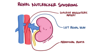
The nutcracker syndrome (NCS) results most commonly from the compression of the left renal vein (LRV) between the abdominal aorta (AA) and superior mesenteric artery (SMA), although other variants exist. The name derives from the fact that, in the sagittal plane and/or transverse plane, the SMA and AA appear to be a nutcracker crushing a nut.

<span class="mw-page-title-main">Angiomyolipoma</span> Medical condition

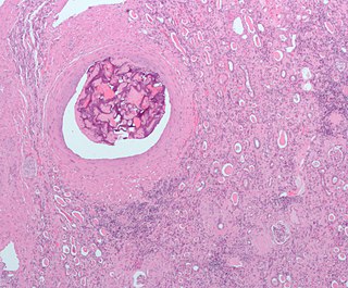
Angiomyolipomas are the most common benign tumour of the kidney. Although regarded as benign, angiomyolipomas may grow such that kidney function is impaired or the blood vessels may dilate and burst, leading to bleeding.

<span class="mw-page-title-main">Posterior urethral valve</span> Medical condition

Posterior urethral valve (PUV) disorder is an obstructive developmental anomaly in the urethra and genitourinary system of male newborns. A posterior urethral valve is an obstructing membrane in the posterior male urethra as a result of abnormal in utero development. It is the most common cause of bladder outlet obstruction in male newborns. The disorder varies in degree, with mild cases presenting late due to milder symptoms. More severe cases can have renal and respiratory failure from lung underdevelopment as result of low amniotic fluid volumes, requiring intensive care and close monitoring. It occurs in about one in 8,000 babies.

Pyelogram is a form of imaging of the renal pelvis and ureter.

<span class="mw-page-title-main">Pampiniform plexus</span> Vein network in human males

The pampiniform plexus is a venous plexus – a network of many small veins found in the human male spermatic cord, and the suspensory ligament of the ovary. In the male, it is formed by the union of multiple testicular veins from the back of the testis and tributaries from the epididymis.

<span class="mw-page-title-main">Loin pain hematuria syndrome</span> Medical condition

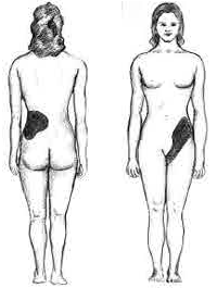
Loin pain hematuria syndrome (LPHS) is the combination of debilitating unilateral or bilateral flank pain and microscopic or macroscopic amounts of blood in the urine that is otherwise unexplained.

Bladder outlet obstruction occurs when urine is unable to flow from the kidneys through the ureters and out of the bladder through the urethra. Decreased flow of urine leads to swelling of the urinary tract, called hydronephrosis. This process of decreased flow of urine through the urinary tract can begin as early as during intrauterine life and it prevents normal development of fetal kidneys and fetal urine. Low levels of fetal urine leads to low amniotic fluid levels and incomplete lung maturation. Older children and adults can also experience bladder outlet obstruction; however, this process is usually reversible and isn't associated with as many poor outcomes as in infants with congenital bladder outlet obstruction.

<span class="mw-page-title-main">Fraley syndrome</span> Medical condition

Fraley syndrome is a condition where the superior infundibulum of the upper calyx of the kidney is obstructed by the crossing renal artery branch, causing distension and dilatation of the calyx and presenting clinically as haematuria and nephralgia. Furthermore, when the renal artery obstructs the proximal collecting system, filling defects can occur anywhere in the calyces, pelvis, or ureter.

<span class="mw-page-title-main">Pelvic congestion syndrome</span> Medical condition

Pelvic congestion syndrome, also known as pelvic vein incompetence, is a long-term condition believed to be due to enlarged veins in the lower abdomen. The condition may cause chronic pain, such as a constant dull ache, which can be worsened by standing or sex. Pain in the legs or lower back may also occur.

Pelvic compression syndrome is characterized by intermittent or persisting pain in the abdomen, which is exacerbated by abdominal pressure. A swelling of the veins in the valveless pampiniform plexus causes it.

References

  1. 1 2 Hacking, Craig (13 October 2022). "Gonadal Vein". Radiopaedia. Retrieved 27 December 2023.
  2. 1 2 Tourné G, Ducroux A, Bourbon M, Blinding H (September 2002). "The ovarian vein syndrome: eight cases and review of the literature". J Gynecol Obstet Biol Reprod (Paris) (in French). 31 (5): 471–7. PMID   12379831.
  3. 1 2 del Valle González N, Estébanez Zarranz J, Escudero Caro T, et al. (January 2006). "Laparoscopic treatment of ovarian vein syndrome". Actas Urol Esp (in Spanish). 30 (1): 85–9. doi:10.1016/s0210-4806(06)73402-7. PMID   16703736.
  4. 1 2 Maubon A, Ferru JM, Thiebaut C, Berger V, Hoche N, Rouanet JP (March 1997). "Left ovarian vein syndrome". J Radiol (in French). 78 (3): 223–5. PMID   9113150.
  5. Elashry OM, Nakada SY, Wolf JS, Figenshau RS, McDougall EM, Clayman RV (October 1996). "Ureterolysis for extrinsic ureteral obstruction: a comparison of laparoscopic and open surgical techniques". J. Urol. 156 (4): 1403–10. doi:10.1016/S0022-5347(01)65601-5. PMID   8808882.
  6. Badger WJ, De EJ, Kaufman RP (2008). "Robotically assisted excision of ovarian vein for intermittent ureteral obstruction". JSLS. 12 (2): 166–8. PMC   3016184 . PMID   18435891.
  7. Kwon SH, Oh JH, Ko KR, Park HC, Huh JY (2007). "Transcatheter ovarian vein embolization using coils for the treatment of pelvic congestion syndrome". Cardiovasc Intervent Radiol. 30 (4): 655–61. doi:10.1007/s00270-007-9040-7. PMID   17468903. S2CID   11622743.
  8. Moyano Calvo JL, Teba del Pino F, Arellano Gañan R, et al. (November 1993). "[The ovarian vein syndrome. Its treatment by percutaneous embolization and a review of the same]". Arch. Esp. Urol. (in Spanish). 46 (9): 802–6. PMID   8304795.
  9. Clark JC. The right ovarian vein syndrome. Emmett J.J. ed. Clinical urography: An Atlas and Textbook of Roentgenologic Diagnosis. 2ª ed. W.B. Saunders Company. Philadelphia 1964;1227-1236.
  10. Dure-Smith P (April 1979). "Ovarian syndrome: is it a myth?". Urology. 13 (4): 355–64. doi:10.1016/0090-4295(79)90330-3. PMID   107642.
  11. Derrick FC, Rosenblum R, Frensilli FJ (May 1973). "Right ovarian vein syndrome. Six-year critique". Urology. 1 (5): 383–5. doi:10.1016/0090-4295(73)90364-6. PMID   4204069.