A myxoma is a rare benigntumor of the heart. Myxomata are the most common primary cardiac tumor in adults, and are most commonly found within the left atrium near the valve of the fossa ovalis. Myxoma may also develop in the other heart chambers.[1] The tumor is derived from multipotent mesenchymal cells.[1] Cardiac myxoma can affect adults between 30 and 60 years of age.[2]
Symptoms may occur at any time, but most often they accompany a change of body position. Pedunculated myxomata can have a "wrecking ball effect", as they lead to stasis and may eventually embolize themselves. Symptoms may include:[3]
Myxomata are the most common type of adult primary heart tumor.[1][5] Most myxomata arise sporadically (90%), and only about 10% are thought to arise due to inheritance.[6]
About 10% of myxomata are inherited, as in Carney syndrome. Such tumors are called familial myxomata. They tend to occur in more than one part of the heart at a time, and often cause symptoms at a younger age than other myxomata. Other abnormalities are observed in people with Carney syndrome include skin myxomata, pigmentation, endocrine hyperactivity, schwannomas and epithelioid blue nevi.[1] Myxomata are more common in women than men.[1][3]
Diagnosis
A doctor will listen to the heart with a stethoscope. A "tumor plop" (a sound related to movement of the tumor), abnormal heart sounds, or a murmur similar to the mid-diastolic rumble of mitral stenosis may be heard. These sounds may change when the patient changes position.[7]
Right atrial myxomata rarely produce symptoms until they have grown to be at least 13cm (about 5inches) wide.[citation needed]
The surgery is treatment of choice,[10] tumor must be surgically removed. Some patients will also need their mitral valve replaced. This can be done during the same surgery. Usually, inadequate excision of the tumor, development from a secondary focus, or intracardiac implantation from the primary tumor are the attributable explanation for recurrence,[11] and it is more likely to occur in the first 10 postoperative years, especially in younger patients.[12]
Prognosis
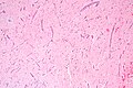
An embolized fragment of an atrial myxoma in the iliac bifurcation.
Although a myxoma is not malignant with risk of metastasis,[3] complications are common. Untreated, a myxoma can lead to an embolism (tumor cells breaking off and traveling with the bloodstream). Myxoma fragments can move to the brain, eye, or limbs.[citation needed]
If the tumor continues to enlarge inside the heart, it can block blood flow through the mitral valve and cause symptoms of mitral stenosis or mitral regurgitation. This may require emergency surgery to prevent sudden death.[13]
↑Masters, Barry R. (2012-05-25). "Harrisons's Principles of Internal Medicine, 18th Edition, two volumes and DVD. Eds: Dan L. Longo, Anthony S. Fauci, Dennis L. Kasper, Stephen L. Hauser, J. Larry Jameson and Joseph Loscalzo, ISBN 9780071748896 McGraw Hill". Graefe's Archive for Clinical and Experimental Ophthalmology. 250 (9): 1407–1408. doi:10.1007/s00417-012-1940-9. ISSN0721-832X. S2CID11647732.
↑"Cardiac Myxoma". The Lecturio Medical Concept Library. Retrieved 6 July 2021.
↑"Cardiac Myxoma". The Lecturio Medical Concept Library. Retrieved 6 July 2021.
↑Lone, R. A.; Ahanger, A. G.; Singh, S.; Mehmood, W.; Shah, S.; Lone, G.; Dar, A.; Bhat, M.; Sharma, M.; Lateef, W. (2008). "Atrial myxoma: Trends in management". International Journal of Health Sciences. 2 (2): 141–151. PMC3068734. PMID21475496.
↑Sheng, W. B.; Luo, B. E.; Liu, Y.; Zhang, H.; Zou, L. J.; Xu, Z. Y.; Zhang, H. Y.; Ji, G. Y. (2012). "Risk factors for postoperative recurrence of cardiac myxoma and the clinical managements: A report of 5 cases in one center and review of literature". Chinese Medical Journal. 125 (16): 2914–2918. PMID22932090.
↑Shah, I. K.; Dearani, J. A.; Daly, R. C.; Suri, R. M.; Park, S. J.; Joyce, L. D.; Li, Z.; Schaff, H. V. (2015). "Cardiac Myxomas: A 50-Year Experience with Resection and Analysis of Risk Factors for Recurrence". The Annals of Thoracic Surgery. 100 (2): 495–500. doi:10.1016/j.athoracsur.2015.03.007. PMID26070596.
This page is based on this Wikipedia article Text is available under the CC BY-SA 4.0 license; additional terms may apply. Images, videos and audio are available under their respective licenses.