
Chrysosporium is a genus of hyaline hyphomycetes fungi in the family Onygenaceae.
Polytolypa is a monotypic genus of fungus containing the single species Polytolypa hystricis. First classified in the Onygenaceae family, as of 2008 it is considered to be in the Ajellomycetaceae, although there is still uncertainty as to its phylogenetic relationships with other similar genera. This species is only known from a single specimen derived in the laboratory from a specimen of dung of the North American porcupine, Erethizon dorsatum, collected in Ontario, Canada. Polytolypa hystricis contains bioactive compounds that have antifungal activity.
Arachnomyces is a genus of cleistothecial ascomycete fungi described in 1902, of which the anamorph (asexual) stage is the genus Onychocola. Although morphologically similar to members of other families, the fungus now belongs to its own monotypic family Arachnomycetaceae, which is the only family in the monotypic order Arachnomycetales.
Massarina carolinensis is a species of fungus in the Lophiostomataceae family. The species is found exclusively on the lower parts of the culms of the saltmarsh Juncus roemerianus on the Atlantic Coast of North Carolina.
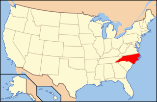
Paraphaeosphaeria pilleata is a species of fungus in the Lophiostomataceae family. The species fruits exclusively in the lower parts of the culms of the black needlerush. It is found on the Atlantic Coast of North Carolina.

Onygena equina, commonly known as the horn stalkball, is a species of fungus in the family Onygenaceae. The fungus grows on putrefying hooves and horns, and can digest the keratin in those substrates. Fruit bodies are small and white, with thick stipes supporting a "head" shaped like a flattened sphere. The skin, or peridium, of the head appears powdery or like a white crust, and breaks open in maturity, falling off in irregular pieces to expose the pale reddish-brown powdery spores within. The fungus is known from Europe and North America.

Aphanoascus fulvescens is a mould fungus that behaves as a keratinophilic saprotroph and belongs to the Ascomycota. It is readily isolated from soil and dung containing keratin-rich tissues that have been separated from their animal hosts. This organism, distributed worldwide, is most commonly found in areas of temperate climate, in keeping with its optimal growth temperature of 28 °C (82 °F). While A. fulvescens is recognized as a geophilic fungal species, it is also a facultative opportunistic pathogen. Although it is not a dermatophyte, A. fulvescens has occasionally been shown to cause onychomycosis infections in humans. Its recognition in the laboratory is clinically important for correct diagnosis and treatment of human dermal infections.
Nannizziopsis vreisii is a keratinophilic microfungus in the Family Onygenaceae of the order Onygenales. Also included in this family are dematophytes and saprophytic species. While the ecology of N. vriessi is not well known, there has been several studies which identifies the Chrysosporium anamorph of N. vriesii as a causal agent of skin lesions in reptiles across several regions. This species is usually identified under a microscope by its white ascomata, and hyaline and globose ascospores. Like many other fungi, N. vreisii has a sexual and asexual state, the asexual states are classified as the genus Chryososporium, Malbranchea or Sporendonema.

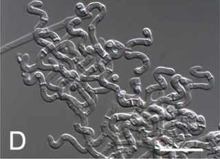
Ophidiomyces ophidiicola is the cause of ophidiomycosis also known as snake fungal disease or SFD in some species of snakes. It is a keratinophilic fungus from the family Onygenaceae of the order Onygenales. O. ophidiicola is an emerging pathogen of captive and wild snakes in North America and Europe. Clinical signs include skin swelling, crusts, and nodules of the skin. The mode of transmission is unknown, but is speculated to occur with direct contact between snakes or with the contaminated environment. Currently no treatment for O. ophidiicola is available. O. ophidiicola was identified by Sigler, Hambleton & Paré in 2013. O. ophidiicola is the only species in the genus Ophidiomyces. It was previously known as Chrysosporium ophiodiicola and is closely related to Chrysosporium anamorph Nannizziopsis vriesii (CANV).
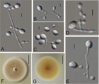
Nannizziopsis guarroi was first documented in 2006 on a variety of lizards then described in Spain in 2010 and was classified as Chrysosporium guarroi, a member of the anamorphic genus Chrysosporium in the family Onygenaceae. Etymologically, the species epithet "guarroi" honours Professor Josep Guarro in recognition of his extensive mycological work including on the genus Chrysosporium. Skin samples taken from pet green iguanas suffering from dermatomycosis were sent to a laboratory for analysis. Five species were isolated and morphologic studies identified the fungus causing the mycoses as a member of the anamorphic species of Chrysosporium. Further investigation of these species using a combination of morphological, cultural and molecular studies showed that they were not identical to any previously described species within the genus Chrysosporium so they were classified as a new species Chrysosporium guarroisp. nov. The delineation of species in the genus Chrysosporium and their assignment to higher taxonomic levels can be challenging due to the marked morphological simplicity of these fungi. Increased scrutiny of strains of these fungi using molecular genetic tools has revealed numerous hidden species and unexpected relationships.
Amauroascus kuehnii is a fungus in the phylum Ascomycota, class Eurotiomycetes. It is keratinophilic but not known to cause any human disease. It has been isolated from animal dungs, soil, and keratinous surfaces of live or deceased animals.

Keratinophyton durum is a keratinophilic fungus, that grows on keratin found in decomposing or shed animal hair and bird feathers. Various studies conducted in Canada, Japan, India, Spain, Poland, Ivory Coast and Iraq have isolated this fungus from decomposing animal hair and bird feathers using SDA and hair-bait technique. Presence of fungus in soil sediments and their ability to decompose hairs make them a potential human pathogen.
Botryotrichum piluliferum is a fungal species first identified in 1885 by Saccardo and Marchal. It was discovered to be the asexual state of a member of the ascomycete genus, Chaetomium. The name B. piluliferum now applies to the fungus in all its states. B. piluliferum has been found worldwide in a wide range of habitats such as animal dung and vegetation. The colonies of this fungus start off white and grow rapidly to a brown colour. The conidia are smooth and white. B. piluliferum grows optimally at a temperature of 25–30 °C and a pH of 5.5.
Ctenomyces serratus is a keratinophilic fungal soil saprotroph classified by the German mycologist, Michael Emil Eduard Eidam in 1880, who found it growing on an old decayed feather. Many accounts have shown that it has a global distribution, having been isolated in select soils as well as on feathers and other substrates with high keratin content. It has also been found in indoor dust of hospitals and houses in Kanpur, Northern India and as a common keratinophilic soil fungus in urban Berlin. This species has been associated with nail infections in humans as well as skin lesions and slower hair growth in guinea pigs.
Microascus manginii is a filamentous fungal species in the genus Microascus. It produces both sexual (teleomorph) and asexual (anamorph) reproductive stages known as M. manginii and Scopulariopsis candida, respectively. Several synonyms appear in the literature because of taxonomic revisions and re-isolation of the species by different researchers. M. manginii is saprotrophic and commonly inhabits soil, indoor environments and decaying plant material. It is distinguishable from closely related species by its light colored and heart-shaped ascospores used for sexual reproduction. Scopulariopsis candida has been identified as the cause of some invasive infections, often in immunocompromised hosts, but is not considered a common human pathogen. There is concern about amphotericin B resistance in S. candida.
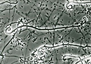
Uncinocarpus reesii is a species of saprotrophic microfungi that grows in soil and on keratinous materials such as hair, feathers and skin. It was the first species to be designated as part of the genus Uncinocarpus, owing in part to its characteristic development of hooked (uncinate) appendages. As the closest non-pathogenic relative of Coccidioides immitis and C. posadasii, it has become a subject of research interest.

Myxotrichum chartarum is a psychrophilic and cellulolytic fungus first discovered in Germany by Gustav Kunze in 1823. Its classification has changed many times over its history to better reflect the information available at the time. Currently, M. chartarum is known to be an ascomycete surrounded by a gymnothecium composed of ornate spines and releases asexual ascospores. The presence of cellulolytic processes are common in fungi within the family Myxotrichaceae. M. chartarum is one of many Myxotrichum species known to degrade paper and paper products. Evidence of M. chartarum "red spot" mold formation, especially on old books, can be found globally. As a result, this fungal species and other cellulolytic molds are endangering old works of art and books. Currently, there is no evidence that suggests that species within the family Myxotrichaceae are pathogenic.
Arachniotus ruber is a species of fungus belonging to the genus Arachniotus in the family Gymnoascaceae. This fungus is a mesophile that reproduces both sexually and asexually. So far, there have been no reports of the fungus being pathogenic.
Auxarthron californiense is a fungus within the family Onygenaceae family and one of the type species of the genus Auxarthron. A. californiense is generally distributed around the world and it is frequently found on dung and in soil near the entrances of animal burrows.
Scytalidium ganodermophthorum is an anthroconidial ascomycete fungus in the Scytalidium genus. It is also known by its teleomorph name Xylogone ganodermophthora. It is the cause of yellow rot in lingzhi mushrooms and it is used in spalting as a pigmenting fungi.
