Comet assay

Last updated • 6 min readFrom Wikipedia, The Free Encyclopedia
CaspLab comet assay application screenshot Casplabapp.jpg
CaspLab comet assay application screenshot

The single cell gel electrophoresis assay (SCGE, also known as comet assay) is an uncomplicated and sensitive technique for the detection of DNA damage at the level of the individual eukaryotic cell. It was first developed by Östling & Johansson in 1984 and later modified by Singh et al. in 1988. [1] It has since increased in popularity as a standard technique for evaluation of DNA damage/repair, biomonitoring and genotoxicity testing. It involves the encapsulation of cells in a low-melting-point agarose suspension, lysis of the cells in neutral or alkaline (pH>13) conditions, and electrophoresis of the suspended lysed cells. The term "comet" refers to the pattern of DNA migration through the electrophoresis gel, which often resembles a comet. [2] [3]

Contents

The comet assay (single-cell gel electrophoresis) is a simple method for measuring deoxyribonucleic acid (DNA) strand breaks in eukaryotic cells. Cells embedded in agarose on a microscope slide are lysed with detergent and high salt to form nucleoids containing supercoiled loops of DNA linked to the nuclear matrix. Electrophoresis at high pH results in structures resembling comets, observed by fluorescence microscopy; the intensity of the comet tail relative to the head reflects the number of DNA breaks. The likely basis for this is that loops containing a break lose their supercoiling and become free to extend toward the anode. This is followed by visual analysis with staining of DNA and calculating fluorescence to determine the extent of DNA damage. This can be performed by manual scoring or automatically by imaging software. [4] [5]

Procedure

Encapsulation

A sample of cells, either derived from an in vitro cell culture or from an in vivo test subject is dispersed into individual cells and suspended in molten low-melting-point agarose at 37 °C. This mono-suspension is cast on a microscope slide. A glass cover slip is held at an angle and the mono-suspension is applied to the point of contact between the coverslip and the slide. As the coverslip is lowered onto the slide the molten agarose spreads to form a thin layer. The agarose is gelled at 4 °C and the coverslip removed.

The agarose forms a matrix of carbohydrate fibres that encapsulate the cells, anchoring them in place. The agarose is considered to be osmotic-neutral, therefore solutions can penetrate the gel and affect the cells without cells shifting position.

In an in vitro study the cells would be exposed to a test agent – typically UV light, ionising radiation, or a genotoxic chemical – to induce DNA damage in the encapsulated cells. For calibration, hydrogen peroxide is usually used to provide a standardized level of DNA damage.

Lysis

The slides are then immersed in a solution that cause the cells to lyse. The lysis solution often used in the comet assay consists of a highly concentrated aqueous salt (often, common table salt can be used) and a detergent (such as Triton X-100 or sarcosinate). The pH of the lysis solution can be adjusted (usually between neutral and alkaline pH) depending upon the type of damage the researcher is investigating.

The aqueous salt disrupts proteins and their bonding patterns within the cell as well as disrupting the RNA content of the cell. The detergent dissolves the cellular membranes. Through the action of the lysis solution the cells are destroyed. All proteins, RNA, membranes and cytoplasmic and nucleoplasmic constituents are disrupted and diffuse into the agarose matrix. Only the DNA of the cell remains, and unravels to fill the cavity in the agarose that the whole cell formerly filled. This structure is called nucleoid (a general term for a structure in which DNA is concentrated).

Electrophoresis

After lysis of the cells (typically 1 to 2 hours at 4 °C) the slides are washed in distilled water to remove all salts and immersed in a second solution – an electrophoresis solution. Again this solution can have its pH adjusted depending upon the type of damage that is being investigated.

The slides are left for ~20 minutes in the electrophoresis solution prior to an electric field being applied. In alkaline conditions the DNA double helix is denatured and the nucleoid becomes single stranded.

An electric field is applied (typically 1 V/cm) for ~20 minutes. The slides are then neutralised to pH 7, stained with a DNA-specific fluorescent stain and analysed using a microscope with an attached CCD (charge-coupled device – essentially a digital camera) that is connected to a computer with image analysis software.

Background

The concept underlying the SCGE assay is that undamaged DNA retains a highly organized association with matrix proteins in the nucleus. When damaged, this organization is disrupted. The individual strands of DNA lose their compact structure and relax, expanding out of the cavity into the agarose. When the electric field is applied the DNA, which has an overall negative charge, is drawn towards the positively charged anode. Undamaged DNA strands are too large and do not leave the cavity, whereas the smaller the fragments, the farther they are free to move in a given period of time. Therefore, the amount of DNA that leaves the cavity is a measure of the amount of DNA damage in the cell.

The image analysis measures the overall intensity of the fluorescence for the whole nucleoid and the fluorescence of the migrated DNA and compares the two signals. The stronger the signal from the migrated DNA the more damage there is present. The overall structure resembles a comet (hence "comet assay") with a circular head corresponding to the undamaged DNA that remains in the cavity and a tail of damaged DNA. The brighter and longer the tail, the higher the level of damage.

The comet assay is a versatile technique for detecting damage and with adjustments to the protocol can be used to quantify the presence of a wide variety of DNA altering lesions (damage). The damage usually detected are single strand breaks and double strand breaks. It is sometimes stated [6] [7] that alkaline conditions and complete denaturating of the DNA is necessary to detect single strand breaks. However this is not true, both single- and double strand breaks are also detected in neutral conditions. [8] [9] [10] [11] In alkaline conditions, however, additional DNA structures are detected as DNA damage: AP sites (abasic sites missing either a pyrimidine or purine nucleotide) and sites where excision repair is taking place. [12] [13]

The comet assay is an extremely sensitive DNA damage assay. This sensitivity needs to be handled carefully as it is also vulnerable to physical changes which can affect the reproducibility of results. Essentially, anything that can cause DNA damage or denaturation except the factor(s) being researched is to be avoided. [14] The most common form of the assay is the alkaline version although there is as yet no definitive alkaline assay protocol. Due to its simple and inexpensive setup, it can be used in conditions where more complex assays are not available.

Applications

These include genotoxicity testing, human biomonitoring and molecular epidemiology, ecogenotoxicology, as well as fundamental research in DNA damage and repair. [15] For example, Swain and Rao, using the comet assay [16] reported marked increases in several types of DNA damages in rat brain neurons and astrocytes during aging, including single-strand breaks, double-strand breaks and modified bases (8-OHdG and uracil).

Sperm DNA fragmentation

A comet assay can determine the degree of DNA fragmentation in sperm cells. The degree of DNA fragmentation has been associated with outcomes of in vitro fertilization. [17] [18]

The comet has been modified for use with sperm cells as a tool for male infertility diagnosis [19] [20] [21]

To break down these tightly bound protamine proteins in order to use the comet for sperm, additional steps in the de-condensation protocol are required. [20]

Related Research Articles

<span class="mw-page-title-main">Agarose gel electrophoresis</span> Method for separation and analysis of biomolecules using agarose gel

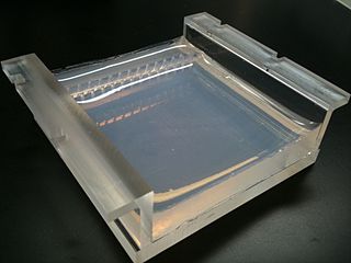
Agarose gel electrophoresis is a method of gel electrophoresis used in biochemistry, molecular biology, genetics, and clinical chemistry to separate a mixed population of macromolecules such as DNA or proteins in a matrix of agarose, one of the two main components of agar. The proteins may be separated by charge and/or size, and the DNA and RNA fragments by length. Biomolecules are separated by applying an electric field to move the charged molecules through an agarose matrix, and the biomolecules are separated by size in the agarose gel matrix.

<span class="mw-page-title-main">Agarose</span> Heteropolysaccharide found in red algae

Agarose is a heteropolysaccharide, generally extracted from certain red algae. It is a linear polymer made up of the repeating unit of agarobiose, which is a disaccharide made up of D-galactose and 3,6-anhydro-L-galactopyranose. Agarose is one of the two principal components of agar, and is purified from agar by removing agar's other component, agaropectin.

<span class="mw-page-title-main">Gel electrophoresis</span> Method for separation and analysis of biomolecules

Gel electrophoresis is a method for separation and analysis of biomacromolecules and their fragments, based on their size and charge. It is used in clinical chemistry to separate proteins by charge or size and in biochemistry and molecular biology to separate a mixed population of DNA and RNA fragments by length, to estimate the size of DNA and RNA fragments or to separate proteins by charge.

Molecular biology is a branch of biology that seeks to understand the molecular basis of biological activity in and between cells, including biomolecular synthesis, modification, mechanisms, and interactions.

<span class="mw-page-title-main">Gel electrophoresis of nucleic acids</span>

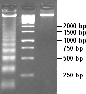
Gel electrophoresis of nucleic acids is an analytical technique to separate DNA or RNA fragments by size and reactivity. Nucleic acid molecules are placed on a gel, where an electric field induces the nucleic acids to migrate toward the positively charged anode. The molecules separate as they travel through the gel based on the each molecule's size and shape. Longer molecules move more slowly because they the gel resists their movement more forcefully than it resists shorter molecules. After some time, the electricity is turned off and the positions of the different molecules are analyzed.

<span class="mw-page-title-main">Ethidium bromide</span> DNA gel stain and veterinary drug

Ethidium bromide is an intercalating agent commonly used as a fluorescent tag in molecular biology laboratories for techniques such as agarose gel electrophoresis. It is commonly abbreviated as EtBr, which is also an abbreviation for bromoethane. To avoid confusion, some laboratories have used the abbreviation EthBr for this salt. When exposed to ultraviolet light, it will fluoresce with an orange colour, intensifying almost 20-fold after binding to DNA. Under the name homidium, it has been commonly used since the 1950s in veterinary medicine to treat trypanosomiasis in cattle. The high incidence of antimicrobial resistance makes this treatment impractical in some areas, where the related isometamidium chloride is used instead. Despite its reputation as a mutagen, tests have shown it to have low mutagenicity without metabolic activation.

Deoxyribonuclease refers to a group of glycoprotein endonucleases which are enzymes that catalyze the hydrolytic cleavage of phosphodiester linkages in the DNA backbone, thus degrading DNA. The role of the DNase enzyme in cells includes breaking down extracellular DNA (ecDNA) excreted by apoptosis, necrosis, and neutrophil extracellular traps (NET) of cells to help reduce inflammatory responses that otherwise are elicited. A wide variety of deoxyribonucleases are known and fall into one of two families, which differ in their substrate specificities, chemical mechanisms, and biological functions. Laboratory applications of DNase include purifying proteins when extracted from prokaryotic organisms. Additionally, DNase has been applied as a treatment for diseases that are caused by ecDNA in the blood plasma. Assays of DNase are emerging in the research field as well.

Site-directed mutagenesis is a molecular biology method that is used to make specific and intentional mutating changes to the DNA sequence of a gene and any gene products. Also called site-specific mutagenesis or oligonucleotide-directed mutagenesis, it is used for investigating the structure and biological activity of DNA, RNA, and protein molecules, and for protein engineering.

Genotoxicity is the property of chemical agents that damage the genetic information within a cell causing mutations, which may lead to cancer. While genotoxicity is often confused with mutagenicity, all mutagens are genotoxic, but some genotoxic substances are not mutagenic. The alteration can have direct or indirect effects on the DNA: the induction of mutations, mistimed event activation, and direct DNA damage leading to mutations. The permanent, heritable changes can affect either somatic cells of the organism or germ cells to be passed on to future generations. Cells prevent expression of the genotoxic mutation by either DNA repair or apoptosis; however, the damage may not always be fixed leading to mutagenesis.

Biomedicine is a branch of medical science that applies biological and physiological principles to clinical practice. Biomedicine stresses standardized, evidence-based treatment validated through biological research, with treatment administered via formally trained doctors, nurses, and other such licensed practitioners.

The first isolation of deoxyribonucleic acid (DNA) was done in 1869 by Friedrich Miescher. DNA extraction is the process of isolating DNA from the cells of an organism isolated from a sample, typically a biological sample such as blood, saliva, or tissue. It involves breaking open the cells, removing proteins and other contaminants, and purifying the DNA so that it is free of other cellular components. The purified DNA can then be used for downstream applications such as PCR, sequencing, or cloning. Currently, it is a routine procedure in molecular biology or forensic analyses.

DNA fragmentation is the separation or breaking of DNA strands into pieces. It can be done intentionally by laboratory personnel or by cells, or can occur spontaneously. Spontaneous or accidental DNA fragmentation is fragmentation that gradually accumulates in a cell. It can be measured by e.g. the Comet assay or by the TUNEL assay.

<span class="mw-page-title-main">Electrophoretic mobility shift assay</span> Technique used to study DNA and RNA

An electrophoretic mobility shift assay (EMSA) or mobility shift electrophoresis, also referred as a gel shift assay, gel mobility shift assay, band shift assay, or gel retardation assay, is a common affinity electrophoresis technique used to study protein–DNA or protein–RNA interactions. This procedure can determine if a protein or mixture of proteins is capable of binding to a given DNA or RNA sequence, and can sometimes indicate if more than one protein molecule is involved in the binding complex. Gel shift assays are often performed in vitro concurrently with DNase footprinting, primer extension, and promoter-probe experiments when studying transcription initiation, DNA gang replication, DNA repair or RNA processing and maturation, as well as pre-mRNA splicing. Although precursors can be found in earlier literature, most current assays are based on methods described by Garner and Revzin and Fried and Crothers.

<span class="mw-page-title-main">DNA laddering</span>

DNA laddering is a feature that can be observed when DNA fragments, resulting from Apoptosis DNA fragmentation are visualized after separation by gel electrophoresis the first described in 1980 by Andrew Wyllie at the University Edinburgh medical school DNA fragments can also be detected in cells that underwent necrosis, but when these DNA fragments after separation are subjected to gel electrophoresis no clear "ladder" pattern is apparent.

DNA footprinting is a method of investigating the sequence specificity of DNA-binding proteins in vitro. This technique can be used to study protein-DNA interactions both outside and within cells.

<span class="mw-page-title-main">Clastogen</span> Substance that can cause breaks in chromosomes

A clastogen is a mutagenic agent that disturbs normal DNA related processes or directly causes DNA strand breakages, thus causing the deletion, insertion, or rearrangement of entire chromosome sections. These processes are a form of mutagenesis which if left unrepaired, or improperly repaired, can lead to cancer. Known clastogens include acridine yellow, benzene, ethylene oxide, arsenic, phosphine, mimosine, actinomycin D, camptothecin, methotrexate, methyl acrylate, resorcinol and 5-fluorodeoxyuridine. Additionally, 1,2-dimethylhydrazine is a known colon carcinogen and shows signs of possessing clastogenic activity. There are many clastogens not listed here and research is ongoing to discover new clastogens. Some known clastogens only exhibit clastogenic activity in certain cell types, such as caffeine which exhibits clastogenic activity in plant cells. Researchers are interested in clastogens for researching cancer, as well as for other human health concerns such as the inheritability of clastogen effected paternal germ cells that lead to fetus developmental defects.

<span class="mw-page-title-main">Electrophoretic color marker</span>

An electrophoretic color marker is a chemical used to monitor the progress of agarose gel electrophoresis and polyacrylamide gel electrophoresis (PAGE) since DNA, RNA, and most proteins are colourless. The color markers are made up of a mixture of dyes that migrate through the gel matrix alongside the sample of interest. They are typically designed to have different mobilities from the sample components and to generate colored bands that can be used to assess the migration and separation of sample components.

DNA damage is an alteration in the chemical structure of DNA, such as a break in a strand of DNA, a nucleobase missing from the backbone of DNA, or a chemically changed base such as 8-OHdG. DNA damage can occur naturally or via environmental factors, but is distinctly different from mutation, although both are types of error in DNA. DNA damage is an abnormal chemical structure in DNA, while a mutation is a change in the sequence of base pairs. DNA damages cause changes in the structure of the genetic material and prevents the replication mechanism from functioning and performing properly. The DNA damage response (DDR) is a complex signal transduction pathway which recognizes when DNA is damaged and initiates the cellular response to the damage.

<span class="mw-page-title-main">Sperm Chromatin Structure Assay</span>

Sperm Chromatin Structure Assay (SCSA) is a diagnostic approach that detects sperm abnormality with a large extent of DNA fragmentation. First described by Evenson in 1980, the assay is a flow cytometric test that detects the vulnerability of sperm DNA to acid-induced denaturation DNA in situ. SCSA measures sperm DNA fragmentation attributed to intrinsic and extrinsic factors and reports the degree of fragmentation in terms of DNA Fragmentation Index (DFI). The use of SCSA expands from evaluation of male infertility and subfertility, toxicology studies and evaluation of quality of laboratory semen samples. Notably, SCSA outcompetes other convention sperm DNA fragmentation (sDF) assays such as TUNEL and COMET in terms of efficiency, objectivity, and repeatability.

Diana Anderson was a British biomedical scientist who is a professor at the University of Bradford. Her research has focussed on early cancer detection and genome stability. She was appointed an Order of the British Empire in 2022 for her services to cancer detection.

References

  1. Singh, Narendra P.; McCoy, Michael T.; Tice, Raymond R.; Schneider, Edward L. (1988-03-01). "A simple technique for quantitation of low levels of DNA damage in individual cells". Experimental Cell Research. 175 (1): 184–191. doi:10.1016/0014-4827(88)90265-0. ISSN   0014-4827. PMID   3345800.
  2. Tice, R. R. (2000). "Single Cell Gel/Comet Assay: Guidelines for in vitro and in vivo Genetic Toxicology Testing". Environmental and Molecular Mutagenesis. 35 (3): 206–21. doi:10.1002/(SICI)1098-2280(2000)35:3<206::AID-EM8>3.0.CO;2-J. PMID   10737956.
  3. Nandhakumar, S; Parasuraman, S; Shanmugam, M M; Rao, K R; Chand, P; Bhat, B V (2011). "Evaluation of DNA damage using single-cell gel electrophoresis (Comet Assay)". J Pharmacol Pharmacother. 2 (2): 107–11. doi: 10.4103/0976-500X.81903 . PMC   3127337 . PMID   21772771.
  4. Collins, A. R. (March 2004). "The comet assay for DNA damage and repair: principles, applications, and limitations". Mol. Biotechnol. 26 (3): 249–61. doi:10.1385/MB:26:3:249. PMID   15004294. S2CID   34355649.
  5. Nandhakumar, S; Parasuraman, S; Shanmugam, MM; Rao, KR; Chand, P; Bhat, BV (Apr 2011). "Evaluation of DNA damage using single-cell gel electrophoresis (Comet Assay)". J Pharmacol Pharmacother. 2 (2): 107–11. doi: 10.4103/0976-500X.81903 . PMC   3127337 . PMID   21772771. Archived from the original on 2018-10-26. Retrieved 2013-12-05.
  6. Pu, Xinzhu; Wang, Zemin; Klaunig, James E. (2015-08-06). "Alkaline Comet Assay for Assessing DNA Damage in Individual Cells". Current Protocols in Toxicology. 65: 3.12.1–11. doi:10.1002/0471140856.tx0312s65. ISBN   978-0471140856. ISSN   1934-9262. PMID   26250399. S2CID   6105193.
  7. Møller, Peter (April 2006). "The alkaline comet assay: towards validation in biomonitoring of DNA damaging exposures". Basic & Clinical Pharmacology & Toxicology. 98 (4): 336–345. doi: 10.1111/j.1742-7843.2006.pto_167.x . ISSN   1742-7835. PMID   16623855.
  8. Belyaev, Igor Y; Eriksson, Stefan; Nygren, Jonas; Torudd, Jesper; Harms-Ringdahl, Mats (1999-08-05). "Effects of ethidium bromide on DNA loop organisation in human lymphocytes measured by anomalous viscosity time dependence and single cell gel electrophoresis". Biochimica et Biophysica Acta (BBA) - General Subjects. 1428 (2–3): 348–356. doi:10.1016/S0304-4165(99)00076-8. ISSN   0304-4165. PMID   10434054.
  9. Olive, P. L.; Banáth, J. P.; Durand, R. E. (April 1990). "Heterogeneity in radiation-induced DNA damage and repair in tumor and normal cells measured using the "comet" assay". Radiation Research. 122 (1): 86–94. Bibcode:1990RadR..122...86O. doi:10.2307/3577587. ISSN   0033-7587. JSTOR   3577587. PMID   2320728.
  10. Powell, S.; McMillan, T.J. (1990-10-01). "DNA damage and repair following treatment with ionizing radiation". Radiotherapy and Oncology. 19 (2): 95–108. doi:10.1016/0167-8140(90)90123-E. ISSN   0167-8140. PMID   2255771.
  11. Wojewódzka, Maria; Buraczewska, Iwona; Kruszewski, Marcin (2002-06-27). "A modified neutral comet assay: elimination of lysis at high temperature and validation of the assay with anti-single-stranded DNA antibody". Mutation Research. 518 (1): 9–20. doi:10.1016/s1383-5718(02)00070-0. ISSN   0027-5107. PMID   12063063.
  12. Collins, Andrew R (1997-04-29). "The comet assay: what can it really tell us?". Mutation Research/Fundamental and Molecular Mechanisms of Mutagenesis. 375 (2): 183–193. doi:10.1016/S0027-5107(97)00013-4. ISSN   0027-5107. PMID   9202728.
  13. Gedik, C. M.; Ewen, S. W.; Collins, A. R. (September 1992). "Single-cell gel electrophoresis applied to the analysis of UV-C damage and its repair in human cells". International Journal of Radiation Biology. 62 (3): 313–320. doi:10.1080/09553009214552161. ISSN   0955-3002. PMID   1356133.
  14. Theoretical and practical limitations to the assay are discussed e.g. in Klaude et al. (1996) and Collins et al. (1997).
  15. https://www2.le.ac.uk/departments/csmm/.../SCG%20Electrophoresis.pdf%5B%5D
  16. Swain, U; Subba Rao, K (Aug 2011). "Study of DNA damage via the comet assay and base excision repair activities in rat brain neurons and astrocytes during aging". Mech Ageing Dev. 132 (8–9): 374–81. doi:10.1016/j.mad.2011.04.012. PMID 21600238. S2CID 22466782
  17. Simon L, Brunborg G, Stevenson M, Lutton D, McManus J, Lewis SE (May 2010). "Clinical significance of sperm DNA damage in assisted reproduction outcome". Hum Reprod. 25 (7): 1594–1608. doi: 10.1093/humrep/deq103 . PMID   20447937.
  18. Nagrajappa; Ganguly, Anutosh; Goswami, Usha (2006). "DNA damage in male gonad cells of Green mussel (Perna viridis) upon exposure to tobacco products". Ecotoxicology. 15 (4): 365–369. doi:10.1007/s10646-006-0077-1. PMID   16673160. S2CID   13956778.
  19. Hughes CM, Lewis SEM, McKelvey-Martin V, Thompson W. Reproducibility of human sperm DNA measurements using a single cell gel electrophoresis assay. Mutation Research 1997 374:261-268.
  20. 1 2 Donnelly, ET; McClure, N; Lewis, SEM (1999). "The effects of ascorbate and @-tocopherol supplementation in vitro on DNA integrity and hydrogen peroxide-induced DNA damage in human spermatozoa". Mutagenesis. 14 (5): 505–511. doi: 10.1093/mutage/14.5.505 . PMID   10473655.
  21. Ribas-Maynou Jordi, Agustı´n Garcı´a-Peiro´ , Alba Fernandez-Encinas, Maria Jose´ Amengual and Benet Jordi ,Double Stranded Sperm DNA Breaks, Measured by Comet Assay, Are Associated with Unexplained Recurrent Miscarriage in Couples without a Female Factor

Further reading