Cyclase-associated protein family

Last updated
CAP N-terminal
PDB 1s0p EBI.jpg
structure of the n-terminal domain of the adenylyl cyclase-associated protein (cap) from dictyostelium discoideum.
Identifiers
SymbolCAP_N
Pfam PF01213
InterPro IPR013992
PROSITE PDOC00835
SCOP2 1s0p / SCOPe / SUPFAM
Available protein structures:
Pfam   structures / ECOD  
PDB RCSB PDB; PDBe; PDBj
PDBsum structure summary
The crystal structure of CAP bound to actin monomers. PDB accession = 6fm2 Cap-actin.gif
The crystal structure of CAP bound to actin monomers. PDB accession = 6fm2
CAP C-terminal
PDB 1kq5 EBI.jpg
c-terminal domain of cyclase associated protein with pro 505 replaced by ser (p505s)
Identifiers
SymbolCAP_C
Pfam PF08603
Pfam clan CL0391
InterPro IPR013912
PROSITE PDOC00835
SCOP2 1kq5 / SCOPe / SUPFAM
Available protein structures:
Pfam   structures / ECOD  
PDB RCSB PDB; PDBe; PDBj
PDBsum structure summary

In molecular biology, the cyclase-associated protein family (CAP) is a family of highly conserved actin-binding proteins present in a wide range of organisms including yeast, flies, plants, and mammals. CAPs are multifunctional proteins that contain several structural domains. CAP is involved in species-specific signalling pathways. [2] [3] [4] [5] In Drosophila , CAP functions in Hedgehog-mediated eye development and in establishing oocyte polarity. In Dictyostelium discoideum (social amoeba), CAP is involved in microfilament reorganisation near the plasma membrane in a PIP2-regulated manner and is required to perpetuate the cAMP relay signal to organise fruitbody formation. In plants, CAP is involved in plant signalling pathways required for co-ordinated organ expansion. In yeast, CAP is involved in adenylate cyclase activation, as well as in vesicle trafficking and endocytosis. In both yeast and mammals, CAPs appear to be involved in recycling G-actin monomers from ADF/cofilins for subsequent rounds of filament assembly. [6] [7] In mammals, there are two different CAPs (CAP1 and CAP2) that share 64% amino acid identity.

Contents

Function

All CAPs appear to contain a C-terminal actin-binding domain that regulates actin remodelling in response to cellular signals and is required for normal cellular morphology, cell division, growth and locomotion in eukaryotes. CAP directly regulates actin filament dynamics and has been implicated in a number of complex developmental and morphological processes, including mRNA localisation and the establishment of cell polarity. Actin exists both as globular (G) (monomeric) actin subunits and assembled into filamentous (F) actin. In cells, actin cycles between these two forms. Proteins that bind F-actin often regulate F-actin assembly and its interaction with other proteins, while proteins that interact with G-actin often control the availability of unpolymerised actin.

The most conserved domain of CAPs appears to be the ADP-G-actin binding CARP domain which promotes nucleotide exchange of actin monomers from ADP-state back to polymerisable ATP-form. [8] Recently, the crystal structure of CARP domain bound to ADP-actin was determined, which revealed that CAPs have a unique dimeric binding mode to backside of ADP-G-actin monomers. [1] Based on the crystal structure and biochemical work, the C-terminus of CARP domain appears to be important in regulating the binding to ADP-G-actin monomers to CAP, and has a conserved role for nucleotide exchange on actin monomers. Genetic work in Saccharomyches ceravisae revealed that nucleotide exchange performed by CAP is critical for normal organisation of actin cytoskeleton.

A second unique function of CAP in regulating actin dynamics is its ability to specifically bind to pointed ends of cofilin-decorated actin filaments, [9] and rapidly drive their depolymerisation. [9] [10] CAP thus is a specialised protein in recycling of actin monomers, by first rapidly dissociating ADP-actin monomers from the pointed end of actin filaments, then simultaneously driving ADP-to-ATP nucleotide exchange to make actin monomers assembly-competent.

In addition to actin-binding, CAPs can have additional roles, and may act as bifunctional proteins. In Saccharomyces cerevisiae (Baker's yeast), CAP is a component of the adenylyl cyclase complex (Cyr1p) that serves as an effector of Ras during normal cell signalling. S. cerevisiae CAP functions to expose adenylate cyclase binding sites to Ras, thereby enabling adenylate cyclase to be activated by Ras regulatory signals. In Schizosaccharomyces pombe (Fission yeast), CAP is also required for adenylate cyclase activity, but not through the Ras pathway. In both organisms, the N-terminal domain is responsible for adenylate cyclase activation, but the S. cerevisiae and S. pombe N-termini cannot complement one another. Yeast CAPs are unique among the CAP family of proteins, because they are the only ones to directly interact with and activate adenylate cyclase. [11] S. cerevisiae CAP has four major domains. In addition to the N-terminal adenylate cyclase-interacting domain, and the C-terminal actin-binding domain, it possesses two other domains: a proline-rich domain that interacts with Src homology 3 (SH3) domains of specific proteins, and a domain that is responsible for CAP oligomerisation to form multimeric complexes (although oligomerisation appears to involve the N- and C-terminal domains as well). The proline-rich domain interacts with profilin, a protein that catalyses nucleotide exchange on G-actin monomers and promotes addition to barbed ends of filamentous F-actin. [6] Since CAP can bind profilin via a proline-rich domain, and G-actin via a C-terminal domain, it has been suggested that a ternary G-actin/CAP/profilin complex could be formed.

Structure

The N-terminal domain has an all-alpha structure consisting of six helices in a bundle with a left-handed twist and an up-and-down topology. [12]

The C-terminal domain is responsible for G-actin-binding. This domain has a superhelical structure, where the superhelix turns are made of two beta-strands each. [13]

Related Research Articles

<span class="mw-page-title-main">Cyclic adenosine monophosphate</span> Cellular second messenger

Cyclic adenosine monophosphate is a second messenger, or cellular signal occurring within cells, that is important in many biological processes. cAMP is a derivative of adenosine triphosphate (ATP) and used for intracellular signal transduction in many different organisms, conveying the cAMP-dependent pathway.

<span class="mw-page-title-main">Microfilament</span> Filament in the cytoplasm of eukaryotic cells

Microfilaments, also called actin filaments, are protein filaments in the cytoplasm of eukaryotic cells that form part of the cytoskeleton. They are primarily composed of polymers of actin, but are modified by and interact with numerous other proteins in the cell. Microfilaments are usually about 7 nm in diameter and made up of two strands of actin. Microfilament functions include cytokinesis, amoeboid movement, cell motility, changes in cell shape, endocytosis and exocytosis, cell contractility, and mechanical stability. Microfilaments are flexible and relatively strong, resisting buckling by multi-piconewton compressive forces and filament fracture by nanonewton tensile forces. In inducing cell motility, one end of the actin filament elongates while the other end contracts, presumably by myosin II molecular motors. Additionally, they function as part of actomyosin-driven contractile molecular motors, wherein the thin filaments serve as tensile platforms for myosin's ATP-dependent pulling action in muscle contraction and pseudopod advancement. Microfilaments have a tough, flexible framework which helps the cell in movement.

<span class="mw-page-title-main">Actin</span> Family of proteins

Actin is a family of globular multi-functional proteins that form microfilaments in the cytoskeleton, and the thin filaments in muscle fibrils. It is found in essentially all eukaryotic cells, where it may be present at a concentration of over 100 μM; its mass is roughly 42 kDa, with a diameter of 4 to 7 nm.

<span class="mw-page-title-main">Profilin</span>

Profilin is an actin-binding protein involved in the dynamic turnover and reconstruction of the actin cytoskeleton. It is found in most eukaryotic organisms. Profilin is important for spatially and temporally controlled growth of actin microfilaments, which is an essential process in cellular locomotion and cell shape changes. This restructuring of the actin cytoskeleton is essential for processes such as organ development, wound healing, and the hunting down of infectious intruders by cells of the immune system.

<span class="mw-page-title-main">ADF/Cofilin family</span>

ADF/cofilin is a family of actin-binding proteins associated with the rapid depolymerization of actin microfilaments that give actin its characteristic dynamic instability. This dynamic instability is central to actin's role in muscle contraction, cell motility and transcription regulation.

<span class="mw-page-title-main">Gelsolin</span> Mammalian protein found in Homo sapiens

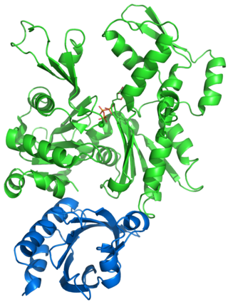
Gelsolin is an actin-binding protein that is a key regulator of actin filament assembly and disassembly. Gelsolin is one of the most potent members of the actin-severing gelsolin/villin superfamily, as it severs with nearly 100% efficiency.

<span class="mw-page-title-main">Treadmilling</span> Simultaneous growth and breakdown on opposite ends of a protein filament

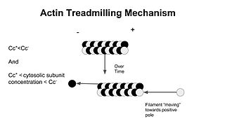
In molecular biology, treadmilling is a phenomenon observed within protein filaments of the cytoskeletons of many cells, especially in actin filaments and microtubules. It occurs when one end of a filament grows in length while the other end shrinks, resulting in a section of filament seemingly "moving" across a stratum or the cytosol. This is due to the constant removal of the protein subunits from these filaments at one end of the filament, while protein subunits are constantly added at the other end. Treadmilling was discovered by Wegner, who defined the thermodynamic and kinetic constraints. Wegner recognized that: “The equilibrium constant (K) for association of a monomer with a polymer is the same at both ends, since the addition of a monomer to each end leads to the same polymer.”; a simple reversible polymer can’t treadmill; ATP hydrolysis is required. GTP is hydrolyzed for microtubule treadmilling.

<span class="mw-page-title-main">Destrin</span> Protein found in humans

Destrin or DSTN is a protein which in humans is encoded by the DSTN gene. Destrin is a component protein in microfilaments.

<span class="mw-page-title-main">Formins</span>

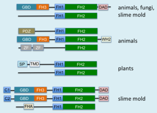
Formins (formin homology proteins) are a group of proteins that are involved in the polymerization of actin and associate with the fast-growing end (barbed end) of actin filaments. Most formins are Rho-GTPase effector proteins. Formins regulate the actin and microtubule cytoskeleton and are involved in various cellular functions such as cell polarity, cytokinesis, cell migration and SRF transcriptional activity. Formins are multidomain proteins that interact with diverse signalling molecules and cytoskeletal proteins, although some formins have been assigned functions within the nucleus.

<span class="mw-page-title-main">CAP1</span> Gene of the species Homo sapiens

Adenylyl cyclase-associated protein 1 is an enzyme that in humans is encoded by the CAP1 gene.

<span class="mw-page-title-main">Stress fiber</span> Contractile actin bundles found in non-muscle cells

Stress fibers are contractile actin bundles found in non-muscle cells. They are composed of actin (microfilaments) and non-muscle myosin II (NMMII), and also contain various crosslinking proteins, such as α-actinin, to form a highly regulated actomyosin structure within non-muscle cells. Stress fibers have been shown to play an important role in cellular contractility, providing force for a number of functions such as cell adhesion, migration and morphogenesis.

<span class="mw-page-title-main">Cordon-bleu protein</span> Protein found in humans

Protein cordon-bleu is a protein that in humans is encoded by the COBL gene.

<span class="mw-page-title-main">Actin assembly-inducing protein</span>

The Actin assembly-inducing protein (ActA) is a protein encoded and used by Listeria monocytogenes to propel itself through a mammalian host cell. ActA is a bacterial surface protein comprising a membrane-spanning region. In a mammalian cell the bacterial ActA interacts with the Arp2/3 complex and actin monomers to induce actin polymerization on the bacterial surface generating an actin comet tail. The gene encoding ActA is named actA or prtB.

Actin remodeling is the biochemical process that allows for the dynamic alterations of cellular organization. The remodeling of actin filaments occurs in a cyclic pattern on cell surfaces and exists as a fundamental aspect to cellular life. During the remodeling process, actin monomers polymerize in response to signaling cascades that stem from environmental cues. The cell's signaling pathways cause actin to affect intracellular organization of the cytoskeleton and often consequently, the cell membrane. Again triggered by environmental conditions, actin filaments break back down into monomers and the cycle is completed. Actin-binding proteins (ABPs) aid in the transformation of actin filaments throughout the actin remodeling process. These proteins account for the diverse structure and changes in shape of Eukaryotic cells. Despite its complexity, actin remodeling may result in complete cytoskeletal reorganization in under a minute.

<span class="mw-page-title-main">Rho-associated protein kinase</span>

Rho-associated protein kinase (ROCK) is a kinase belonging to the AGC family of serine-threonine specific protein kinases. It is involved mainly in regulating the shape and movement of cells by acting on the cytoskeleton.

<span class="mw-page-title-main">MDia1</span> Protein

mDia1 is a member of the protein family called the formins and is a Rho effector. It is the mouse version of the diaphanous homolog 1 of Drosophila. mDia1 localizes to cells' mitotic spindle and midbody, plays a role in stress fiber and filopodia formation, phagocytosis, activation of serum response factor, formation of adherens junctions, and it can act as a transcription factor. mDia1 accelerates actin nucleation and elongation by interacting with barbed ends of actin filaments. The gene encoding mDia1 is located on Chromosome 18 of Mus musculus and named Diap1.

<span class="mw-page-title-main">ADF-H domain</span>

In molecular biology, ADF-H domain is an approximately 150 amino acid motif that is present in three phylogenetically distinct classes of eukaryotic actin-binding proteins.

Ras2 is a Saccharomyces cerevisiae guanine nucleotide-binding protein which becomes activated by binding GTP when glucose is present in the environment. It affects growth regulation and starvation response.

<span class="mw-page-title-main">David G. Drubin</span> American biologist, academic, and researcher

David G. Drubin is an American biologist, academic, and researcher. He is a Distinguished Professor of Cell and Developmental Biology at the University of California, Berkeley where he holds the Ernette Comby Chair in Microbiology.

References

  1. 1 2 Kotila T, Kogan K, Enkavi G, Guo S, Vattulainen I, Goode BL, Lappalainen P (May 2018). "Structural basis of actin monomer re-charging by cyclase-associated protein". Nature Communications. 9 (1): 1892. Bibcode:2018NatCo...9.1892K. doi:10.1038/s41467-018-04231-7. PMC   5951797 . PMID   29760438.
  2. Hubberstey AV, Mottillo EP (April 2002). "Cyclase-associated proteins: CAPacity for linking signal transduction and actin polymerization". FASEB Journal. 16 (6): 487–99. doi:10.1096/fj.01-0659rev. PMID   11919151. S2CID   24648371.
  3. Deeks MJ, Rodrigues C, Dimmock S, Ketelaar T, Maciver SK, Malhó R, Hussey PJ (August 2007). "Arabidopsis CAP1 - a key regulator of actin organisation and development". Journal of Cell Science. 120 (Pt 15): 2609–18. doi:10.1242/jcs.007302. PMID   17635992. S2CID   24219034.
  4. Freeman NL, Field J (February 2000). "Mammalian homolog of the yeast cyclase associated protein, CAP/Srv2p, regulates actin filament assembly". Cell Motility and the Cytoskeleton. 45 (2): 106–20. doi: 10.1002/(SICI)1097-0169(200002)45:2<106::AID-CM3>3.0.CO;2-3 . PMID   10658207.
  5. Hofmann A, Hess S, Noegel AA, Schleicher M, Wlodawer A (October 2002). "Crystallization of cyclase-associated protein from Dictyostelium discoideum". Acta Crystallographica D. 58 (Pt 10 Pt 2): 1858–61. doi: 10.1107/S0907444902013306 . PMID   12351838.
  6. 1 2 Bertling E, Quintero-Monzon O, Mattila PK, Goode BL, Lappalainen P (April 2007). "Mechanism and biological role of profilin-Srv2/CAP interaction". Journal of Cell Science. 120 (Pt 7): 1225–34. doi:10.1242/jcs.000158. PMID   17376963. S2CID   23970944.
  7. Bertling E, Hotulainen P, Mattila PK, Matilainen T, Salminen M, Lappalainen P (May 2004). "Cyclase-associated protein 1 (CAP1) promotes cofilin-induced actin dynamics in mammalian nonmuscle cells". Molecular Biology of the Cell. 15 (5): 2324–34. doi:10.1091/mbc.E04-01-0048. PMC   404026 . PMID   15004221.
  8. Moriyama K, Yahara I (April 2002). "Human CAP1 is a key factor in the recycling of cofilin and actin for rapid actin turnover". Journal of Cell Science. 115 (Pt 8): 1591–601. doi:10.1242/jcs.115.8.1591. PMID   11950878.
  9. 1 2 Kotila T, Wioland H, Enkavi G, Kogan K, Vattulainen I, Jégou A, Romet-Lemonne G, Lappalainen P (22 November 2019). "Mechanism of synergistic actin filament pointed end depolymerization by cyclase-associated protein and cofilin". Nature Communications. 10 (1). 5320. Bibcode:2019NatCo..10.5320K. doi: 10.1038/s41467-019-13213-2 . PMC   6876575 . PMID   31757941.
  10. Shekhar S, Chung J, Kondev J, Gelles J, Goode BL (22 November 2019). "Synergy between Cyclase-associated protein and Cofilin accelerates actin filament depolymerization by two orders of magnitude". Nature Communications. 10 (1). 5319. Bibcode:2019NatCo..10.5319S. doi: 10.1038/s41467-019-13268-1 . PMC   6876572 . PMID   31757952.
  11. Shima F, Okada T, Kido M, Sen H, Tanaka Y, Tamada M, Hu CD, Yamawaki-Kataoka Y, Kariya K, Kataoka T (January 2000). "Association of yeast adenylyl cyclase with cyclase-associated protein CAP forms a second Ras-binding site which mediates its Ras-dependent activation". Molecular and Cellular Biology. 20 (1): 26–33. doi:10.1128/mcb.20.1.26-33.2000. PMC   85033 . PMID   10594005.
  12. Ksiazek D, Brandstetter H, Israel L, Bourenkov GP, Katchalova G, Janssen KP, Bartunik HD, Noegel AA, Schleicher M, Holak TA (September 2003). "Structure of the N-terminal domain of the adenylyl cyclase-associated protein (CAP) from Dictyostelium discoideum". Structure. 11 (9): 1171–8. doi: 10.1016/S0969-2126(03)00180-1 . PMID   12962635.
  13. Dodatko T, Fedorov AA, Grynberg M, Patskovsky Y, Rozwarski DA, Jaroszewski L, Aronoff-Spencer E, Kondraskina E, Irving T, Godzik A, Almo SC (August 2004). "Crystal structure of the actin binding domain of the cyclase-associated protein". Biochemistry. 43 (33): 10628–41. doi:10.1021/bi049071r. PMID   15311924.
This article incorporates text from the public domain Pfam and InterPro: IPR013992
This article incorporates text from the public domain Pfam and InterPro: IPR013912