Fauces (throat)

Last updated
Fauces
Gray1014.png
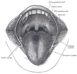
A view of the fauces through the mouth cavity. The cheeks have been slit transversely and the tongue pulled forward. (Fauces labelled as Isthmus faucium at center right.)
Illu pharynx.jpg
Pharynx
Details
System Lymphoid system
Artery Faucial artery
Vein Faucial vein
Nerve Brachial plexus
Lymph Cervical nodes
Identifiers
Latin fauces
TA98 A05.2.01.002
TA2 2846
FMA 55006
Anatomical terminology

The fauces, isthmus of fauces, or the oropharyngeal isthmus is the opening at the back of the mouth into the throat. [1] It is a narrow passage between the velum and the base of the tongue. [2]

Contents

The fauces is a part of the oropharynx directly behind the oral cavity as a subdivision, bounded superiorly by the soft palate, laterally by the palatoglossal and palatopharyngeal arches, and inferiorly by the tongue. The arches form the pillars of the fauces. The anterior pillar is the palatoglossal arch formed of the palatoglossus muscle. The posterior pillar is the palatopharyngeal arch formed of the palatopharyngeus muscle. Between these two arches on the lateral walls of the oropharynx is the tonsillar fossa which is the location of the palatine tonsil. [3] The arches are also known together as the palatine arches.

Each arch runs downwards, laterally and forwards, from the soft palate to the side of the tongue. The approximation of the arches due to the contraction of the palatoglossal muscles constricts the fauces, and is essential to swallowing.

Faucitis

Faucitis in a 12-year-old cat that presented with inappetence, tenderness of the mouth, and inability to groom itself. Gingivitis, erosive lingual ulcers and faucitis were evident clinically Faucitis01.jpg
Faucitis in a 12-year-old cat that presented with inappetence, tenderness of the mouth, and inability to groom itself. Gingivitis, erosive lingual ulcers and faucitis were evident clinically

Inflammation of the fauces, known as faucitis, is seen in animals. In cats, faucitis is usually a secondary disease to gingivitis but can be a primary disease. In this species faucitis is usually caused by bacterial and viral infections although food allergies need to be excluded in any diagnosis. Treatment is symptomatic and includes broad-spectrum antibiotics and in severe cases where cats are inappetant, corticosteroids (often given as depot forms, e.g. depomedrol) or chemotherapy (e.g. chlorambucil).

See also

Related Research Articles

<span class="mw-page-title-main">Tongue</span> Muscular organ in the mouth of most vertebrates

The tongue is a muscular organ in the mouth of a typical tetrapod. It manipulates food for chewing and swallowing as part of the digestive process, and is the primary organ of taste. The tongue's upper surface (dorsum) is covered by taste buds housed in numerous lingual papillae. It is sensitive and kept moist by saliva and is richly supplied with nerves and blood vessels. The tongue also serves as a natural means of cleaning the teeth. A major function of the tongue is the enabling of speech in humans and vocalization in other animals.

Swallowing, also called deglutition or inglutition in scientific contexts, is the process in the body of a human that allows for a substance to pass from the mouth, to the pharynx, and into the esophagus, while shutting the epiglottis. Swallowing is an important part of eating and drinking. If the process fails and the material goes through the trachea, then choking or pulmonary aspiration can occur. In the human body the automatic temporary closing of the epiglottis is controlled by the swallowing reflex.

<span class="mw-page-title-main">Facial nerve</span> Cranial nerve VII, for the face and tasting

The facial nerve, also known as the seventh cranial nerve, cranial nerve VII, or simply CN VII, is a cranial nerve that emerges from the pons of the brainstem, controls the muscles of facial expression, and functions in the conveyance of taste sensations from the anterior two-thirds of the tongue. The nerve typically travels from the pons through the facial canal in the temporal bone and exits the skull at the stylomastoid foramen. It arises from the brainstem from an area posterior to the cranial nerve VI and anterior to cranial nerve VIII.

<span class="mw-page-title-main">Soft palate</span> Flexible part of maxilla

The soft palate is, in mammals, the soft tissue constituting the back of the roof of the mouth. The soft palate is part of the palate of the mouth; the other part is the hard palate. The soft palate is distinguished from the hard palate at the front of the mouth in that it does not contain bone.

<span class="mw-page-title-main">Palatine bone</span> Bone of the facial skeleton

In anatomy, the palatine bones are two irregular bones of the facial skeleton in many animal species, located above the uvula in the throat. Together with the maxilla, they comprise the hard palate.

<span class="mw-page-title-main">Palatine tonsil</span> Lymphoid organs at the back of the throat on both sides

Palatine tonsils, commonly called the tonsils and occasionally called the faucial tonsils, are tonsils located on the left and right sides at the back of the throat in humans and other mammals, which can often be seen as flesh-colored, pinkish lumps. Tonsils only present as "white lumps" if they are inflamed or infected with symptoms of exudates and severe swelling.

<span class="mw-page-title-main">Palatoglossus muscle</span> Muscle of the soft palate

The palatoglossal muscle is a muscle of the soft palate and an extrinsic muscle of the tongue. Its surface is covered by oral mucosa and forms the visible palatoglossal arch.

<span class="mw-page-title-main">Levator veli palatini</span> Muscle of the soft palate

The levator veli palatini is a muscle of the soft palate and pharynx. It is innervated by the vagus nerve via its pharyngeal plexus. During swallowing, it contracts, elevating the soft palate to help prevent food from entering the nasopharynx.

<span class="mw-page-title-main">Palatopharyngeus muscle</span>

The palatopharyngeusmuscle is a small muscle in the roof of the mouth.

<span class="mw-page-title-main">Tensor veli palatini muscle</span> Muscle of the soft palate

The tensor veli palatini muscle is a thin, triangular muscle of the head that tenses the soft palate and opens the Eustachian tube to equalise pressure in the middle ear.

<span class="mw-page-title-main">Musculus uvulae</span> Human muscle

The musculus uvulae is a bilaterally muscle of the soft palate that acts to shorten the uvula when both muscles contract. It forms most of the mass of the uvula. It is innervated by the pharyngeal plexus of vagus nerve.

<span class="mw-page-title-main">Ascending palatine artery</span> Blood vessel

The ascending palatine artery is an artery is a branch of the facial artery which ascends along the neck before splitting into two terminal branches; one branch supplies the soft palate, and the other supplies the palatine tonsil and pharyngotympanic tube.

<span class="mw-page-title-main">Palatal lift prosthesis</span>

A palatal lift prosthesis is a prosthesis that addresses a condition referred to as palatopharyngeal incompetence. Palatopharyngeal incompetence broadly refers to a muscular inability to sufficiently close the port between the nasopharynx and oropharynx during speech and/or swallowing. An inability to adequately close the palatopharyngeal port during speech results in hypernasalance that, depending upon its severity, can render speakers difficult to understand or unintelligible. The potential for compromised intelligibility secondary to hypernasalance is underscored when consideration is given to the fact that only three English language phonemes – /m/, /n/, and /ng/ – are pronounced with an open palatopharyngeal port. Furthermore, an impaired ability to effect a closure of the palatopharyngeal port while swallowing can result in the nasopharyngeal regurgitation of liquid or solid boluses.

<span class="mw-page-title-main">Palatopharyngeal arch</span>

The palatopharyngeal arch is larger and projects further toward the middle line than the palatoglossal arch; it runs downward, lateralward, and backward to the side of the pharynx, and is formed by the projection of the palatopharyngeal muscle, covered by mucous membrane.

<span class="mw-page-title-main">Palatoglossal arch</span>

The palatoglossal arch on either side runs downward, lateral, and forward to the side of the base of the tongue, and is formed by the projection of the glossopalatine muscle with its covering mucous membrane. It is the anterior border of the isthmus of the fauces and marks the border between the mouth and the palatopharyngeal arch. The latter marks the beginning of the pharynx.

<span class="mw-page-title-main">Mallampati score</span> Scoring metric for airway anatomic features

In anesthesia, the Mallampati score or Mallampati classification, named after the Indian anaesthesiologist Seshagiri Mallampati, is used to predict the ease of endotracheal intubation. The test comprises a visual assessment of the distance from the tongue base to the roof of the mouth, and therefore the amount of space in which there is to work. It is an indirect way of assessing how difficult an intubation will be; this is more definitively scored using the Cormack–Lehane classification system, which describes what is actually seen using direct laryngoscopy during the intubation process itself. A high Mallampati score is associated with more difficult intubation as well as a higher incidence of sleep apnea.

<span class="mw-page-title-main">Outline of human anatomy</span> Overview of and topical guide to human anatomy

The following outline is provided as an overview of and topical guide to human anatomy:

<span class="mw-page-title-main">Pharynx</span> Part of the throat that is behind the mouth and nasal cavity

The pharynx is the part of the throat behind the mouth and nasal cavity, and above the esophagus and trachea. It is found in vertebrates and invertebrates, though its structure varies across species. The pharynx carries food to the esophagus and air to the larynx. The flap of cartilage called the epiglottis stops food from entering the larynx.

<span class="mw-page-title-main">Human mouth</span> Part of human anatomy

In human anatomy, the mouth is the first portion of the alimentary canal that receives food and produces saliva. The oral mucosa is the mucous membrane epithelium lining the inside of the mouth.

Passavant's ridge is a mucous elevation situated behind the floor of the naso-pharynx.

References

  1. Saladin, Kenneth (2011-01-13). Anatomy & Physiology: The Unity of Form and Function. McGraw-Hill Education. p. 958. ISBN   9780073378251.
  2. "Definition of FAUCES".
  3. Singh, Vishram (2014). Textbook of anatomy head, neck and brain. Volume 3 (Second ed.). Elsevier. pp. 208–210. ISBN   9788131237274.