Related Research Articles

White matter refers to areas of the central nervous system (CNS) that are mainly made up of myelinated axons, also called tracts. Long thought to be passive tissue, white matter affects learning and brain functions, modulating the distribution of action potentials, acting as a relay and coordinating communication between different brain regions.

The insular cortex is a portion of the cerebral cortex folded deep within the lateral sulcus within each hemisphere of the mammalian brain.

The angular gyrus is a region of the brain lying mainly in the posteroinferior region of the parietal lobe, occupying the posterior part of the inferior parietal lobule. It represents the Brodmann area 39.
Neuroplasticity, also known as neural plasticity or brain plasticity, is the ability of neural networks in the brain to change through growth and reorganization. It is when the brain is rewired to function in some way that differs from how it previously functioned. These changes range from individual neuron pathways making new connections, to systematic adjustments like cortical remapping or neural oscillation. Other forms of neuroplasticity include homologous area adaptation, cross modal reassignment, map expansion, and compensatory masquerade. Examples of neuroplasticity include circuit and network changes that result from learning a new ability, information acquisition, environmental influences, pregnancy, caloric intake, practice/training, and psychological stress.
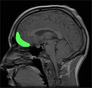
The orbitofrontal cortex (OFC) is a prefrontal cortex region in the frontal lobes of the brain which is involved in the cognitive process of decision-making. In non-human primates it consists of the association cortex areas Brodmann area 11, 12 and 13; in humans it consists of Brodmann area 10, 11 and 47.
Denise Manahan-Vaughan is an Irish neuroscientist and neurophysiologist. She is head of the Department of Neurophysiology, dean of studies and director of the International Graduate School of Neuroscience and co-founder of the Research Department of Neuroscience of the Ruhr University Bochum. Her research focuses on elucidation of the cellular and synaptic mechanisms underlying the acquisition and long-term maintenance of associative memories. She uses a multidisciplinary approach to study how spatial experiences, sensory input, neuromodulation, or brain disease impacts on, and provide insight into, the function of the hippocampus in enabling long-term memory.
Connectomics is the production and study of connectomes: comprehensive maps of connections within an organism's nervous system. More generally, it can be thought of as the study of neuronal wiring diagrams with a focus on how structural connectivity, individual synapses, cellular morphology, and cellular ultrastructure contribute to the make up of a network. The nervous system is a network made of billions of connections and these connections are responsible for our thoughts, emotions, actions, memories, function and dysfunction. Therefore, the study of connectomics aims to advance our understanding of mental health and cognition by understanding how cells in the nervous system are connected and communicate. Because these structures are extremely complex, methods within this field use a high-throughput application of functional and structural neural imaging, most commonly magnetic resonance imaging (MRI), electron microscopy, and histological techniques in order to increase the speed, efficiency, and resolution of these nervous system maps. To date, tens of large scale datasets have been collected spanning the nervous system including the various areas of cortex, cerebellum, the retina, the peripheral nervous system and neuromuscular junctions.

Environmental enrichment is the stimulation of the brain by its physical and social surroundings. Brains in richer, more stimulating environments have higher rates of synaptogenesis and more complex dendrite arbors, leading to increased brain activity. This effect takes place primarily during neurodevelopment, but also during adulthood to a lesser degree. With extra synapses there is also increased synapse activity, leading to an increased size and number of glial energy-support cells. Environmental enrichment also enhances capillary vasculation, providing the neurons and glial cells with extra energy. The neuropil expands, thickening the cortex. Research on rodent brains suggests that environmental enrichment may also lead to an increased rate of neurogenesis.
Psychophysiological interaction (PPI) is a brain connectivity analysis method for functional brain imaging data, mainly functional magnetic resonance imaging (fMRI). It estimates context-dependent changes in effective connectivity (coupling) between brain regions. Thus, PPI analysis identifies brain regions whose activity depends on an interaction between psychological context and physiological state of the seed region.

Cross modal plasticity is the adaptive reorganization of neurons to integrate the function of two or more sensory systems. Cross modal plasticity is a type of neuroplasticity and often occurs after sensory deprivation due to disease or brain damage. The reorganization of the neural network is greatest following long-term sensory deprivation, such as congenital blindness or pre-lingual deafness. In these instances, cross modal plasticity can strengthen other sensory systems to compensate for the lack of vision or hearing. This strengthening is due to new connections that are formed to brain cortices that no longer receive sensory input.
Sensory stimulation therapy (SST) is an experimental therapy that aims to use neural plasticity mechanisms to aid in the recovery of somatosensory function after stroke or cognitive ageing. Stroke and cognitive ageing are well known sources of cognitive loss, the former by neuronal death, the latter by weakening of neural connections. SST stimulates a specific sense at a specific frequency. Research suggests that this technique may reverse cognitive ageing by up to 30 years, and may selectively improve or impair two point discrimination thresholds.

Eleanor Anne Maguire is an Irish neuroscientist. Since 2007, she has been Professor of Cognitive Neuroscience at University College London where she is also a Wellcome Trust Principal Research Fellow.

Resting state fMRI is a method of functional magnetic resonance imaging (fMRI) that is used in brain mapping to evaluate regional interactions that occur in a resting or task-negative state, when an explicit task is not being performed. A number of resting-state brain networks have been identified, one of which is the default mode network. These brain networks are observed through changes in blood flow in the brain which creates what is referred to as a blood-oxygen-level dependent (BOLD) signal that can be measured using fMRI.
Yi Zuo is a neuroscience professor and researcher born in China. She studies molecular, cellular and developmental biology. Zuo is currently an associate professor of Molecular, Cell and Developmental Biology at the University of California, Santa Cruz (UCSC), where she also heads a research lab.
Gait variability seen in Parkinson's Disorders arise due to cortical changes induced by pathophysiology of the disease process. Gait rehabilitation is focused to harness the adapted connections involved actively to control these variations during the disease progression. Gait variabilities seen are attributed to the defective inputs from the Basal Ganglia. However, there is altered activation of other cortical areas that support the deficient control to bring about a movement and maintain some functional mobility.
Anelis Kaiser is professor of gender studies at MINT, University of Freiburg, Germany. She is also on the lecturer within the social psychology and social neuroscience department at the University of Bern, Switzerland. Along with Isabelle Dussauge, Kaiser was a guest editor of a special issue on Neuroscience and sex/gender of the journal Neuroethics, they also co-founded The NeuroGenderings Network together.
Kathryn Emma Watkins is an experimental psychologist in the Wellcome Trust centre for integrative neuroimaging at the University of Oxford and a tutorial fellow at St Anne's College, Oxford. Her research investigates the brain processes that underlie speech, language and development.
Catherine J. "Cathy" Price is a British neuroscientist and academic. She is Professor of Cognitive Neuroscience and director of the Wellcome Trust Centre for Neuroimaging at University College London.
Matthew F. S. Rushworth is Watts Professor of Experimental Psychology at the University of Oxford where his laboratory is funded by the Wellcome Trust and Medical Research Council.
Charlotte Stagg is a British neurophysiologist who is a professor at the University of Oxford. She leads the Physiological Neuroimaging Group.
References
- ↑ "Heidi Johansen-Berg". Nuffield Department of Clinical Neuroscience.
- ↑ "Past Officers of OHBM - Organization for Human Brain Mapping". humanbrainmapping.org. Retrieved 2019-09-08.
- ↑ "Wellcome Trust Award for work on plasticity". Nuffield Department of Clinical Neuroscience.
- ↑ Johansen-Berg H, Rushworth MF, Bogdanovic MD, Kischka U, Wimalaratna S, Matthews PM (October 2002). "The role of ipsilateral premotor cortex in hand movement after stroke". Proceedings of the National Academy of Sciences of the United States of America. 99 (22): 14518–23. Bibcode:2002PNAS...9914518J. doi: 10.1073/pnas.222536799 . PMC 137915 . PMID 12376621.
- ↑ Johansen-Berg H, Dawes H, Guy C, Smith SM, Wade DT, Matthews PM (December 2002). "Correlation between motor improvements and altered fMRI activity after rehabilitative therapy". Brain. 125 (Pt 12): 2731–42. doi: 10.1093/brain/awf282 . PMID 12429600.
- ↑ Johansen-Berg H, Behrens TE, Robson MD, Drobnjak I, Rushworth MF, Brady JM, Smith SM, Higham DJ, Matthews PM (September 2004). "Changes in connectivity profiles define functionally distinct regions in human medial frontal cortex". Proc. Natl. Acad. Sci. U.S.A. 101 (36): 13335–40. Bibcode:2004PNAS..10113335J. doi: 10.1073/pnas.0403743101 . PMC 516567 . PMID 15340158.
- ↑ Scholz J, Klein MC, Behrens TE, Johansen-Berg H (November 2009). "Training induces changes in white-matter architecture". Nat. Neurosci. 12 (11): 1370–1. doi:10.1038/nn.2412. PMC 2770457 . PMID 19820707.
- ↑ Sampaio-Baptista C, Khrapitchev AA, Foxley S, Schlagheck T, Scholz J, Jbabdi S, DeLuca GC, Miller KL, Taylor A, Thomas N, Kleim J, Sibson NR, Bannerman D, Johansen-Berg H (December 2013). "Motor skill learning induces changes in white matter microstructure and myelination". J. Neurosci. 33 (50): 19499–503. doi:10.1523/JNEUROSCI.3048-13.2013. PMC 3858622 . PMID 24336716.
| International | |
|---|---|
| National | |
| Academics | |