Jugular foramen syndrome

Last updated
Jugular foramen syndrome
Brain human normal inferior view with labels en-2.svg
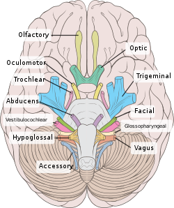
Human brain inferior view showing cranial nerves

Jugular foramen syndrome, or Vernet's syndrome, is characterized by paresis of the glossopharyngeal, vagal, and accessory (with or without the hypoglossal) nerves. [1] [2]

Contents

Symptoms

Symptoms of this syndrome are consequences of this paresis. As such, an affected patient may show:[ citation needed ]

Causes

Diagnosis

Related Research Articles

<span class="mw-page-title-main">Neurofibromatosis</span> Medical condition

Neurofibromatosis (NF) is a group of three conditions in which tumors grow in the nervous system. The three types are neurofibromatosis type I (NF1), neurofibromatosis type II (NF2), and schwannomatosis. In NF1 symptoms include light brown spots on the skin, freckles in the armpit and groin, small bumps within nerves, and scoliosis. In NF2, there may be hearing loss, cataracts at a young age, balance problems, flesh colored skin flaps, and muscle wasting. In schwannomatosis there may be pain either in one location or in wide areas of the body. The tumors in NF are generally non-cancerous.

<span class="mw-page-title-main">Glossopharyngeal nerve</span> Cranial nerve IX, for the tongue and pharynx

The glossopharyngeal nerve, also known as the ninth cranial nerve, cranial nerve IX, or simply CN IX, is a cranial nerve that exits the brainstem from the sides of the upper medulla, just anterior to the vagus nerve. Being a mixed nerve (sensorimotor), it carries afferent sensory and efferent motor information. The motor division of the glossopharyngeal nerve is derived from the basal plate of the embryonic medulla oblongata, whereas the sensory division originates from the cranial neural crest.

<span class="mw-page-title-main">Occipital bone</span> Saucer-shaped membrane bone situated at the back and lower part of the cranium

The occipital bone is a cranial dermal bone and the main bone of the occiput. It is trapezoidal in shape and curved on itself like a shallow dish. The occipital bone overlies the occipital lobes of the cerebrum. At the base of the skull in the occipital bone, there is a large oval opening called the foramen magnum, which allows the passage of the spinal cord.

<span class="mw-page-title-main">Temporal bone</span> Sides and base of skull, connecting to the jaw and occipital bone

The temporal bones are situated at the sides and base of the skull, and lateral to the temporal lobes of the cerebral cortex.

<span class="mw-page-title-main">Internal jugular vein</span> Blood vessel that drains the head

The internal jugular vein is a paired jugular vein that collects blood from the brain and the superficial parts of the face and neck. This vein runs in the carotid sheath with the common carotid artery and vagus nerve.

<span class="mw-page-title-main">Posterior cranial fossa</span> Area of the cranium containing the brainstem and cerebellum

The posterior cranial fossa is the part of the cranial cavity located between the foramen magnum, and tentorium cerebelli. It is formed by the sphenoid bones, temporal bones, and occipital bone. It lodges the cerebellum, and parts of the brainstem.

<span class="mw-page-title-main">Superior orbital fissure</span> Foramen in the skull allowing for passage of cranial nerves

The superior orbital fissure is a foramen or cleft of the skull between the lesser and greater wings of the sphenoid bone. It gives passage to multiple structures, including the oculomotor nerve, trochlear nerve, ophthalmic nerve, abducens nerve, ophthalmic veins, and sympathetic fibres from the cavernous plexus.

<span class="mw-page-title-main">Cavernous sinus</span> Sinus in the human head

The cavernous sinus within the human head is one of the dural venous sinuses creating a cavity called the lateral sellar compartment bordered by the temporal bone of the skull and the sphenoid bone, lateral to the sella turcica.

<span class="mw-page-title-main">Hypoglossal canal</span> Hole in the occipital bone of the skull

The hypoglossal canal is a foramen in the occipital bone of the skull. It is hidden medially and superiorly to each occipital condyle. It transmits the hypoglossal nerve.

<span class="mw-page-title-main">Jugular foramen</span> Opening in the base of the skull allowing many structures to pass

A jugular foramen is one of the two large foramina (openings) in the base of the skull, located behind the carotid canal. It is formed by the temporal bone and the occipital bone. It allows many structures to pass, including the inferior petrosal sinus, three cranial nerves, the sigmoid sinus, and meningeal arteries.

<span class="mw-page-title-main">Frontal nerve</span> Nerve supplying the forehead, frontal sinus, and upper eyelid

The frontal nerve is the largest branch of the ophthalmic nerve (V1), itself a branch of the trigeminal nerve (CN V). It supplies sensation to the skin of the forehead, the mucosa of the frontal sinus, and the skin of the upper eyelid. It may be affected by schwannoma.

<span class="mw-page-title-main">Petrous part of the temporal bone</span> Feature at the base of the human skull

The petrous part of the temporal bone is pyramid-shaped and is wedged in at the base of the skull between the sphenoid and occipital bones. Directed medially, forward, and a little upward, it presents a base, an apex, three surfaces, and three angles, and houses in its interior, the components of the inner ear. The petrous portion is among the most basal elements of the skull and forms part of the endocranium. Petrous comes from the Latin word petrosus, meaning "stone-like, hard". It is one of the densest bones in the body. In other mammals, it is a separate bone, the petrosal bone.

<span class="mw-page-title-main">Lateral parts of occipital bone</span>

The lateral parts of the occipital bone are situated at the sides of the foramen magnum; on their under surfaces are the condyles for articulation with the superior facets of the atlas.

<span class="mw-page-title-main">Jugular tubercle</span>

The jugular tubercle is a rounded prominence/oval elevation upon the superior surface of the occipital condyle at the junction of the basilar part and lateral part of the occipital bone, just medial to the jugular foramen on either side of the foramen magnum.

<span class="mw-page-title-main">Schwannoma</span> Medical condition

A schwannoma is a usually benign nerve sheath tumor composed of Schwann cells, which normally produce the insulating myelin sheath covering peripheral nerves.

<span class="mw-page-title-main">Occipital condyles</span> Undersurface protuberances of the occipital bone in vertebrates

The occipital condyles are undersurface protuberances of the occipital bone in vertebrates, which function in articulation with the superior facets of the atlas vertebra.

<span class="mw-page-title-main">Nasopharyngeal carcinoma</span> Type of throat cancer; most common to occur in the nasopharynx

Nasopharyngeal carcinoma (NPC), or nasopharynx cancer, is the most common cancer originating in the nasopharynx, most commonly in the postero-lateral nasopharynx or pharyngeal recess, accounting for 50% of cases. NPC occurs in children and adults. NPC differs significantly from other cancers of the head and neck in its occurrence, causes, clinical behavior, and treatment. It is vastly more common in certain regions of East Asia and Africa than elsewhere, with viral, dietary and genetic factors implicated in its causation. It is most common in males. It is a squamous cell carcinoma of an undifferentiated type. Squamous epithelial cells are a flat type of cell found in the skin and the membranes that line some body cavities. Undifferentiated cells are cells that do not have their mature features or functions.

<span class="mw-page-title-main">Inferior petrosal sulcus</span>

The inferior petrosal sulcus is the groove containing the inferior petrosal sinus.

The cerebellopontine angle syndrome is a distinct neurological syndrome of deficits that can arise due to the closeness of the cerebellopontine angle to specific cranial nerves. Indications include unilateral hearing loss (85%), speech impediments, disequilibrium, tremors or other loss of motor control. The cerebellopontine angle cistern is a subarachnoid cistern formed by the cerebellopontine angle that lies between the cerebellum and the pons. It is filled with cerebrospinal fluid and is a common site for the growth of acoustic neuromas or schwannomas.

Villaret's syndrome combines ipsilateral paralysis of the last four cranial nerves and Horner syndrome. Sometimes cranial nerve VII is also involved. It may also involve the cervical ganglia of the sympathetic trunk. Paralysis is caused by a lesion in the retroparotid space, which is bounded posteriorly by the cervical vertebrae, superiorly by the skull near the jugular foramen, anteriorly by the parotid gland, laterally by the sternocleidomastoid muscle, and medially by the pharynx.

References

  1. Erol FS, Kaplan M, Kavakli A, Ozveren MF.Jugular foramen syndrome caused by choleastatoma. Clin Neurol Neurosurg. 2005 Jun;107(4):342-6.
  2. Quinones-Hinojosa, Alfredo, ed. (2012). Schmidek and Sweet Indications, Methods and Results (Expert Consult - Online and Print) (6th ed.). London: Elsevier Health Sciences. p. 2337. ISBN   9781455723287.