Laminin 111

Last updated • 6 min readFrom Wikipedia, The Free Encyclopedia
A schematic diagram of the structure of laminin 111 and several hypothesized binding sites of cell-surface receptors. Schematic Diagram of Laminin 111.jpg
A schematic diagram of the structure of laminin 111 and several hypothesized binding sites of cell-surface receptors.

Laminin–111 (also "laminin–1") is a protein of the type known as laminin isoforms. It was among the first of the laminin isoforms to be discovered. [2] The "111" identifies the isoform's chain composition of α1β1γ1. [2] This protein plays an important role in embryonic development. Injections of this substance are used in treatment for Duchenne muscular dystrophy, and its cellular action may potentially become a focus of study in cancer research.

Contents

Distribution

The distribution of the different laminin isoforms is tissue-specific. [3] Laminin–111 is predominantly expressed in the embryonic epithelium, but can also be found in some adult epithelium such as the kidney, liver, testis, ovaries, and brain blood vessels. [3] [4] Different levels of expression of α chains have a large influence on the differential expression of laminin, thereby determining the isoform produced. [3] From studying a mouse model, it was found that transcription factors present in the parietal endoderm regulate the expression of the α1 and large amounts of laminin-111 are produced. [3]

Functions

The synthesized laminin–111 formed in an embryo contributes to the formation of Reichert’s membrane, a thick extra-embryonic basement membrane. [5] When the laminin α1 chain is deficient in an organism, an embryo dies, likely as a result of a defective Reichert’s membrane due to a lack of laminin–111 being produced. [4] Laminin-111 has been identified as a crucial molecule for development of the embryo as shown by the consequences that occur when laminin-111 is lacking.

Laminin-111 is expressed very early on in development and is present in the blastocyst. [1] When various parts of the trimer chains are knocked out by mutations, devastating consequences occur in the embryo. If the β1 or γ1 chains of laminin-111 are absent the basement membrane fails to form. [1] Without a basement membrane cells have nowhere to attach and all dependent activities such as cell migration and epithelial formation can no longer occur. [5] [1] The self-assembly and tight network formation by laminin-111 are essential for holding the basement membrane together.

Although it is expressed abundantly during the early embryonic stage, laminin-111 is mostly absent in adults. [5] The injection of laminin-111, however, helps with Duchenne muscular dystrophy, a neuromuscular disease in which the connection between the extracellular matrix and cell cytoskeleton is lost. [6] [7] Increased levels of laminin-111 triggered an increase in the expression of α7-integrin receptor and this prevented onset of the disease. [7] Additionally, the presence of laminin-111 increased muscle strength and protected it from injury. [7] When injected with myoblast transplants, laminin–111 decreased degeneration and inflammatory reactions and increased the success of the transplantation. [6] The experiments utilizing laminin–111 as a source of therapy for Duchenne muscular dystrophy suggest that it has protective qualities in addition to its association with muscle tissue.

Mechanisms of action

Cell adhesion

A schematic diagram of the laminin 111-a6b4 integrin interaction in hemidesmosomes for cell adhesion in epithelial tissue. Laminin Basement Layer.jpg
A schematic diagram of the laminin 111-α6β4 integrin interaction in hemidesmosomes for cell adhesion in epithelial tissue.

In cell adhesion laminin-111 and other isoforms are important proteins that anchor cells to the extracellular matrix (ECM). [8] The linkage between cells and the ECM is formed by binding cell surface receptors to one end of the laminin α chain and binding ECM components to another region of the laminin. [8] Globular domains (G-Domain) of the α chain are the regions on laminin-111 that allow the binding of integrins, glycoproteins, sulfated glycolipids and dystroglycan. [8]

Cell signaling

Besides anchoring cells to the ECM, laminins are also involved in the signalling of cells and other components of the ECM. [8] Even though there is not a general mechanism that applies to all laminins in signalling, there are some common pathways that can be seen in more than one isoform of laminin. [8] For example, the PI3K/AKT pathway is used by laminin-111 (promotes cell-survival), 511 (prevents apoptosis with laminin 521), and 521 (stabilizes pluripotency of human embryonic stem cells). [8] The pathway begins with the adhesion of the cell to the ECM for activation of the lipid-associated PI3K. [9] Once PI3K is activated, it will localize AKT that is in the cytoplasm to the cell membrane where AKT is then phosphorylated to promote cell survival. [8]

Neurite outgrowth

When α chains [10] of laminin-111 bind to cell surface receptors integrins α1β1, α3β1, α4β1, α6β1 and Cdc42 GTPase are activated. [11] The activated GTPase then activates Cdc42 which further activates c-Jun kinases and phosphorylation of Jun. [11] Activation of c-Jun kinases leads to high levels of c-Jun expression which results in neurite outgrowth. [12] The synthesis of Nitric Oxides resides somewhere in the pathway and is yet to be determined. [10] Weston et al. (2000) proposed that the synthesis of Nitric Oxide may be upstream to the activation of Cdc42. [11] Nonetheless, Nitric Oxide synthesis is shown to be an important element in laminin-mediated neurite outgrowth. [10]

Future applications

Dynamic reciprocity theory

The dynamic reciprocity theory states that a cell’s fate depends on the exchange of chemical signals between the extracellular matrix and the nucleus of the cell. [13] Focussing on connections between laminin-111 and other proteins involved in cell-to-cell communication could spark further research that may help to further our current understanding of cancer and how to slow down or stop its process.

Actin plays a role in nuclear activity which is an important process with regard to cell signalling influencing cell differentiation and replication. It has been suggested that actin interactions directly influence gene transcription as it interacts with chromatin remodeling complexes as well as RNA polymerases I, II and III. [14] However, the exact role that actin plays in transcription has not yet been determined.

Implications for cancer research

A group of distinguished scientists [15] from the U.S. Department of Energy’s (DOE) Lawrence Berkeley National Laboratory (Berkeley Lab) did a recent study on how laminin-111 interacts with the cytoplasmic protein, actin. Their study gave the following conclusions:

The biological process in which a cell ceases to continue growing and dividing is called quiescence (the opposite of cancer). ECM laminin-111 sends chemical signals that promotes adhesion of a cell and its ECM. Although the mechanism is unknown, these signals have also been linked to cell quiescence. Adding laminin-111 to breast epithelial cells leads to quiescence by altering nuclear actin. High levels of laminin-111 deplete nuclear actin which induces quiescence of cells. However, when an isoform of actin, that cannot exit a cell’s nucleus, is active, cells continue to grow and divide even when laminin levels are high. ECM laminin-111 levels in a normal breast cell are significantly higher than laminin-111 levels in tissues of cancerous breast tissue. Simply increasing laminin levels in the ECM of cancerous breast cells is not enough to lead to quiescence. Therefore, it is implied that there are multiple factors working together influencing cell-to-cell communication. How laminin-111 and nuclear actin communicate is one of these factors. Laminin-111 could be the physiological regulator of nuclear actin which would suggest that depleting nuclear actin could be a key to achieving cell quiescence and returning to homeostatic operating conditions. Decreased expression of laminin-111 and the growth-inhibitory signals that it produces in malignant myoepithelial cells begs for further investigation with regard to cancer research. Therefore, further exploration of laminin-111 and nuclear actin interaction could be a target for future experimental therapeutic investigations. [15]

Related Research Articles

Integrin

Integrins are transmembrane receptors that facilitate cell-cell and cell-extracellular matrix (ECM) adhesion. Upon ligand binding, integrins activate signal transduction pathways that mediate cellular signals such as regulation of the cell cycle, organization of the intracellular cytoskeleton, and movement of new receptors to the cell membrane. The presence of integrins allows rapid and flexible responses to events at the cell surface.

Smooth muscle Involuntary non-striated muscle

Smooth muscle is an involuntary non-striated muscle, so-called because it has no sarcomeres and therefore no striations. It is divided into two subgroups, single-unit and multiunit smooth muscle. Within single-unit muscle, the whole bundle or sheet of smooth muscle cells contracts as a syncytium.

Dystrophin Rod-shaped cytoplasmic protein

Dystrophin is a rod-shaped cytoplasmic protein, and a vital part of a protein complex that connects the cytoskeleton of a muscle fiber to the surrounding extracellular matrix through the cell membrane. This complex is variously known as the costamere or the dystrophin-associated protein complex (DAPC). Many muscle proteins, such as α-dystrobrevin, syncoilin, synemin, sarcoglycan, dystroglycan, and sarcospan, colocalize with dystrophin at the costamere. It has a molecular weight of 427 kDa

Hemidesmosome

Hemidesmosomes are very small stud-like structures found in keratinocytes of the epidermis of skin that attach to the extracellular matrix. They are similar in form to desmosomes when visualized by electron microscopy, however, desmosomes attach to adjacent cells. Hemidesmosomes are also comparable to focal adhesions, as they both attach cells to the extracellular matrix. Instead of desmogleins and desmocollins in the extracellular space, hemidesmosomes utilize integrins. Hemidesmosomes are found in epithelial cells connecting the basal epithelial cells to the lamina lucida, which is part of the basal lamina. Hemidesmosomes are also involved in signaling pathways, such as keratinocyte migration or carcinoma cell intrusion.

Laminin Protein in the extracellular matrix

Laminins are high-molecular weight proteins of the extracellular matrix. They are a major component of the basal lamina, a protein network foundation for most cells and organs. The laminins are an important and biologically active part of the basal lamina, influencing cell differentiation, migration, and adhesion.

Perlecan

Perlecan (PLC) also known as basement membrane-specific heparan sulfate proteoglycan core protein (HSPG) or heparan sulfate proteoglycan 2 (HSPG2), is a protein that in humans is encoded by the HSPG2 gene.

Dystroglycan

Dystroglycan is a protein that in humans is encoded by the DAG1 gene.

Originally identified as Kirsten ras associated gene (krag), Sarcospan (SSPN) (is a 25-kDa transmembrane protein located in the dystrophin-associated protein complex of skeletal muscle cells, where it is most abundant. It contains four transmembrane spanning helices with both N- and C-terminal domains located intracellularly. Loss of SSPN expression occurs in patients with Duchenne muscular dystrophy. Dystrophin is required for proper localization of SSPN. SSPN is also an essential regulator of Akt signaling pathways. Without SSPN, Akt signaling pathways will be hindered and muscle regeneration will not occur.

Dystrobrevin is a protein that binds to dystrophin in the costamere of skeletal muscle cells. In humans, there are at least two isoforms of dystrobrevin, α-dystrobrevin and β-dystrobrevin.

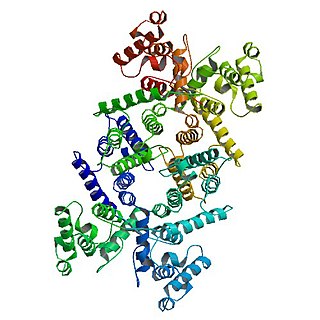
The syntrophins are a family of five 60-kiloDalton proteins that are associated with dystrophin, the protein associated with Duchenne muscular dystrophy and Becker muscular dystrophy. The name comes from the Greek word syntrophos, meaning "companion." The five syntrophins are encoded by separate genes and are termed α, β1, β2, γ1, and γ2. Syntrophin was first identified as a dystrophin-associated protein present in the Torpedo electric organ. Subsequently, α-syntrophin was shown to be the predominant isoform in skeletal muscle where it is localized on the sarcolemma and enriched at the neuromuscular junction. The β-syntrophins and γ2-syntrophin are also present in skeletal muscle but also are in most other tissues. The expression of γ1-syntrophin is mostly confined to brain. The syntrophins are adaptor proteins that use their multiple protein interaction domains to localize a variety of signaling proteins to specific intracellular locations. α-Syntrophin binds to nNOS in the dystrophin-associated glycoprotein complex in skeletal muscle cells. There it produces NO upon muscle contraction leading to dilation of the arteries in the local area.

Laminin, gamma 2

Laminin subunit gamma-2 is a protein that in humans is encoded by the LAMC2 gene.

Laminin, alpha 3

Laminin subunit alpha-3 is a protein that in humans is encoded by the LAMA3 gene.

Laminin, gamma 1

Laminin subunit gamma-1 is a protein that in humans is encoded by the LAMC1 gene.

Laminin, beta 1

Laminin subunit beta-1 is a protein that in humans is encoded by the LAMB1 gene.

Laminin, alpha 2

Laminin subunit alpha-2 is a protein that in humans is encoded by the LAMA2 gene.

Integrin alpha 7

Alpha-7 integrin is a protein that in humans is encoded by the ITGA7 gene. Alpha-7 integrin is critical for modulating cell-matrix interactions. Alpha-7 integrin is highly expressed in cardiac muscle, skeletal muscle and smooth muscle cells, and localizes to Z-disc and costamere structures. Mutations in ITGA7 have been associated with congenital myopathies and noncompaction cardiomyopathy, and altered expression levels of alpha-7 integrin have been identified in various forms of muscular dystrophy.

Laminin, alpha 4

Laminin subunit alpha-4 is a protein that in humans is encoded by the LAMA4 gene.

Laminin, beta 2

Laminin subunit beta-2 is a protein that in humans is encoded by the LAMB2 gene.

The Akt signaling pathway or PI3K-Akt signaling pathway is a signal transduction pathway that promotes survival and growth in response to extracellular signals. Key proteins involved are PI3K and Akt.

Laminin, gamma 3

Laminin subunit gamma-3 also known as LAMC3 is a protein that in humans is encoded by the LAMC3 gene.

References

  1. 1 2 3 4 Colognato, H., & Yurchenco, P. D. (2000). Form and function: The laminin family of heterotrimers. Developmental Dynamics, 218(2): 213-234.
  2. 1 2 Aumailley, M., Bruckner-Tuderman, L., Carter, W. G., Deutzmann, R., Edgar, D., Ekblom, P., & Yurchenco, P. D. (2005). A simplified laminin nomenclature. Matrix biology, 24(5): 326-332.
  3. 1 2 3 4 Durbeej, M. (2010). Laminins. Cell and Tissue Research, 339(1): 259-268.
  4. 1 2 Ekblom, M., Falk, M., Salmivirta, K., Durbeej, M., & Ekblom, P. (1998). Laminin isoforms and epithelial development. Annals of the New York Academy of Sciences, 857(1): 194-211.
  5. 1 2 3 Minler, J.H. and Yurchenco P.D. (2004). Laminin functions in tissue morphogenesis. Annual Review of Cell and Developmental Biology, 20:255-284.
  6. 1 2 Goudenege, S., Lamarre, Y., Dumont, N., Rousseau, J., Frenette , J., Skuk, D. and Tremblay, J.P. (2010). Laminin-111: A potential therapeutic agent for Duchenne muscular dystrophy. Molecular Therapy, 18(12): 2155-2163. doi:10.1038/mt.2010.165.
  7. 1 2 3 Rooney, J. E., Gurpur, P. B., & Burkin, D. J. (2009). Laminin-111 protein therapy prevents muscle disease in the mdx mouse model for Duchenne muscular dystrophy. Proceedings of the National Academy of Sciences, 106(19): 7991-7996.
  8. 1 2 3 4 5 6 7 Domogatskaya, An., Rodin, S., and Tryggvason, K. (2012). Functional diversity of laminins. Annual Review of Cell and Developmental Biology, 28: 523-553.
  9. Langenbach, K.J. and Rando, T.A. (2002). Inhibition of dystroglycan binding to laminin disrupts the PI3K/AKT pathway and cell-survival signalling in muscle cells. Muscle and Nerve, 26: 644-653.
  10. 1 2 3 Rialas, C. M., Nomizu, M., Patterson, M., Kleinman, H.K., Weston, C.A., and Weeks, B.S. (2000). Nitric Oxide mediates laminin-induced neurite outgrowth in PC12 cells. Experimental Cell Research, 260(2): 268-276.
  11. 1 2 3 Weston, C.A., Anova, L., Rialas, C., Prives, J.M., and Weeks, B.S. (2000). Laminin-1 activates Cdc42 in the mechanism of laminin-1-mediated neurite outgrowth. Experimental Cell Research, 260: 374-378.
  12. Dragunow, M., Xu, R., Walkton, M., Woodgate, A., Lawlor, P., MacGibbon, G.A., Young, D., Gibbons, H., Lipski, J., Muravlev, A., Pearson, A., and During, M. (2000). Thus, C-Jun promotes neurite outgrowth and survival in PC12 cells. Molecular Brain Research, 83(1-2): 20-33.
  13. Schultz GS, Davidson JM, Kirsner RS, Bornstein P, Herman IM. (2011). Dynamic reciprocity in the wound microenvironment. Wound Repair Regen., 19: 134–148.
  14. Zheng B, Han M, Bernier M, Wen JK. (2009). Nuclear actin and actin-binding proteins in the regulation of transcription and gene expression. FEBS J., 276: 2669–2685.
  15. 1 2 Spencer VA, Costes S, Inman JL, Xu R, Chen J, Hendzel MJ, Bissell MJ. (2011). Depletion of nuclear actin is a key mediator of quiescence in epithelial cells. J Cell Sci., 124: 123–132.