Lomentospora prolificans

Last updated

Lomentospora prolificans
Scedosporium prolificans.jpg
Photomicrograph of colony growing on Modified Leonian's agar
Scientific classification
Kingdom:
Division:
Class:
Order:
Family:
Genus:
Species:
L. prolificans
Binomial name
Lomentospora prolificans
Hennebert & B.G.Desai(1974)
Type strain
CBS 467.74; UAMH 7149
Synonyms
  • Scedosporium prolificans(Hennebert & B.G.Desai) E.Guého & de Hoog (1991)
  • Scedosporium inflatumMalloch & Salkin (1984)

Lomentospora prolificans is an emerging opportunistic fungal pathogen that causes a wide variety of infections in immunologically normal and immunosuppressed people and animals. [1] [2] [3] It is resistant to most antifungal drugs and infections are often fatal. [3] Drugs targeting the Class II dihydroorotate dehydrogenase (DHODH) proteins of L. prolificans, Scedosporium, Aspergillus and other deadly moulds are the basis for at least one new therapy, Olorofim, which is currently in phase 2b clinical trials and has received breakthrough status by FDA. [4] For information on all DHODH proteins, please see Dihydroorotate dehydrogenase.

Contents

History

The genus Lomentospora was erected by G. Hennebert and B.G. Desai in 1974 to accommodate a culture obtained from greenhouse soil originating from a forest in Belgium. [5] The fungus, which they named Lomentospora prolificans, was thought incorrectly to be related to the genus Beauveria - a group of insect-pathogenic soil fungi affiliated with the order Hypocreales. [5] [6] The genus name "Lomentospora" referred to the shape of the apex of the spore-bearing cell, which the authors interpreted to be a rachis resembling a bean pod of the sort constricted at each seed. The species epithet "prolificans" derived from the prolific nature of the mold's sporulation. The fungus was later independently described as Scedosporium inflatum by Malloch and Salkin in 1984 from a bone biopsy of the foot of a boy who had stepped on a nail. [lower-alpha 1] [8] The species epithet "inflatum" referred to the characteristically swollen base of the spore-bearing cell which they recognized correctly to be an annelide. Malloch and Salkin did not observe a sexual state, however they recognized the fungus to be associated with the family Microascaceae, and suspected it to be allied with the genus Pseudallescheria . [8] In 1991, Guého and De Hoog re-examined a set of cultures of Scedosporium-like fungi from clinical cases by careful morphological examination and the evaluation of DNA-DNA reassociation complementarity. Along with two strains from their own work, they found the cultures of Hennebert & Desai and Malloch & Salkin to constitute a single species which they confirmed to belong in the genus Scedosporium . [9] Lomentospora prolificans was then transferred to Scedosporium as S. prolificans, and Scedosporium inflatum became a synonym. This synonymy has since been confirmed by phylogenetic analysis of the nuclear ribosomal internal transcribed spacer regions. [10] Despite this change, and even as recently as 2012, the name Scedosporium inflatum has continued to appear in the medical literature. [11] [12]

In 2014 Lackner et al. proposed a move back to L. prolificans. [13]

Appearance

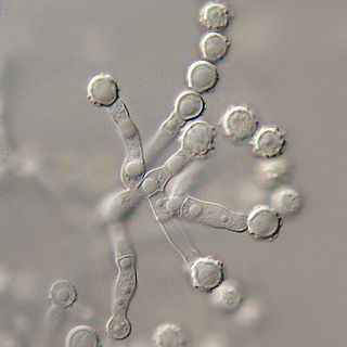
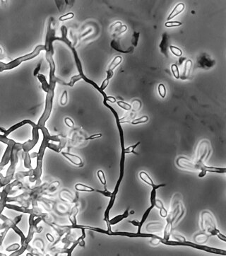
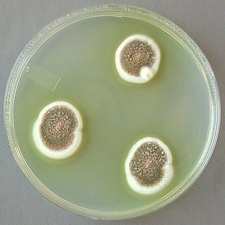
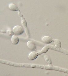
Lomentospora prolificans produces small, delicate annellides with a distinct basal swelling peculiar to this species and absent in the closely related species, Scedosporium apiospermum . Annellides necks become long and distinctly annellated with age. Annellides occur individually or in clusters irregularly along undifferentiated hyphae, frequently exhibiting a pronounced penicillate arrangement in older cultures. Conidia are smooth-walled, light to dark brown, 3–7 x 2–3 μm, accumulating in slime droplets at annelide apices. Colonies of Lomentospora prolificans are grey to brownish, spreading, with scant, cobweb-like aerial mycelium recalling a moth-eaten woolen garment. This species is sensitive to cycloheximide. As this species may be slow to emerge from clinical materials, specimens in which this agent is suspected often require an extended period of culture incubation (e.g., up to 4 weeks). [14]

Ecology

Lomentospora prolificans is a soil fungus, and has been found in the soils of ornamental plants [lower-alpha 2] and greenhouse plants. [lower-alpha 3] Along with other fungi, Lomentospora prolificans has been isolated from soils of Ficus benjamina and Heptapleurum actinophyllum plantings in hospitals, suggesting that these materials have potential to serve as reservoirs of nosocomial fungal pathogens. [15]

Human disease

Lomentospora prolificans has been recognized as an agent of opportunistic human disease since the 1990s. This species is primarily associated with subcutaneous lesions arising from injury following traumatic implantation of the agent via contaminated splinters or plant thorns. The majority of Lomentospora prolificans infections in immunologically normal people remain localized, characteristically with bone or joint involvement. [16] Disseminated infections from Lomentospora prolificans are largely limited to people with pre-existing immune impairment. Notably, Lomentospora prolificans exhibits varying tolerance to all currently available antifungal agents. This is particularly true of strains recovered from disseminated infections, and these infections carry a high mortality. Lomentospora prolificans has also been known to cause disseminated disease secondary to myeloblastic leukemia [17] and following lung transplant. [18] In otherwise healthy people, it was recorded as a cause of corneal infection following a lawn trimmer mishap, [lower-alpha 4] and bone infection following trauma. [8]

Drug resistance

Infections caused by Lomentospora prolificans are recognized to be difficult to treat due to the tendency of this species to exhibit resistance to many commonly used antifungal agents. [14] [16] [17] [19] Successful control of disseminated Lomentospora prolificans infection can be obtained with a combination of voriconazole and terbinafine, [20] but some strains are resistant to this treatment. Drugs that might also be of help are posaconazole, miltefosine and albaconazole. Albaconazole is in phase III clinical trials.

Notes

  1. Osteomyelitis UAMH 5819 [7]
  2. Potted plant soil: UAMH 5735, UAMH 5736 [7]
  3. Greenhouse soil: UAMH 7149 [7]
  4. Corneal scraping: UAMH 8524 [7]

Related Research Articles

<span class="mw-page-title-main">Eumycetoma</span> Human and animal fungal infection

Eumycetoma, also known as Madura foot, is a persistent fungal infection of the skin and the tissues just under the skin, affecting most commonly the feet, although it can occur in hands and other body parts. It starts as a painless wet nodule, which may be present for years before ulceration, swelling, grainy discharge and weeping from sinuses and fistulae, followed by bone deformity.

<i>Aspergillus terreus</i> Species of fungus

Aspergillus terreus, also known as Aspergillus terrestris, is a fungus (mold) found worldwide in soil. Although thought to be strictly asexual until recently, A. terreus is now known to be capable of sexual reproduction. This saprotrophic fungus is prevalent in warmer climates such as tropical and subtropical regions. Aside from being located in soil, A. terreus has also been found in habitats such as decomposing vegetation and dust. A. terreus is commonly used in industry to produce important organic acids, such as itaconic acid and cis-aconitic acid, as well as enzymes, like xylanase. It was also the initial source for the drug mevinolin (lovastatin), a drug for lowering serum cholesterol.

<i>Cochliobolus lunatus</i> Fungal plant pathogen

Cochliobolus lunatus is a fungal plant pathogen that can cause disease in humans and other animals. The anamorph of this fungus is known as Curvularia lunata, while C. lunatus denotes the teleomorph or sexual stage. They are, however, the same biological entity. C. lunatus is the most commonly reported species in clinical cases of reported Cochliobolus infection.

Acremonium strictum is an environmentally widespread saprotroph species found in soil, plant debris, and rotting mushrooms. Isolates have been collected in North and Central America, Asia, Europe and Egypt. A. strictum is an agent of hyalohyphomycosis and has been identified as an increasingly frequent human pathogen in immunosuppressed individuals, causing localized, disseminated and invasive infections. Although extremely rare, A. strictum can infect immunocompetent individuals, as well as neonates. Due to the growing number of infections caused by A. strictum in the past few years, the need for new medical techniques in the identification of the fungus as well as for the treatment of human infections has risen considerably.

<i>Acrophialophora fusispora</i> Species of ascomycete fungus found in soil, air and various plants

Acrophialophora fusispora is a poorly studied ascomycete fungus found in soil, air and various plants. A. fusispora is morphologically similar to the genera Paecilomyces and Masonia, but differ in the presence of pigmented conidiophores, verticillate phialides, and frequent sympodial proliferation. Moreover, A. fusispora is distinguished by its pigmented spindle-shaped conidia, covered with spiral bands. The fungus is naturally found in soils of tropical to temperate regions. The fungus has been identified as a plant and animal pathogen, and has recently been recognized as an emerging opportunistic human pathogen. A. fusispora infection in human is rare and has few documented clinical cases, but due to the rarity of the fungus and potential misidentification, the infections may be underdiagnosed. Clinical cases of A. fusispora include cases of keratitis, pulmonary colonization and infection, and cerebral infections. The fungus also has two documented cases of infection in dogs.

Exophiala jeanselmei is a saprotrophic fungus in the family Herpotrichiellaceae. Four varieties have been discovered: Exophiala jeanselmei var. heteromorpha, E. jeanselmei var. lecanii-corni, E. jeanselmei var. jeanselmei, and E. jeanselmei var. castellanii. Other species in the genus Exophiala such as E. dermatitidis and E. spinifera have been reported to have similar annellidic conidiogenesis and may therefore be difficult to differentiate.

<i>Pseudallescheria boydii</i> Species of fungus

Pseudallescheria boydii is a species of fungus classified in the Ascomycota. It is associated with some forms of eumycetoma/maduromycosis and is the causative agent of pseudallescheriasis. Typically found in stagnant and polluted water, it has been implicated in the infection of immunocompromised and near-drowned pneumonia patients. Treatment of infections with P. boydii is complicated by resistance to many of the standard antifungal agents normally used to treat infections by filamentous fungi.
<span class="mw-page-title-main">Microascaceae</span> Family of fungi

The Microascaceae are a family of fungi in the class Sordariomycetes, subclass Hypocreomycetidae. The family was published by David Malloch in 1970, an emended description based on Everet Stanley Luttrell's original 1951 publication. Family was updated in 2020.

<i>Exophiala dermatitidis</i> Species of fungus

Exophiala dermatitidis is a thermophilic black yeast, and a member of the Herpotrichiellaceae. While the species is only found at low abundance in nature, metabolically active strains are commonly isolated in saunas, steam baths, and dish washers. Exophiala dermatitidis only rarely causes infection in humans, however cases have been reported around the world. In East Asia, the species has caused lethal brain infections in young and otherwise healthy individuals. The fungus has been known to cause cutaneous and subcutaneous phaeohyphomycosis, and as a lung colonist in people with cystic fibrosis in Europe. In 2002, an outbreak of systemic E. dermatitidis infection occurred in women who had received contaminated steroid injections at North Carolina hospitals.
Myceliophthora thermophila is an ascomycete fungus that grows optimally at 45–50 °C (113–122 °F). It efficiently degrades cellulose and is of interest in the production of biofuels. The genome has recently been sequenced, revealing the full range of enzymes used by this organism for the degradation of plant cell wall material.

<i>Ochroconis gallopava</i> Species of fungus

Ochroconis gallopava, also called Dactylaria gallopava or Dactylaria constricta var. gallopava, is a member of genus Dactylaria. Ochroconis gallopava is a thermotolerant, darkly pigmented fungus that causes various infections in fowls, turkeys, poults, and immunocompromised humans first reported in 1986. Since then, the fungus has been increasingly reported as an agent of human disease especially in recipients of solid organ transplants. Ochroconis gallopava infection has a long onset and can involve a variety of body sites. Treatment of infection often involves a combination of antifungal drug therapy and surgical excision.

<i>Apophysomyces variabilis</i> Species of fungus

Apophysomyces variabilis is an emerging fungal pathogen that can cause serious and sometimes fatal infection in humans. This fungus is a soil-dwelling saprobe with tropical to subtropical distribution. It is a zygomycete that causes mucormycosis, an infection in humans brought about by fungi in the order Mucorales. Infectious cases have been reported globally in locations including the Americas, Southeast Asia, India, and Australia. Apophysomyces variabilis infections are not transmissible from person to person.

Coniochaeta hoffmannii, also known as Lecythophora hoffmannii, is an ascomycete fungus that grows commonly in soil. It has also been categorized as a soft-rot fungus capable of bringing the surface layer of timber into a state of decay, even when safeguarded with preservatives. Additionally, it has pathogenic properties, although it causes serious infection only in rare cases. A plant pathogen lacking a known sexual state, C. hoffmannii has been classified as a "dematiaceous fungus" despite its contradictory lack of pigmentation; both in vivo and in vitro, there is no correlation between its appearance and its classification.

Phaeohyphomycosis is a diverse group of fungal infections, caused by dematiaceous fungi whose morphologic characteristics in tissue include hyphae, yeast-like cells, or a combination of these. It can be associated an array of melanistic filamentous fungi including Alternaria species,Exophiala jeanselmei, and Rhinocladiella mackenziei.

<i>Lichtheimia corymbifera</i> Species of fungus

Lichtheimia corymbifera is a thermophilic fungus in the phylum Zygomycota. It normally lives as a saprotrophic mold, but can also be an opportunistic pathogen known to cause pulmonary, CNS, rhinocerebral, or cutaneous infections in animals and humans with impaired immunity.
<i>Aspergillus ustus</i> Species of fungus

Aspergillus ustus is a microfungus and member of the division Ascomycota. It is commonly found in indoor environments and soil. Isolated cases of human infection resulting from A. ustus have been described; however the majority of these are nail infections.

Scedosporiosis is the general name for any mycosis - i.e., fungal infection - caused by a fungus from the genus Scedosporium. Current population-based studies suggest Scedosporium prolificans and Scedosporium apiospermum to be among the most common infecting agents from the genus, although infections caused by other members thereof are not unheard of. The latter is an asexual form (anamorph) of another fungus, Pseudallescheria boydii. The former is a “black yeast”, currently not characterized as well, although both of them have been described as saprophytes.

<i>Scedosporium</i> Genus of fungi

Scedosporium is a genus of fungi in the family Microascaceae.

Petriella setifera is a fungus commonly found in soil and feces. The fungus has also been located on wood rot, plant species, and compost. A significant portion of P. setifera reports are found on sources with no previous association with the fungus. There are no known human cases of fungal infection, but one reported case of a dolphin infection. The fungus may have immunosuppressive characteristics, but it has not been confirmed. Many properties of the fungus are unknown, requiring further research.

Cladophialophora arxii is a black yeast shaped dematiaceous fungus that is able to cause serious phaeohyphomycotic infections. C. arxii was first discovered in 1995 in Germany from a 22-year-old female patient suffering multiple granulomatous tracheal tumours. It is a clinical strain that is typically found in humans and is also capable of acting as an opportunistic fungus of other vertebrates Human cases caused by C. arxii have been reported from all parts of the world such as Germany and Australia.
References

  1. Cortez KJ, Roilides E, Quiroz-Telles F, et al. (2008). "Infections caused by Scedosporium spp". Clinical Microbiology Reviews. 21 (1): 157–97. doi:10.1128/CMR.00039-07. PMC   2223844 . PMID   18202441.
  2. Elad, Daniel (1 January 2011). "Infections caused by fungi of the Scedosporium/Pseudallescheria complex in veterinary species". The Veterinary Journal. 187 (1): 33–41. doi:10.1016/j.tvjl.2010.05.028. PMID   20580291.
  3. 1 2 Rodriguez-Tudela, Juan Luis; Rodriguez-Tudela, Juan Luis; Berenguer, Juan; Guarro, Josep; Kantarcioglu, A. Serda; Horre, Regine; Sybren De Hoog, G.; Cuenca-Estrella, Manuel (1 January 2009). "Epidemiology and outcome of Scedosporium prolificans infection, a review of 162 cases". Medical Mycology. 47 (4): 359–370. doi: 10.1080/13693780802524506 . PMID   19031336.
  4. "Evaluate F901318 Treatment of Invasive Fungal Infections in Patients Lacking Treatment Options - Tabular View - ClinicalTrials.gov". clinicaltrials.gov. Retrieved 15 February 2020.
  5. 1 2 Hennebert, G. L.; Desai, B.G. (1974). "Lomentospora prolificans, a new hyphomycete from greenhouse soil". Mycotaxon. 1 (1): 45–50.
  6. MycoBank. "Beauveria". Centraalbureau voor Schimmelcultures. Retrieved 10 October 2013.
  7. 1 2 3 4 Sigler, L. "UAMH Culture Collection Catalogue". UAMH Centre for Global Microfungal Biodiversity. University of Toronto.
  8. 1 2 3 Malloch, D; Salkin IF (1984). "A new species of Scedosporium associated with osteomyelitis in humans". Mycotaxon. 21: 247–255.
  9. Guého E; de Hoog GS (1991). "Taxonomy of the medical species of Pseudallescheria and Scedosporium". Journal de Mycologie Médicale. 1: 3–9.
  10. Lennon, PA; Cooper CR; Salkin IF; Lee SB (1994). "Ribosomal DNA internal transcribed spacer analysis supports synonymy of Scedosporium inflatum and Lomentospora prolificans". Journal of Clinical Microbiology. 32 (10): 2413–2416. doi: 10.1128/JCM.32.10.2413-2416.1994 . PMC   264076 .
  11. Cetrulo, Curtis L.; Barone, Angelo A. Leto; Jordan, Kathleen; Chang, David S.; Louie, Kevin; Buntic, Rudolf F.; Brooks, Darrell (1 February 2012). "A multi-disciplinary approach to the management of fungal osteomyelitis: Current concepts in post-traumatic lower extremity reconstruction: A case report". Microsurgery. 32 (2): 144–147. doi:10.1002/micr.20956. PMID   22389900.
  12. Taylor, Alexandra; Wiffen, Steven J; Kennedy, Christopher J (1 February 2002). "Post-traumatic Scedosporium inflatum endophthalmitis". Clinical and Experimental Ophthalmology. 30 (1): 47–48. doi:10.1046/j.1442-9071.2002.00475.x. PMID   11885796.
  13. Lackner, Michaela; de Hoog, G. Sybren; Yang, Liyue; Ferreira Moreno, Leandro; Ahmed, Sarah A.; Andreas, Fritz; Kaltseis, Josef; Nagl, Markus; Lass-Flörl, Cornelia; Risslegger, Brigitte; Rambach, Günter (1 July 2014). "Proposed nomenclature for Pseudallescheria, Scedosporium and related genera". Fungal Diversity. 67 (1): 1–10. doi:10.1007/s13225-014-0295-4. ISSN   1878-9129.
  14. 1 2 Sigler, L (2003). "Chapter 11: Miscellaneous opportunistic fungi: Microascaceae and other Ascomycetes, Hyphomycetes, Coelomycetes and Basidiomycetes". In Howard DH (ed.). Pathogenic fungi in humans and animals (2nd ed.). New York: Marcel Dekker. pp. 637–676. ISBN   978-0824706838.
  15. Summerbell, RC; Krajden S; Kane J (1989). "Potted plants in hospitals as reservoirs of pathogenic fungi". Mycopathologia. 106 (1): 13–22. doi:10.1007/bf00436921. PMID   2671744.
  16. 1 2 Matlani, M; Kaur, R; Shweta (January 2013). "A case of Scedosporium prolificans osteomyelitis in an immunocompetent child, misdiagnosed as tubercular osteomyelitis". Indian Journal of Dermatology. 58 (1): 80–1. doi: 10.4103/0019-5154.105319 . PMC   3555384 . PMID   23372223.
  17. 1 2 Roberto, Reinoso; et al. (2013). "Fatal disseminated Scedosporium prolificans infection initiated by ophthalmic involvement in a patient with acute myeloblastic leukemia". Diagnostic Microbiology and Infectious Disease. 76 (3): 375–378. doi:10.1016/j.diagmicrobio.2013.03.006. PMID   23602787.
  18. Sayah, D.M.; Schwartz, B.S.; Kukreja, J.; Singer, J.P.; Golden, J.A.; Leard, L.E. (1 April 2013). "Scedosporium prolificans pericarditis and mycotic aortic aneurysm in a lung transplant recipient receiving voriconazole prophylaxis". Transplant Infectious Disease. 15 (2): E70–E74. doi:10.1111/tid.12056. PMID   23387799.
  19. Salkin, I. F.; McGinnis, M. R.; Dykstra, M. J.; Rinaldi, M. G. (1988). "Scedosporium inflatum, an emerging pathogen". J Clin Microbiol. 26 (3): 498–503. doi:10.1128/JCM.26.3.498-503.1988. PMC   266320 . PMID   3356789.
  20. Howden, BP; Slavin, MA; Schwarer, AP; Mijch, AM (February 2003). "Successful control of disseminated Scedosporium prolificans infection with a combination of voriconazole and terbinafine". European Journal of Clinical Microbiology & Infectious Diseases. 22 (2): 111–113. doi:10.1007/s10096-002-0877-z. PMID   12627286.