
The sievert is a derived unit of ionizing radiation dose in the International System of Units (SI) and is a measure of the health effect of low levels of ionizing radiation on the human body. The sievert is important in dosimetry and radiation protection, and is named after Rolf Maximilian Sievert, a Swedish medical physicist renowned for work on radiation dose measurement and research into the biological effects of radiation.
The becquerel is the SI derived unit of radioactivity. One becquerel is defined as the activity of a quantity of radioactive material in which one nucleus decays per second. For applications relating to human health this is a small quantity, and SI multiples of the unit are commonly used.
Medical physics deals with the application of the concepts and methods of physics to the prevention, diagnosis and treatment of human diseases with a specific goal of improving human health and well-being. Since 2008, medical physics has been included as a health profession according to International Standard Classification of Occupation of the International Labour Organization.

Mammography is the process of using low-energy X-rays to examine the human breast for diagnosis and screening. The goal of mammography is the early detection of breast cancer, typically through detection of characteristic masses or microcalcifications.
The gray is a derived unit of ionizing radiation dose in the International System of Units (SI). It is defined as the absorption of one joule of radiation energy per kilogram of matter.
Radiation dosimetry in the fields of health physics and radiation protection is the measurement, calculation and assessment of the ionizing radiation dose absorbed by an object, usually the human body. This applies both internally, due to ingested or inhaled radioactive substances, or externally due to irradiation by sources of radiation.
Absorbed dose is a dose quantity which is the measure of the energy deposited in matter by ionizing radiation per unit mass. Absorbed dose is used in the calculation of dose uptake in living tissue in both radiation protection, and radiology. It is also used to directly compare the effect of radiation on inanimate matter such as in radiation hardening.
In radiation physics, Kerma is an acronym for "kinetic energy released per unit mass", defined as the sum of the initial kinetic energies of all the charged particles liberated by uncharged ionizing radiation in a sample of matter, divided by the mass of the sample. It is defined by the quotient .
A material's half-value layer (HVL), or half-value thickness, is the thickness of the material at which the intensity of radiation entering it is reduced by one half. HVL can also be expressed in terms of air kerma rate (AKR), rather than intensity: the half-value layer is the thickness of specified material that, "attenuates the beam of radiation to an extent such that the AKR is reduced to one-half of its original value. In this definition the contribution of all scattered radiation, other than any [...] present initially in the beam concerned, is deemed to be excluded." Rather than AKR, measurements of air kerma, exposure, or exposure rate can be used to determine half value layer, as long as it is given in the description.
Radiobiology is a field of clinical and basic medical sciences that involves the study of the action of ionizing radiation on living things, especially health effects of radiation. Ionizing radiation is generally harmful and potentially lethal to living things but can have health benefits in radiation therapy for the treatment of cancer and thyrotoxicosis. Its most common impact is the induction of cancer with a latent period of years or decades after exposure. High doses can cause visually dramatic radiation burns, and/or rapid fatality through acute radiation syndrome. Controlled doses are used for medical imaging and radiotherapy.

Tomosynthesis, also digital tomosynthesis (DTS), is a method for performing high-resolution limited-angle tomography at radiation dose levels comparable with projectional radiography. It has been studied for a variety of clinical applications, including vascular imaging, dental imaging, orthopedic imaging, mammographic imaging, musculoskeletal imaging, and chest imaging.
Dose area product (DAP) is a quantity used in assessing the radiation risk from diagnostic X-ray examinations and interventional procedures. It is defined as the absorbed dose multiplied by the area irradiated, expressed in gray-centimetres squared. Manufacturers of DAP meters usually calibrate them in terms of absorbed dose to air. DAP reflects not only the dose within the radiation field but also the area of tissue irradiated. Therefore, it may be a better indicator of the overall risk of inducing cancer than the dose within the field. It also has the advantages of being easily measured, with the permanent installation of a DAP meter on the X-ray set. Due to the divergence of a beam emitted from a "point source", the area irradiated (A) increases with the square of distance from the source, while radiation intensity (I) decreases according to the inverse square of distance. Consequently, the product of intensity and area, and therefore DAP, is independent of distance from the source.

The roentgen or röntgen is a legacy unit of measurement for the exposure of X-rays and gamma rays, and is defined as the electric charge freed by such radiation in a specified volume of air divided by the mass of that air . In 1928, it was adopted as the first international measurement quantity for ionising radiation to be defined for radiation protection, as it was then the most easily replicated method of measuring air ionization by using ion chambers. It is named after the German physicist Wilhelm Röntgen, who discovered X-rays.
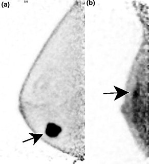
Positron emission mammography (PEM) is a nuclear medicine imaging modality used to detect or characterise breast cancer. Mammography typically refers to x-ray imaging of the breast, while PEM uses an injected positron emitting isotope and a dedicated scanner to locate breast tumors. Scintimammography is another nuclear medicine breast imaging technique, however it is performed using a gamma camera. Breasts can be imaged on standard whole-body PET scanners, however dedicated PEM scanners offer advantages including improved resolution.

Astronauts are exposed to approximately 50-2,000 millisieverts (mSv) while on six-month-duration missions to the International Space Station (ISS), the Moon and beyond. The risk of cancer caused by ionizing radiation is well documented at radiation doses beginning at 100mSv and above.

Photon counting is a technique in which individual photons are counted using a single-photon detector (SPD). A single-photon detector emits a pulse of signal for each detected photon, in contrast to a normal photodetector, which generates an analog signal proportional to the photon flux. The number of pulses is counted, giving an integer number of photons detected per measurement interval. The counting efficiency is determined by the quantum efficiency and the system's electronic losses.

In medicine, breast imaging is a sub-speciality of diagnostic radiology that involves imaging of the breasts for screening or diagnostic purposes. There are various methods of breast imaging using a variety of technologies as described in detail below. Traditional screening and diagnostic mammography uses x-ray technology. Breast tomosynthesis is a new digital mammography technique that produces 3D images of the breast using x-rays. Xeromammography and Galactography also use x-ray technology and are also used infrequently in the detection of breast cancer. Breast ultrasound is another technology employed in diagnosis & screening and specifically can help differentiate between fluid filled and solid lumps that can help determine if cancerous. Breast MRI is, yet, another technology reserved for high-risk patients and can help determine the extent of cancer if diagnosed. Lastly, scintimammography is used in a subgroup of patients who have abnormal mammograms or whose screening is not reliable on the basis of using traditional mammography or ultrasound.
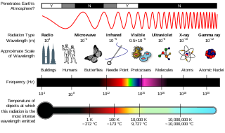
Radiation is a moving form of energy, classified into ionizing and non-ionizing type. Ionizing radiation is further categorized into electromagnetic radiation and particulate radiation. Electromagnetic radiation consists of photons, which can be thought of as energy packets, traveling in the form of a wave. Examples of electromagnetic radiation includes X-rays and gamma rays. These types of radiation can easily penetrate the human body because of high energy. Radiation exposure is a measure of the ionization of air due to ionizing radiation from photons. It is defined as the electric charge freed by such radiation in a specified volume of air divided by the mass of that air. Medical exposure is defined by the International Commission on Radiological Protection as exposure incurred by patients as part of their own medical or dental diagnosis or treatment; by persons, other than those occupationally exposed, knowingly, while voluntarily helping in the support and comfort of patients; and by volunteers in a programme of biomedical research involving their exposure. Common medical tests and treatments involving radiation include X-rays, CT scans, mammography, lung ventilation and perfusion scans, bone scans, cardiac perfusion scan, angiography, radiation therapy, and more. Each type of test carries its own amount of radiation exposure. There are two general categories of adverse health effects caused by radiation exposure: deterministic effects and stochastic effects. Deterministic effects are due to the killing/malfunction of cells following high doses; and stochastic effects involve either cancer development in exposed individuals caused by mutation of somatic cells, or heritable disease in their offspring from mutation of reproductive (germ) cells.
Spectral imaging is an umbrella term for energy-resolved X-ray imaging in medicine. The technique makes use of the energy dependence of X-ray attenuation to either increase the contrast-to-noise ratio, or to provide quantitative image data and reduce image artefacts by so-called material decomposition. Dual-energy imaging, i.e. imaging at two energy levels, is a special case of spectral imaging and is still the most widely used terminology, but the terms "spectral imaging" and "spectral CT" have been coined to acknowledge the fact that photon-counting detectors have the potential for measurements at a larger number of energy levels.
Photon-counting mammography was introduced commercially in 2003 and was the first widely available application of photon-counting detector technology in medical x-ray imaging. Photon-counting mammography improves dose efficiency compared to conventional technologies, and enables spectral imaging.


