Molecular lesion

Last updated
Ball and Stick Model of Double Helical DNA DNA orbit animated.gif
Ball and Stick Model of Double Helical DNA

A molecular lesion or point lesion is damage to the structure of a biological molecule such as DNA, RNA, or protein. This damage may result in the reduction or absence of normal function, and in rare cases the gain of a new function. Lesions in DNA may consist of breaks or other changes in chemical structure of the helix, ultimately preventing transcription. Meanwhile, lesions in proteins consist of both broken bonds and improper folding of the amino acid chain. While many nucleic acid lesions are general across DNA and RNA, some are specific to one, such as thymine dimers being found exclusively in DNA. Several cellular repair mechanisms exist, ranging from global to specific, in order to prevent lasting damage resulting from lesions.

Contents

Causes

There are two broad causes of nucleic acid lesions, endogenous and exogenous factors. Endogenous factors, or endogeny, refer to the resulting conditions that develop within an organism. This is in contrast with exogenous factors which originate from outside the organism. DNA and RNA lesions caused by endogenous factors generally occur more frequently than damage caused by exogenous ones. [1]

Endogenous Factors

Endogenous sources of specific DNA damage include pathways like hydrolysis, oxidation, alkylation, mismatch of DNA bases, depurination, depyrimidination, double-strand breaks (DSS), and cytosine deamination. DNA lesions can also naturally occur from the release of specific compounds such as reactive oxygen species (ROS), reactive nitrogen species (RNS), reactive carbonyl species (RCS), lipid peroxidation products, adducts, and alkylating agents through metabolic processes. ROS is one of the major endogenous sources of DNA damage and the most studied oxidative DNA adduct is 8-oxo-dG. Other adducts known to form are etheno-, propano-, and malondialdehyde-derived DNA adducts. The aldehydes formed from lipid peroxidation also pose another threat to DNA. [2] Proteins such as "damage-up" proteins (DDPs) can promote endogenous DNA lesions by either increasing the amount of reactive oxygen by transmembrane transporters, losing chromosomes by replisome binding, and stalling replication by transcription factors. [3] For RNA lesions specifically, the most abundant types of endogenous damage include oxidation, alkylation, and chlorination. [4] Phagocytic cells produce radical species that include hypochlorous acid (HOCl), nitric oxide (NO•), and peroxynitrite (ONOO−) to fight infections, and many cell types use nitric oxide as a signaling molecule. However, these radical species can also cause the pathways that form RNA lesions. [5]

Thymine Photodimer Caused by UV Light Thymine photodimer.svg
Thymine Photodimer Caused by UV Light

Exogenous Factors

Ultraviolet Radiation

UV light, specifically non-ionizing shorter-wavelength radiation such as UVC and UVB, causes direct DNA damage by initiating a synthesis reaction between two thymine molecules. The resulting dimer is very stable. Although they can be removed through excision repairs, when UV damage is extensive, the entire DNA molecule breaks down and the cell dies. If the damage is not too extensive, precancerous or cancerous cells are created from healthy cells. [6]

Chemotherapy drugs

Chemotherapeutics, by design, induce DNA damage and are targeted towards rapidly dividing cancer cells. [7] However, these drugs can not tell the difference between sick and healthy cells, resulting in the damage of normal cells. [8]

Alkylating agents

Alkylating agents are a type of chemotherapeutic drug which keeps the cell from undergoing mitosis by damaging its DNA. They work in all phases of the cell cycle. The use of alkylating agents may result in leukemia due to them being able to target the cells of the bone marrow. [8]

Cancer causing agents

Carcinogens are known to cause a number of DNA lesions, such as single-strand breaks, double- strand breaks, and covalently bound chemical DNA adducts. Tobacco products are one of the most prevalent cancer-causing agents of today. [9] Other DNA damaging, cancer-causing agents include asbestos, which can cause damage through physical interaction with DNA or by indirectly setting off a reactive oxygen species, [10] excessive nickel exposure, which can repress the DNA damage-repair pathways, [11] aflatoxins, which are found in food, [9] and many more.

Lesions of Nucleic Acids

Types of Structural DNA Molecular Lesion Damage Types of DNA Damage.jpg
Types of Structural DNA Molecular Lesion Damage

Oxidative lesions

Oxidative lesions are an umbrella category of lesions caused by reactive oxygen species (ROS), reactive nitrogen species (RNS), other byproducts of cellular metabolism, and exogenous factors such as ionizing or ultraviolet radiation. [12] Byproducts of oxidative respiration are the main source of reactive species which cause a background level of oxidative lesions in the cell. DNA and RNA are both affected by this, and it has been found that RNA oxidative lesions are more abundant in humans compared to DNA. This may be due cytoplasmic RNA having closer proximity to the electron transport chain. [13] Known oxidative lesions characterized in DNA and RNA are many in number, as oxidized products are unstable and may resolve quickly. The hydroxyl radical and singlet oxygen are common reactive oxygen species responsible for these lesions. [14] 8-oxo-guanine (8-oxoG) is the most abundant and well characterized oxidative lesion, found in both RNA and DNA. Accumulation of 8-oxoG may cause dire damage within the mitochondria and is thought to be a key player in the aging process. [15] RNA oxidation has direct consequences in the production of proteins. mRNA affected by oxidative lesions is still recognized by ribosome, but the ribosome will undergo stalling and dysfunction. This results in proteins having either decreased expression or truncation, leading to aggregation and general dysfunction. [16]

Structural rearrangements

Single and Double Stranded Breaks

Single-strand breaks (SSBs) occur when one strand of the DNA double helix experiences breakage of a single nucleotide accompanied by damaged 5’- and/or 3’-termini at this point. One common source of SSBs is due to oxidative attack by physiological reactive oxygen species (ROS) such as hydrogen peroxide. H2O2 causes SSBs three times more frequently than double-strand breaks (DSBs). Alternative methods of SSB acquisition include direct disintegration of the oxidized sugar or through DNA base-excision repair (BER) of damaged bases. Additionally, cellular enzymes may perform erroneous activity leading to SSBs or DSBs by a variety of mechanisms. One such example would be when the cleavage complex formed by DNA topoisomerase 1 (TOP1) relaxes DNA during transcription and replication through the transient formation of a nick. While TOP1 normally reseals this nick shortly after, these cleavage complexes may collide with RNA or DNA polymerases or be proximal to other lesions, leading to TOP1-linked SSBs or TOP1-linked DSBs. [20]

Chemical Adducts

A DNA adduct is a segment of DNA that binds to a chemical carcinogen. Some adducts that cause lesions to DNA included oxidatively modified bases, propano-, etheno-, and MDA-induced adducts. [2] 5‐Hydroxymethyluracil is an example of an oxidatively modified base where oxidation of the methyl group of thymine occurs. [21] This adduct interferes with the binding of transcription factors to DNA which can trigger apoptosis or result in deletion mutations. [21] Propano adducts are derived by species generated by lipid peroxidation. For example, HNE is a major toxic product of the process. [22] It regulates the expression of genes that are involved in cell cycle regulation and apoptosis. Some of the aldehydes from lipid peroxidation can be converted to epoxy aldehydes by oxidation reactions. [23] These epoxy aldehydes can damage DNA by producing etheno adducts. An increase in this type of DNA lesion exhibits conditions resulting in oxidative stress which is known to be associated with an increased risk of cancer. [24] Malondialdehyde (MDA) is another highly toxic product from lipid peroxidation and also in the synthesis of prostaglandin. MDA reacts with DNA to form the M1dG adduct which causes DNA lesions. [2]

Disease Effects

Many systems are in place to repair DNA and RNA lesions but it is possible for lesions to escape these measures. This may lead to mutations or large genome abnormalities, which can threaten the cell or organism's ability to live. Several cancers are a result of DNA lesions. Even repair mechanisms to heal the damage may end up causing more damage. Mismatch repair defects, for example, cause instability that predisposes to colorectal and endometrial carcinomas. [9]

DNA lesions in neurons may lead to neurodegenerative disorders such as Alzheimer's, Huntington's, and Parkinson's diseases. These come as a result of neurons generally being associated with high mitochondrial respiration and redox species production, which can damage nuclear DNA. Since these cells often cannot be replaced after being damaged, the damage done to them leads to dire consequences. Other disorders stemming from DNA lesions and their association with neurons include but are not limited to Fragile X syndrome, Friedreich's ataxia, and Spinocerebellar ataxias. [9]

During replication, usually DNA polymerases are unable to go past the lesioned area, however, some cells are equipped with special polymerases which allow for translesion synthesis (TLS). TLS polymerases allow for the replication of DNA past lesions and risk generating mutations at a high frequency. Common mutations that occur after undergoing this process are point mutations and frameshift mutations. Several diseases come as a result of this process including several cancers and Xeroderma pigmentosum. [25]

The effect of oxidatively damaged RNA has resulted in a number of human diseases and is especially associated with chronic degeneration. This type of damage has been observed in many neurodegenerative diseases such as Amyotrophic lateral sclerosis, [9] Alzheimer's, Parkinson's, dementia with Lewy bodies, and several prion diseases. [26] It is important to note that this list is rapidly growing and data suggests that RNA oxidation occurs early in the development of these diseases, rather than as an effect of cellular decay. [9] RNA and DNA lesions are both associated with the development of diabetes mellitus type 2. [9]

Repair Mechanisms

DNA Damage Response

When DNA is damaged such as due to a lesion, a complex signal transduction pathway is activated which is responsible for recognizing the damage and instigating the cell's response for repair. Compared to the other lesion repair mechanisms, DDR is the highest level of repair and is employed for the most complex lesions. DDR consists of various pathways, the most common of which are the DDR kinase signaling cascades. These are controlled by phosphatidylinositol 3-kinase-related kinases (PIKK), and range from DNA-dependent protein kinase (DNA-PKcs) and ataxia telangiectasia-mutated (ATM) most involved in repairing DSBs to the more versatile Rad3-related (ATR). ATR is crucial to human cell viability, while ATM mutations cause the severe disorder ataxia-telangiectasia leading to neurodegeneration, cancer, and immunodeficiency. These three DDR kinases all recognize damage via protein-protein interactions which localize the kinases to the areas of damage. Next, further protein-protein interactions and posttranslational modifications (PTMs) complete the kinase activation, and a series of phosphorylation events takes place. DDR kinases perform repair regulation at three levels - via PTMs, at the level of chromatin, and at the level of the nucleus. [27]

BER Pathway BER basic pathway.svg
BER Pathway

Base Excision Repair

Base excision repair (BER) is responsible for removing damaged bases in DNA. This mechanism specifically works on excising small base lesions which do not distort the DNA double helix, in contrast to the nucleotide excision repair pathway which is employed in correcting more prominent distorting lesions. DNA glycosylases initiate BER by both recognizing the faulty or incorrect bases and then removing them, forming AP sites lacking any purine or pyrimidine. AP endonuclease then cleaves the AP site, and the single-strand break is either processed by short-patch BER to replace a single nucleotide long-patch BER to create 2-10 replacement nucleotides. [28]

Single Stranded Break Repair

DNA DSB Repair System Recombinational repair of DNA double-strand damage.jpg
DNA DSB Repair System

Single stranded breaks (SSBs) can severely threaten genetic stability and cell survival if not quickly and properly repaired, so cells have developed fast and efficient SSB repair (SSBR) mechanisms. While global SSBR systems extract SSBs throughout the genome and during interphase, S-phase specific SSBR processes work together with homologous recombination at the replication forks. [29]

Double Stranded Break Repair

Double stranded breaks (DSB) are a threat to all organisms as they can cause cell death and cancer. They can be caused exogenously as a result of radiation and endogenously from errors in replication or encounters with DNA lesions by the replication fork. [30] DSB repair occurs through a variety of different pathways and mechanisms in order to correctly repair these errors.

Nucleotide Excision and Mismatch Repair

Nucleotide excision repair  is one of the main mechanisms used to remove bulky adducts from DNA lesions caused by chemotherapy drugs, environmental mutagens, and most importantly UV radiation. [9] This mechanism functions by releasing a short damage containing oligonucleotide from the DNA site, and then that gap is filled in and repaired by NER. [9] NER recognizes a variety of structurally unrelated DNA lesions due to the flexibility of the mechanism itself, as NER is highly sensitive to changes in the DNA helical structure. [31] Bulky adducts seem to trigger NER. [31] The XPC-RAD23-CETN2 heterotrimer involved with NER has a critical role in DNA lesion recognition. [32] In addition to other general lesions in the genome, UV damaged DNA binding protein complex (UV-DDB)  also has an important role in both recognition and repair of UV-induced DNA photolesions. [32]

Mismatch repair (MMR) mechanisms within the cell correct base mispairs that occur during replication using a variety of pathways. It has a high affinity for targeting DNA lesions with specificity, as alternations in base pair stacking that occur at DNA lesion sites affect the helical structure. [33] This is likely one of many signals that triggers MMR.

Related Research Articles

Mutagenesis is a process by which the genetic information of an organism is changed by the production of a mutation. It may occur spontaneously in nature, or as a result of exposure to mutagens. It can also be achieved experimentally using laboratory procedures. A mutagen is a mutation-causing agent, be it chemical or physical, which results in an increased rate of mutations in an organism's genetic code. In nature mutagenesis can lead to cancer and various heritable diseases, and it is also a driving force of evolution. Mutagenesis as a science was developed based on work done by Hermann Muller, Charlotte Auerbach and J. M. Robson in the first half of the 20th century.

<span class="mw-page-title-main">Reactive oxygen species</span> Highly reactive molecules formed from diatomic oxygen (O₂)

In chemistry and biology, reactive oxygen species (ROS) are highly reactive chemicals formed from diatomic oxygen (O2), water, and hydrogen peroxide. Some prominent ROS are hydroperoxide (O2H), superoxide (O2-), hydroxyl radical (OH.), and singlet oxygen. ROS are pervasive because they are readily produced from O2, which is abundant. ROS are important in many ways, both beneficial and otherwise. ROS function as signals, that turn on and off biological functions. They are intermediates in the redox behavior of O2, which is central to fuel cells. ROS are central to the photodegradation of organic pollutants in the atmosphere. Most often however, ROS are discussed in a biological context, ranging from their effects on aging and their role in causing dangerous genetic mutations.

<span class="mw-page-title-main">DNA repair</span> Cellular mechanism

DNA repair is a collection of processes by which a cell identifies and corrects damage to the DNA molecules that encode its genome. In human cells, both normal metabolic activities and environmental factors such as radiation can cause DNA damage, resulting in tens of thousands of individual molecular lesions per cell per day. Many of these lesions cause structural damage to the DNA molecule and can alter or eliminate the cell's ability to transcribe the gene that the affected DNA encodes. Other lesions induce potentially harmful mutations in the cell's genome, which affect the survival of its daughter cells after it undergoes mitosis. As a consequence, the DNA repair process is constantly active as it responds to damage in the DNA structure. When normal repair processes fail, and when cellular apoptosis does not occur, irreparable DNA damage may occur. This can eventually lead to malignant tumors, or cancer as per the two-hit hypothesis.

Lipid peroxidation, or lipid oxidation, is a complex chemical process that leads to oxidative degradation of lipids, resulting in the formation of peroxide and hydroperoxide derivatives. It occurs when free radicals, specifically reactive oxygen species (ROS), interact with lipids within cell membranes, typically polyunsaturated fatty acids (PUFAs) as they have carbon–carbon double bonds. This reaction leads to the formation of lipid radicals, collectively referred to as lipid peroxides or lipid oxidation products (LOPs), which in turn react with other oxidizing agents, leading to a chain reaction that results in oxidative stress and cell damage.

<span class="mw-page-title-main">Cockayne syndrome</span> Medical condition

Cockayne syndrome (CS), also called Neill-Dingwall syndrome, is a rare and fatal autosomal recessive neurodegenerative disorder characterized by growth failure, impaired development of the nervous system, abnormal sensitivity to sunlight (photosensitivity), eye disorders and premature aging. Failure to thrive and neurological disorders are criteria for diagnosis, while photosensitivity, hearing loss, eye abnormalities, and cavities are other very common features. Problems with any or all of the internal organs are possible. It is associated with a group of disorders called leukodystrophies, which are conditions characterized by degradation of neurological white matter. There are two primary types of Cockayne syndrome: Cockayne syndrome type A (CSA), arising from mutations in the ERCC8 gene, and Cockayne syndrome type B (CSB), resulting from mutations in the ERCC6 gene.

<span class="mw-page-title-main">Nucleotide excision repair</span> DNA repair mechanism

Nucleotide excision repair is a DNA repair mechanism. DNA damage occurs constantly because of chemicals, radiation and other mutagens. Three excision repair pathways exist to repair single stranded DNA damage: Nucleotide excision repair (NER), base excision repair (BER), and DNA mismatch repair (MMR). While the BER pathway can recognize specific non-bulky lesions in DNA, it can correct only damaged bases that are removed by specific glycosylases. Similarly, the MMR pathway only targets mismatched Watson-Crick base pairs.

<span class="mw-page-title-main">Oxidative stress</span> Free radical toxicity

Oxidative stress reflects an imbalance between the systemic manifestation of reactive oxygen species and a biological system's ability to readily detoxify the reactive intermediates or to repair the resulting damage. Disturbances in the normal redox state of cells can cause toxic effects through the production of peroxides and free radicals that damage all components of the cell, including proteins, lipids, and DNA. Oxidative stress from oxidative metabolism causes base damage, as well as strand breaks in DNA. Base damage is mostly indirect and caused by the reactive oxygen species generated, e.g., O2 (superoxide radical), OH (hydroxyl radical) and H2O2 (hydrogen peroxide). Further, some reactive oxidative species act as cellular messengers in redox signaling. Thus, oxidative stress can cause disruptions in normal mechanisms of cellular signaling.

<span class="mw-page-title-main">Base excision repair</span> DNA repair process

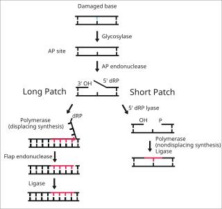
Base excision repair (BER) is a cellular mechanism, studied in the fields of biochemistry and genetics, that repairs damaged DNA throughout the cell cycle. It is responsible primarily for removing small, non-helix-distorting base lesions from the genome. The related nucleotide excision repair pathway repairs bulky helix-distorting lesions. BER is important for removing damaged bases that could otherwise cause mutations by mispairing or lead to breaks in DNA during replication. BER is initiated by DNA glycosylases, which recognize and remove specific damaged or inappropriate bases, forming AP sites. These are then cleaved by an AP endonuclease. The resulting single-strand break can then be processed by either short-patch or long-patch BER.

DNA oxidation is the process of oxidative damage of deoxyribonucleic acid. As described in detail by Burrows et al., 8-oxo-2'-deoxyguanosine (8-oxo-dG) is the most common oxidative lesion observed in duplex DNA because guanine has a lower one-electron reduction potential than the other nucleosides in DNA. The one electron reduction potentials of the nucleosides are guanine 1.29, adenine 1.42, cytosine 1.6 and thymine 1.7. About 1 in 40,000 guanines in the genome are present as 8-oxo-dG under normal conditions. This means that >30,000 8-oxo-dGs may exist at any given time in the genome of a human cell. Another product of DNA oxidation is 8-oxo-dA. 8-oxo-dA occurs at about 1/10 the frequency of 8-oxo-dG. The reduction potential of guanine may be reduced by as much as 50%, depending on the particular neighboring nucleosides stacked next to it within DNA.

Cell damage is a variety of changes of stress that a cell suffers due to external as well as internal environmental changes. Amongst other causes, this can be due to physical, chemical, infectious, biological, nutritional or immunological factors. Cell damage can be reversible or irreversible. Depending on the extent of injury, the cellular response may be adaptive and where possible, homeostasis is restored. Cell death occurs when the severity of the injury exceeds the cell's ability to repair itself. Cell death is relative to both the length of exposure to a harmful stimulus and the severity of the damage caused. Cell death may occur by necrosis or apoptosis.

<span class="mw-page-title-main">Crosslinking of DNA</span> Phenomenon in genetics

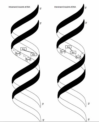
In genetics, crosslinking of DNA occurs when various exogenous or endogenous agents react with two nucleotides of DNA, forming a covalent linkage between them. This crosslink can occur within the same strand (intrastrand) or between opposite strands of double-stranded DNA (interstrand). These adducts interfere with cellular metabolism, such as DNA replication and transcription, triggering cell death. These crosslinks can, however, be repaired through excision or recombination pathways.

<span class="mw-page-title-main">DNA adduct</span> Segment of DNA bound to a cancer-causing chemical

In molecular genetics, a DNA adduct is a segment of DNA bound to a cancer-causing chemical. This process could lead to the development of cancerous cells, or carcinogenesis. DNA adducts in scientific experiments are used as biomarkers of exposure. They are especially useful in quantifying an organism's exposure to a carcinogen. The presence of such an adduct indicates prior exposure to a potential carcinogen, but it does not necessarily indicate the presence of cancer in the subject animal.

<span class="mw-page-title-main">ERCC2</span> Mammalian protein found in humans

ERCC2, or XPD is a protein involved in transcription-coupled nucleotide excision repair.

<span class="mw-page-title-main">XPC (gene)</span> Protein-coding gene in the species Homo sapiens

Xeroderma pigmentosum, complementation group C, also known as XPC, is a protein which in humans is encoded by the XPC gene. XPC is involved in the recognition of bulky DNA adducts in nucleotide excision repair. It is located on chromosome 3.

At its simplest, the adductome is the totality of chemical adducts that are present in particular cellular macromolecules such as DNA, and RNA, or proteins found within the organism. These adducts can detrimentally alter the chemical properties of these macromolecules and are therefore also referred to as damage. Adducts may arise as a consequence of the chemical reaction between a given "physicochemical agent to which an organism is exposed across the lifespan". These physicochemical agents can be exogenous in origin, and include ionizing and non-ionizing radiation, the diet, lifestyle factors, pollution, and xenobiotics. They made damage the macromolecules directly, or indirectly e.g., some xenobiotic substances require metabolism of the xenobiotic to produce a chemically reactive metabolite which can then form a covalent bond with the endogenous macromolecule. Agents that damage macromolecules can also arise from endogenous sources, such as reactive oxygen species that are a side product of normal respiration, leading to the formation of oxidatively damaged DNA etc., or other reactive species e.g., reactive nitrogen, sulphur, carbon, selenium and halogen species.

The DNA damage theory of aging proposes that aging is a consequence of unrepaired accumulation of naturally occurring DNA damage. Damage in this context is a DNA alteration that has an abnormal structure. Although both mitochondrial and nuclear DNA damage can contribute to aging, nuclear DNA is the main subject of this analysis. Nuclear DNA damage can contribute to aging either indirectly or directly.

<span class="mw-page-title-main">8-Oxo-2'-deoxyguanosine</span> Chemical compound

8-Oxo-2'-deoxyguanosine (8-oxo-dG) is an oxidized derivative of deoxyguanosine. 8-Oxo-dG is one of the major products of DNA oxidation. Concentrations of 8-oxo-dG within a cell are a measurement of oxidative stress.

Arsenic biochemistry refers to biochemical processes that can use arsenic or its compounds, such as arsenate. Arsenic is a moderately abundant element in Earth's crust, and although many arsenic compounds are often considered highly toxic to most life, a wide variety of organoarsenic compounds are produced biologically and various organic and inorganic arsenic compounds are metabolized by numerous organisms. This pattern is general for other related elements, including selenium, which can exhibit both beneficial and deleterious effects. Arsenic biochemistry has become topical since many toxic arsenic compounds are found in some aquifers, potentially affecting many millions of people via biochemical processes.

DNA damage is an alteration in the chemical structure of DNA, such as a break in a strand of DNA, a nucleobase missing from the backbone of DNA, or a chemically changed base such as 8-OHdG. DNA damage can occur naturally or via environmental factors, but is distinctly different from mutation, although both are types of error in DNA. DNA damage is an abnormal chemical structure in DNA, while a mutation is a change in the sequence of base pairs. DNA damages cause changes in the structure of the genetic material and prevents the replication mechanism from functioning and performing properly. The DNA damage response (DDR) is a complex signal transduction pathway which recognizes when DNA is damaged and initiates the cellular response to the damage.

<span class="mw-page-title-main">Cynthia Burrows</span> American chemist

Cynthia J. Burrows is an American chemist, currently a distinguished professor in the department of chemistry at the University of Utah, where she is also the Thatcher Presidential Endowed Chair of Biological Chemistry. Burrows was the Senior Editor of the Journal of Organic Chemistry (2001-2013) and became Editor-in-Chief of Accounts of Chemical Research in 2014.,,

References

  1. Chakarov, Stoyan; Petkova, Rumena; Russev, George Ch; Zhelev, Nikolai (2014-02-23). "DNA damage and mutation. Types of DNA damage". BioDiscovery. 11 (11): e8957. Bibcode:2014BioDi..11....1C. doi: 10.7750/BioDiscovery.2014.11.1 . ISSN   2050-2966.
  2. 1 2 3 De Bont, Rinne; van Larebeke, Nik (2004-05-01). "Endogenous DNA damage in humans: a review of quantitative data". Mutagenesis. 19 (3): 169–185. doi: 10.1093/mutage/geh025 . ISSN   0267-8357. PMID   15123782.
  3. Xia, Jun; Chiu, Li-Ya; Nehring, Ralf B.; Núñez, María Angélica Bravo; Mei, Qian; Perez, Mercedes; Zhai, Yin; Fitzgerald, Devon M.; Pribis, John P.; Wang, Yumeng; Hu, Chenyue W. (2019-01-10). "Bacteria-to-human protein networks reveal origins of endogenous DNA damage". Cell. 176 (1–2): 127–143.e24. doi:10.1016/j.cell.2018.12.008. ISSN   0092-8674. PMC   6344048 . PMID   30633903.
  4. Yan, Liewei L.; Zaher, Hani S. (2019-10-11). "How do cells cope with RNA damage and its consequences?". The Journal of Biological Chemistry. 294 (41): 15158–15171. doi: 10.1074/jbc.REV119.006513 . ISSN   0021-9258. PMC   6791314 . PMID   31439666.
  5. Wurtmann, Elisabeth J.; Wolin, Sandra L. (2009). "RNA under attack: Cellular handling of RNA damage". Critical Reviews in Biochemistry and Molecular Biology. 44 (1): 34–49. doi:10.1080/10409230802594043. ISSN   1040-9238. PMC   2656420 . PMID   19089684.
  6. "How does ultraviolet light kill cells?". Scientific American. Retrieved 2020-11-30.
  7. Woods, Derek; Turchi, John J. (2013-05-01). "Chemotherapy induced DNA damage response". Cancer Biology & Therapy. 14 (5): 379–389. doi:10.4161/cbt.23761. ISSN   1538-4047. PMC   3672181 . PMID   23380594.
  8. 1 2 "How Chemotherapy Drugs Work". www.cancer.org. Retrieved 2020-11-30.
  9. 1 2 3 4 5 6 7 8 9 Jackson, Stephen P.; Bartek, Jiri (2009-10-22). "The DNA-damage response in human biology and disease". Nature. 461 (7267): 1071–1078. Bibcode:2009Natur.461.1071J. doi:10.1038/nature08467. ISSN   0028-0836. PMC   2906700 . PMID   19847258.
  10. Okayasu, Ryuichi; Takahashi, Sentaro; Yamada, Shigeru; Hei, Tom K.; Ullrich, Robert L. (1999-01-15). "Asbestos and DNA Double Strand Breaks". Cancer Research. 59 (2): 298–300. ISSN   0008-5472. PMID   9927035.
  11. Guo, Hongrui; Liu, Huan; Wu, Hongbin; Cui, Hengmin; Fang, Jing; Zuo, Zhicai; Deng, Junliang; Li, Yinglun; Wang, Xun; Zhao, Ling (2019-09-21). "Nickel Carcinogenesis Mechanism: DNA Damage". International Journal of Molecular Sciences. 20 (19): 4690. doi: 10.3390/ijms20194690 . ISSN   1422-0067. PMC   6802009 . PMID   31546657.
  12. Cooke, Marcus S.; Evans, Mark D.; Dizdaroglu, Miral; Lunec, Joseph (2003). "Oxidative DNA damage: mechanisms, mutation, and disease". The FASEB Journal. 17 (10): 1195–1214. doi: 10.1096/fj.02-0752rev . ISSN   1530-6860. PMID   12832285. S2CID   1132537.
  13. Weimann, Allan; Belling, Dorthe; Poulsen, Henrik E. (2002-01-15). "Quantification of 8-oxo-guanine and guanine as the nucleobase, nucleoside and deoxynucleoside forms in human urine by high-performance liquid chromatography–electrospray tandem mass spectrometry". Nucleic Acids Research. 30 (2): e7. doi:10.1093/nar/30.2.e7. ISSN   0305-1048. PMC   99846 . PMID   11788733.
  14. Calabretta, Alessandro; Küpfer, Pascal A.; Leumann, Christian J. (2015-05-19). "The effect of RNA base lesions on mRNA translation". Nucleic Acids Research. 43 (9): 4713–4720. doi:10.1093/nar/gkv377. ISSN   1362-4962. PMC   4482091 . PMID   25897124.
  15. Radak, Zsolt; Boldogh, Istvan (2010-08-15). "8-oxo-7,8-dihydroguanine: Link to gene expression, aging and defense against oxidative stress". Free Radical Biology & Medicine. 49 (4): 587–596. doi:10.1016/j.freeradbiomed.2010.05.008. ISSN   0891-5849. PMC   2943936 . PMID   20483371.
  16. Poulsen, Henrik E.; Specht, Elisabeth; Broedbaek, Kasper; Henriksen, Trine; Ellervik, Christina; Mandrup-Poulsen, Thomas; Tonnesen, Morten; Nielsen, Peter E.; Andersen, Henrik U.; Weimann, Allan (2012-04-15). "RNA modifications by oxidation: a novel disease mechanism?". Free Radical Biology & Medicine. 52 (8): 1353–1361. doi:10.1016/j.freeradbiomed.2012.01.009. ISSN   1873-4596. PMID   22306201.
  17. Cavalieri, Ercole; Saeed, Muhammad; Zahid, Muhammad; Cassada, David; Snow, Daniel; Miljkovic, Momcilo; Rogan, Eleanor (February 2012). "Mechanism of DNA Depurination by Carcinogens in Relation to Cancer Initiation". IUBMB Life. 64 (2): 169–179. doi:10.1002/iub.586. ISSN   1521-6543. PMC   4418633 . PMID   22162200.
  18. 1 2 Griffiths, Anthony JF; Miller, Jeffrey H.; Suzuki, David T.; Lewontin, Richard C.; Gelbart, William M. (2000). "Spontaneous mutations". An Introduction to Genetic Analysis. 7th Edition.
  19. "DNA Damage - the major cause of missing pieces from the DNA puzzle | NEB". www.neb.com. Retrieved 2020-12-01.
  20. Caldecott, Keith W. (August 2008). "Single-strand break repair and genetic disease" . Nature Reviews Genetics. 9 (8): 619–631. doi:10.1038/nrg2380. ISSN   1471-0064. PMID   18626472. S2CID   39412478.
  21. 1 2 Rogstad, Daniel K.; Liu, Pingfang; Burdzy, Artur; Lin, Susan S.; Sowers, Lawrence C. (2002-06-25). "Endogenous DNA lesions can inhibit the binding of the AP-1 (c-Jun) transcription factor". Biochemistry. 41 (25): 8093–8102. doi:10.1021/bi012180a. ISSN   0006-2960. PMID   12069602.
  22. Esterbauer, H.; Eckl, P.; Ortner, A. (May 1990). "Possible mutagens derived from lipids and lipid precursors". Mutation Research. 238 (3): 223–233. doi:10.1016/0165-1110(90)90014-3. ISSN   0027-5107. PMID   2342513.
  23. Chung, F. L.; Chen, H. J.; Nath, R. G. (October 1996). "Lipid peroxidation as a potential endogenous source for the formation of exocyclic DNA adducts". Carcinogenesis. 17 (10): 2105–2111. doi: 10.1093/carcin/17.10.2105 . ISSN   0143-3334. PMID   8895475.
  24. Bartsch, H.; Nair, J. (2000-11-16). "Ultrasensitive and specific detection methods for exocylic DNA adducts: markers for lipid peroxidation and oxidative stress". Toxicology. 153 (1–3): 105–114. doi:10.1016/s0300-483x(00)00307-3. ISSN   0300-483X. PMID   11090950.
  25. Pagès, Vincent; Fuchs, Robert PP (December 2002). "How DNA lesions are turned into mutations within cells?". Oncogene. 21 (58): 8957–8966. doi:10.1038/sj.onc.1206006. ISSN   1476-5594. PMID   12483512. S2CID   25226350.
  26. Fimognari, Carmela (2015). "Role of Oxidative RNA Damage in Chronic-Degenerative Diseases". Oxidative Medicine and Cellular Longevity. 2015: 358713. doi: 10.1155/2015/358713 . ISSN   1942-0900. PMC   4452857 . PMID   26078805.
  27. Sirbu, Bianca M.; Cortez, David (August 2013). "DNA Damage Response: Three Levels of DNA Repair Regulation". Cold Spring Harbor Perspectives in Biology. 5 (8): a012724. doi:10.1101/cshperspect.a012724. ISSN   1943-0264. PMC   3721278 . PMID   23813586.
  28. Krokan, Hans E.; Bjørås, Magnar (April 2013). "Base Excision Repair". Cold Spring Harbor Perspectives in Biology. 5 (4): a012583. doi:10.1101/cshperspect.a012583. ISSN   1943-0264. PMC   3683898 . PMID   23545420.
  29. Jackson, Stephen P. (2002-05-01). "Sensing and repairing DNA double-strand breaks". Carcinogenesis. 23 (5): 687–696. doi: 10.1093/carcin/23.5.687 . ISSN   0143-3334. PMID   12016139.
  30. Cannan, Wendy J.; Pederson, David S. (January 2016). "Mechanisms and Consequences of Double-strand DNA Break Formation in Chromatin". Journal of Cellular Physiology. 231 (1): 3–14. doi:10.1002/jcp.25048. ISSN   0021-9541. PMC   4994891 . PMID   26040249.
  31. 1 2 de Boer, Jan; Hoeijmakers, Jan H. J. (2000-03-01). "Nucleotide excision repair and human syndromes". Carcinogenesis. 21 (3): 453–460. doi: 10.1093/carcin/21.3.453 . hdl: 1765/3166 . ISSN   0143-3334. PMID   10688865.
  32. 1 2 Kusakabe, Masayuki; Onishi, Yuki; Tada, Haruto; Kurihara, Fumika; Kusao, Kanako; Furukawa, Mari; Iwai, Shigenori; Yokoi, Masayuki; Sakai, Wataru; Sugasawa, Kaoru (2019-01-25). "Mechanism and regulation of DNA damage recognition in nucleotide excision repair". Genes and Environment. 41 (1): 2. Bibcode:2019GeneE..41....2K. doi: 10.1186/s41021-019-0119-6 . ISSN   1880-7062. PMC   6346561 . PMID   30700997.
  33. Hsieh, Peggy; Yamane, Kazuhiko (2008). "DNA mismatch repair: Molecular mechanism, cancer, and ageing". Mechanisms of Ageing and Development. 129 (7–8): 391–407. doi:10.1016/j.mad.2008.02.012. ISSN   0047-6374. PMC   2574955 . PMID   18406444.