Neisseria flavescens

Last updated

Neisseria flavescens
Neisseria flavescens, gram stain, 1977.jpg
A gram stain of Neisseria flavescens provided by CDC/Dr. W. A. Clark
Scientific classification OOjs UI icon edit-ltr.svg
Domain: Bacteria
Phylum: Pseudomonadota
Class: Betaproteobacteria
Order: Neisseriales
Family: Neisseriaceae
Genus: Neisseria
Species:
N. flavescens
Binomial name
Neisseria flavescens
Branham 1930 [1]

Neisseria flavescens was first isolated from cerebrospinal fluid in the midst of an epidemic meningitis outbreak in Chicago. [2] These gram-negative, aerobic bacteria reside in the mucosal membranes of the upper respiratory tract, functioning as commensals. [3] However, this species can also play a pathogenic role in immunocompromised and diabetic individuals. [4] In rare cases, it has been linked to meningitis, pneumonia, empyema, endocarditis, and sepsis.

Contents

Morphology

These bacteria are gram-negative and diplococcus, rendering them virtually indistinguishable from the other Neisseria species. [2] Yet, Neisseria flavescens remains distinct due to its signature pigmented colonies, yellow-gold in color. [5] And it is through this yellow-gold color that this bacteria earned its name, with flavescens precisely translating as "becoming a golden yellow." [2] This pigmentation also indicates N. flavescens' similarity to saccharolytic Neisseria species, which also exhibit pigmentation. [6] In addition, these pigmented species differ from meningococcus, which lack pigmentation. [2]

Biochemical processes

Similar to saccharolytic species, N. flavescens strains are capable of producing polysaccharides from sucrose and are colistin-susceptible. [6] This bacteria is also catalase and oxidase positive. [3] It is not capable of acid-production from glucose, maltose, fructose, sucrose, mannose, or lactose, in contrast to meningococcus, which are active-fermenters. [2] Furthermore, fundamental differences between these two species are again shown, as serological testing reveals N. flavescens' lack of cross-agglutination. [2] At the same time, biochemical testing distinguishes Neisseria flavescens from other gram-negative diplococci, with N. flavescens being DNase negative, weakly positive to Superoxol, and capable of prolyl aminopeptidase production in an enzyme-substrate test. [5]

Molecular biology

Though it shares many similarities with the saccharolytic species, Neisseria flavescens has a greater genetic relation to pathogenic Neisseria species, [6] as molecular studies have shown. [7] In addition, studies implicate that this species plays a role in penicillin-resistant strains of Neisseria meningitidis. The increasing selective pressure from penicillin treatment has led to N. meningitidis' uptake of an altered penicillin-binding protein gene, penA, from Neisseria flavescens via transformation. [8] This modified penicillin-binding protein, also known as mecA , inhibits Neisseria meningitidis' transpeptidases from binding to the β-lactam portion of penicillin.

Disease

Typically serving as a commensal, Neisseria flavescens has also played a pathogenic role, ever since its origin. Arising from an epidemic meningitis outbreak in Chicago, N. flavescens was isolated from the cerebrospinal fluid of infected individuals. In particular, out of forty-seven total cases of meningitis, fourteen individuals were found to carry N. flavescens, in contrast to carrying one of the typical four meningococci. [2] Additionally, the mortality rate among these fourteen individuals was close to thirty percent, indicating that this bacterium's role as a possible causative agent for meningitis should not be overlooked. [2] Since then, four other cases of meningitis have also found Neisseria flavescens to be the causative agent. [9]

Along with meningitis, this organism has also been linked to sepsis following surgery. [9] A patient presented with clinical signs typical of meningococcal-strains: fever, chills, headache, myalgia, arthralgia, and skin rash. [9] To identify the causative agent, smears from skin lesions and blood cultures were obtained from the patient. [9] Gram-negative, diplococci were present in the smear, narrowing the organism down to a Neisseria species. Ultimately, blood cultures revealed N. flavescens to be the culprit, due to observation of yellow-gold colony formation and no sugar fermentation. [9]

In addition to blood and CSF, Neisseria flavescens can also act as a pathogen in the lower respiratory tract. [4] Isolation via a transthoracic pulmonary fine-needle aspiration identified N. flavescens as the cause of pneumonia and empyema in a diabetic patient. [4] More specifically, the aspirate was sent off to the respiration department, where it underwent acid fast and gram staining, inoculation, and biochemical testing to identify N. flavescens. [4] Next, 16S rRNA sequencing was done, further confirming that Neisseria flavescens was indeed the causative agent. [4]

Lastly, this bacteria has also been the pathogen behind a case of endocarditis. Testing β-lactamase positive, Neisseria flavescens rendered penicillin an ineffective treatment for the patient and, instead, was targeted by cefotaxime. [10]

See also

Related Research Articles

<span class="mw-page-title-main">Gram-negative bacteria</span> Group of bacteria that do not retain the Gram stain used in bacterial differentiation

Gram-negative bacteria are bacteria that do not retain the crystal violet stain used in the Gram staining method of bacterial differentiation. They are characterized by their cell envelopes, which are composed of a thin peptidoglycan cell wall sandwiched between an inner cytoplasmic cell membrane and a bacterial outer membrane.

<span class="mw-page-title-main">Penicillin</span> Group of antibiotics derived from Penicillium fungi

Penicillins are a group of β-lactam antibiotics originally obtained from Penicillium moulds, principally P. chrysogenum and P. rubens. Most penicillins in clinical use are synthesised by P. chrysogenum using deep tank fermentation and then purified. A number of natural penicillins have been discovered, but only two purified compounds are in clinical use: penicillin G and penicillin V. Penicillins were among the first medications to be effective against many bacterial infections caused by staphylococci and streptococci. They are still widely used today for different bacterial infections, though many types of bacteria have developed resistance following extensive use.

<i>Neisseria gonorrhoeae</i> Species of bacterium

Neisseria gonorrhoeae, also known as gonococcus (singular), or gonococci (plural), is a species of Gram-negative diplococci bacteria isolated by Albert Neisser in 1879. It causes the sexually transmitted genitourinary infection gonorrhea as well as other forms of gonococcal disease including disseminated gonococcemia, septic arthritis, and gonococcal ophthalmia neonatorum.

<i>Neisseria</i> Genus of bacteria

Neisseria is a large genus of bacteria that colonize the mucosal surfaces of many animals. Of the 11 species that colonize humans, only two are pathogens, N. meningitidis and N. gonorrhoeae.

<span class="mw-page-title-main">Lipopolysaccharide</span> Class of molecules found in the outer membrane of Gram-negative bacteria

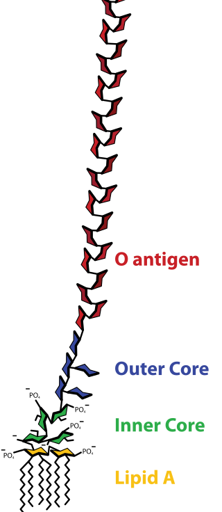
Lipopolysaccharides (LPS) are large molecules consisting of a lipid and a polysaccharide that are bacterial toxins. They are composed of an O-antigen, an outer core, and an inner core all joined by covalent bonds, and are found in the outer membrane of Gram-negative bacteria, such as E. coli and Salmonella. Today, the term endotoxin is often used synonymously with LPS, although there are a few endotoxins that are not related to LPS, such as the so-called delta endotoxin proteins produced by Bacillus thuringiensis.

MeNZB was a vaccine against a specific strain of group B meningococcus, used to control an epidemic of meningococcal disease in New Zealand. Most people are able to carry the meningococcus bacteria safely with no ill effects. However, meningococcal disease can cause meningitis and sepsis, resulting in brain damage, failure of various organs, severe skin and soft-tissue damage, and death.

<span class="mw-page-title-main">Bacterial capsule</span> Polysaccharide layer that lies outside the cell envelope in many bacteria

The bacteria capsule is a large structure common to many bacteria. It is a polysaccharide layer that lies outside the cell envelope, and is thus deemed part of the outer envelope of a bacterial cell. It is a well-organized layer, not easily washed off, and it can be the cause of various diseases.

<i>Neisseria meningitidis</i> Species of bacterium that can cause meningitis

Neisseria meningitidis, often referred to as the meningococcus, is a Gram-negative bacterium that can cause meningitis and other forms of meningococcal disease such as meningococcemia, a life-threatening sepsis. The bacterium is referred to as a coccus because it is round, and more specifically a diplococcus because of its tendency to form pairs.

<span class="mw-page-title-main">Diplococcus</span> Interlocked duo of cocci (e.g. Pneumococcus)

A diplococcus is a round bacterium that typically occurs in the form of two joined cells.

<span class="mw-page-title-main">Meningococcal disease</span> Medical condition

Meningococcal disease describes infections caused by the bacterium Neisseria meningitidis. It has a high mortality rate if untreated but is vaccine-preventable. While best known as a cause of meningitis, it can also result in sepsis, which is an even more damaging and dangerous condition. Meningitis and meningococcemia are major causes of illness, death, and disability in both developed and under-developed countries.

<i>Elizabethkingia meningoseptica</i> Species of bacterium

Elizabethkingia meningoseptica is a Gram-negative, rod-shaped bacterium widely distributed in nature. It may be normally present in fish and frogs; it may be isolated from chronic infectious states, as in the sputum of cystic fibrosis patients. In 1959, American bacteriologist Elizabeth O. King was studying unclassified bacteria associated with pediatric meningitis at the Centers for Disease Control and Prevention in Atlanta, when she isolated an organism that she named Flavobacterium meningosepticum. In 1994, it was reclassified in the genus Chryseobacterium and renamed Chryseobacterium meningosepticum(chryseos = "golden" in Greek, so Chryseobacterium means a golden/yellow rod similar to Flavobacterium). In 2005, a 16S rRNA phylogenetic tree of Chryseobacteria showed that C. meningosepticum along with C. miricola were close to each other but outside the tree of the rest of the Chryseobacteria and were then placed in a new genus Elizabethkingia named after the original discoverer of F. meningosepticum.

Capnocytophaga is a genus of Gram-negative bacteria. Normally found in the oropharyngeal tract of mammals, they are involved in the pathogenesis of some animal bite wounds and periodontal diseases.

<span class="mw-page-title-main">Cefodizime</span> Chemical compound

Cefodizime is a 3rd generation cephalosporin antibiotic with broad spectrum activity against aerobic gram positive and gram negative bacteria. Clinically, it has been shown to be effective against upper and lower respiratory tract infections, urinary tract infections, and gonorrhea. Cefodizime is a bactericidal antibiotic that targets penicillin-binding proteins (PBPs) 1A/B, 2, and 3 resulting in the eventual death of the bacterial cell. In vivo experimental models of infection showed that bacterial clearance by this drug is at least as effective compared with other 3rd generation cephalosporins. It has similar adverse effect profile to other 3rd generation cephalosporins as well, mainly being limited to gastrointestinal or dermatological side effects.

<span class="mw-page-title-main">Thayer–Martin agar</span>

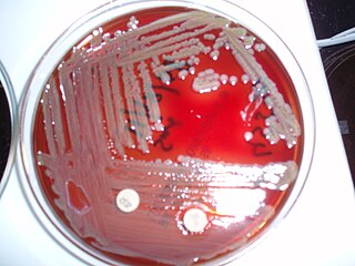
Thayer–Martin agar is a Mueller–Hinton agar with 5% chocolate sheep blood and antibiotics. It is used for culturing and primarily isolating pathogenic Neisseria bacteria, including Neisseria gonorrhoeae and Neisseria meningitidis, as the medium inhibits the growth of most other microorganisms. When growing Neisseria meningitidis, one usually starts with a normally sterile body fluid, so a plain chocolate agar is used. Thayer–Martin agar was initially developed in 1964, with an improved formulation published in 1966.

Neisseria lactamica is a gram-negative diplococcus bacterium. It is strictly a commensal species of the nasopharynx. Uniquely among the Neisseria they are able to produce β-D-galactosidase and ferment lactose.

<span class="mw-page-title-main">Gonococcemia</span> Medical condition

Gonococcemia is a rare complication of mucosal Neisseria gonorrhoeae infection, or Gonorrhea, that occurs when the bacteria invade the bloodstream. It is characterized by fever, tender hemorrhagic pustules on the extremities or the trunk, migratory polyarthritis, and tenosynovitis. It also rarely leads to endocarditis and meningitis. This condition occurs in 0.5-3% of individuals with gonorrhea, and it usually presents 2–3 weeks after acquiring the infection. Risk factors include female sex, sexual promiscuity, and infection with resistant strains of Neisseria gonorrhoeae. This condition is treated with cephalosporin and fluoroquinolone antibiotics.

IgA protease is an enzyme. This enzyme catalyses the following chemical reaction[reaction equation needed]

Neisseria cinerea is a commensal species grouped with the Gram-negative, oxidase-positive, and catalase-positive diplococci. It was first classified as Micrococcus cinereus by Alexander von Lingelsheim in 1906. Using DNA hybridization, N. cinerea exhibits 50% similarity to Neisseria gonorrhoeae.

<span class="mw-page-title-main">New York City agar</span>

The NYC medium or GC medium agar is used for isolating Gonococci.

<i>Neisseria polysaccharea</i> Species of bacterium

Neisseria polysaccharea was described in 1983 and is characterized by its ability to produce acid from glucose and maltose and polysaccharide from sucrose. It is nonpathogenic. Strains of this species were previously identified as nontypable strains of N. meningitidis. Strains of N. polysaccharea also may have been misidentified previously as N. subflava because their ability to produce polysaccharide from sucrose was not determined. Other Neisseria species have been be misidentified as N. polysaccharea by acid production tests and supplemental tests.

References

  1. LPSN lpsn.dsmz.de
  2. 1 2 3 4 5 6 7 8 Branham, Sara E. (1930-04-18). "A New Meningococcus-like Organism (Neisseria flavescens n. sp.) from Epidemic Meningitis". Public Health Reports. 45 (16): 845–849. doi:10.2307/4579618. JSTOR   4579618.
  3. 1 2 "What does Neisseria flavescens mean? Definition, meaning and sense (The Titi Tudorancea Encyclopedia)". www.tititudorancea.com. Retrieved 2015-11-17.
  4. 1 2 3 4 5 Huang, Ling; Ma, Lan; Fan, Kun; Li, Yang; Xie, Le; Xia, Wenying; Gu, Bing; Liu, Genyan (2014-05-01). "Necrotizing pneumonia and empyema caused by Neisseria flavescens infection". Journal of Thoracic Disease. 6 (5): 553–557. doi:10.3978/j.issn.2072-1439.2014.02.16. ISSN   2072-1439. PMC   4015020 . PMID   24822118.
  5. 1 2 "Neisseria flavescens - Gonorrhea - STD Information from CDC". www.cdc.gov. Retrieved 2015-11-17.
  6. 1 2 3 Knapp, J. S. (1988). "Historical perspectives and identification of Neisseria and related species" (PDF). Clinical Microbiology Reviews. 1 (4): 415–431. doi:10.1128/CMR.1.4.415. PMC   358063 . PMID   3069201.
  7. HOKE, C.; VEDROS, N. A. (1982). "Taxonomy of the Neisseriae: Deoxyribonucleic Acid Base Composition, Interspecific Transformation, and Deoxyribonucleic Acid Hybridization". International Journal of Systematic Bacteriology. 32 (1): 57–66. doi: 10.1099/00207713-32-1-57 .
  8. Spratt, B G; Zhang, Q Y; Jones, D M; Hutchison, A; Brannigan, J A; Dowson, C G (1989-11-01). "Recruitment of a penicillin-binding protein gene from Neisseria flavescens during the emergence of penicillin resistance in Neisseria meningitidis". Proceedings of the National Academy of Sciences of the United States of America. 86 (22): 8988–8992. Bibcode:1989PNAS...86.8988S. doi: 10.1073/pnas.86.22.8988 . ISSN   0027-8424. PMC   298417 . PMID   2510173.
  9. 1 2 3 4 5 Wertlake, Paul T.; Williams, Temple W. (1968-07-01). "Septicaemia caused by Neisseria flavescens". Journal of Clinical Pathology. 21 (4): 437–439. doi:10.1136/jcp.21.4.437. ISSN   0021-9746. PMC   473828 . PMID   4972296.
  10. Sinave, Christian P.; Ratzan, Kenneth R. (1987). "Infective endocarditis caused by Neisseria flavescens". The American Journal of Medicine. 82 (1): 163–164. doi:10.1016/0002-9343(87)90399-8. PMID   3799678 . Retrieved 2015-11-28.