
In the fields of molecular biology and genetics, a genome is all the genetic information of an organism. It consists of nucleotide sequences of DNA. The nuclear genome includes protein-coding genes and non-coding genes, other functional regions of the genome such as regulatory sequences, and often a substantial fraction of junk DNA with no evident function. Almost all eukaryotes have mitochondria and a small mitochondrial genome. Algae and plants also contain chloroplasts with a chloroplast genome.
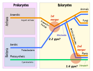
Symbiogenesis is the leading evolutionary theory of the origin of eukaryotic cells from prokaryotic organisms. The theory holds that mitochondria, plastids such as chloroplasts, and possibly other organelles of eukaryotic cells are descended from formerly free-living prokaryotes taken one inside the other in endosymbiosis. Mitochondria appear to be phylogenetically related to Rickettsiales bacteria, while chloroplasts are thought to be related to cyanobacteria.
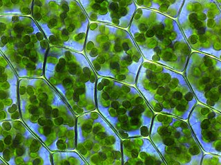
A plastid, pl. plastids, is a membrane-bound organelle found in the cells of plants, algae, and some other eukaryotic organisms. They are considered to be intracellular endosymbiotic cyanobacteria.
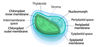
Nucleomorphs are small, vestigial eukaryotic nuclei found between the inner and outer pairs of membranes in certain plastids. They are thought to be vestiges of primitive red and green algal nuclei that were engulfed by a larger eukaryote. Because the nucleomorph lies between two sets of membranes, nucleomorphs support the endosymbiotic theory and are evidence that the plastids containing them are complex plastids. Having two sets of membranes indicate that the plastid, a prokaryote, was engulfed by a eukaryote, an alga, which was then engulfed by another eukaryote, the host cell, making the plastid an example of secondary endosymbiosis.
Phototropins are photoreceptor proteins that mediate phototropism responses in various species of algae, fungi and higher plants. Phototropins can be found throughout the leaves of a plant. Along with cryptochromes and phytochromes they allow plants to respond and alter their growth in response to the light environment. Phototropins may also be important for the opening of stomata and the movement of chloroplasts. These blue light receptors are seen across the entire green plant lineage. When Phototropins are hit with blue light, they induce a signal transduction pathway that alters the plant cells' functions in different ways.

The Archaeplastida are a major group of eukaryotes, comprising the photoautotrophic red algae (Rhodophyta), green algae, land plants, and the minor group glaucophytes. It also includes the non-photosynthetic lineage Rhodelphidia, a predatorial (eukaryotrophic) flagellate that is sister to the Rhodophyta, and probably the microscopic picozoans. The Archaeplastida have chloroplasts that are surrounded by two membranes, suggesting that they were acquired directly through a single endosymbiosis event by phagocytosis of a cyanobacterium. All other groups which have chloroplasts, besides the amoeboid genus Paulinella, have chloroplasts surrounded by three or four membranes, suggesting they were acquired secondarily from red or green algae. Unlike red and green algae, glaucophytes have never been involved in secondary endosymbiosis events.

Photosynthetic picoplankton or picophytoplankton is the fraction of the photosynthetic phytoplankton of cell sizes between 0.2 and 2 µm. It is especially important in the central oligotrophic regions of the world oceans that have very low concentration of nutrients.
Phycodnaviridae is a family of large (100–560 kb) double-stranded DNA viruses that infect marine or freshwater eukaryotic algae. Viruses within this family have a similar morphology, with an icosahedral capsid. As of 2014, there were 33 species in this family, divided among 6 genera. This family belongs to a super-group of large viruses known as nucleocytoplasmic large DNA viruses. Evidence was published in 2014 suggesting that specific strains of Phycodnaviridae might infect humans rather than just algal species, as was previously believed. Most genera under this family enter the host cell by cell receptor endocytosis and replicate in the nucleus. Phycodnaviridae play important ecological roles by regulating the growth and productivity of their algal hosts. Algal species such Heterosigma akashiwo and the genus Chrysochromulina can form dense blooms which can be damaging to fisheries, resulting in losses in the aquaculture industry. Heterosigma akashiwo virus (HaV) has been suggested for use as a microbial agent to prevent the recurrence of toxic red tides produced by this algal species. Phycodnaviridae cause death and lysis of freshwater and marine algal species, liberating organic carbon, nitrogen and phosphorus into the water, providing nutrients for the microbial loop.
Ostreococcus is a genus of unicellular coccoid or spherically shaped green algae belonging to the class Mamiellophyceae. It includes prominent members of the global picoplankton community, which plays a central role in the oceanic carbon cycle.

The prasinophytes are a group of unicellular green algae. Prasinophytes mainly include marine planktonic species, as well as some freshwater representatives. The prasinophytes are morphologically diverse, including flagellates with one to eight flagella and non-motile (coccoid) unicells. The cells of many species are covered with organic body scales; others are naked. Well studied genera include Ostreococcus, considered to be the smallest free-living eukaryote, and Micromonas, both of which are found in marine waters worldwide. Prasinophytes have simple cellular structures, containing a single chloroplast and a single mitochondrion. The genomes are relatively small compared to other eukaryotes . At least one species, the Antarctic form Pyramimonas gelidicola, is capable of phagocytosis and is therefore a mixotrophic algae.
Micromonas is a genus of green algae in the family Mamiellaceae.
Picoeukaryotes are picoplanktonic eukaryotic organisms 3.0 µm or less in size. They are distributed throughout the world's marine and freshwater ecosystems and constitute a significant contribution to autotrophic communities. Though the SI prefix pico- might imply an organism smaller than atomic size, the term was likely used to avoid confusion with existing size classifications of plankton.

Red algae, or Rhodophyta, are one of the oldest groups of eukaryotic algae. The Rhodophyta comprises one of the largest phyla of algae, containing over 7,000 currently recognized species with taxonomic revisions ongoing. The majority of species (6,793) are found in the Florideophyceae (class), and mostly consist of multicellular, marine algae, including many notable seaweeds. Red algae are abundant in marine habitats but relatively rare in freshwaters. Approximately 5% of red algae species occur in freshwater environments, with greater concentrations found in warmer areas. Except for two coastal cave dwelling species in the asexual class Cyanidiophyceae, there are no terrestrial species, which may be due to an evolutionary bottleneck in which the last common ancestor lost about 25% of its core genes and much of its evolutionary plasticity.
The eukaryotes constitute the domain of Eukarya or Eukaryota, organisms whose cells have a membrane-bound nucleus. All animals, plants, fungi, and many unicellular organisms are eukaryotes. They constitute a major group of life forms alongside the two groups of prokaryotes: the Bacteria and the Archaea. Eukaryotes represent a small minority of the number of organisms, but given their generally much larger size, their collective global biomass is much larger than that of prokaryotes.

Cyanidioschyzon merolae is a small (2μm), club-shaped, unicellular haploid red alga adapted to high sulfur acidic hot spring environments. The cellular architecture of C. merolae is extremely simple, containing only a single chloroplast and a single mitochondrion and lacking a vacuole and cell wall. In addition, the cellular and organelle divisions can be synchronized. For these reasons, C. merolae is considered an excellent model system for study of cellular and organelle division processes, as well as biochemistry and structural biology. The organism's genome was the first full algal genome to be sequenced in 2004; its plastid was sequenced in 2000 and 2003, and its mitochondrion in 1998. The organism has been considered the simplest of eukaryotic cells for its minimalist cellular organization.
Bathycoccus prasinos is a picoplankton belonging to the order Mamiellales, along with Ostreococcus and Micromonas. The cells are around 1–2 μm in length, are non-motile and are covered in scales.

Picozoa, Picobiliphyta, Picobiliphytes, or Biliphytes are protists of a phylum of marine unicellular heterotrophic eukaryotes with a size of less than about 3 micrometers. They were formerly treated as eukaryotic algae and the smallest member of photosynthetic picoplankton before it was discovered they do not perform photosynthesis. The first species identified therein is Picomonas judraskeda. They probably belong in the Archaeplastida as sister of the Rhodophyta.

Prasinovirus is a genus of large double-stranded DNA viruses, in the family Phycodnaviridae that infect phytoplankton in the Prasinophyceae. There are three groups in this genus, including Micromonas pusilla virus SP1, which infects the cosmopolitan photosynthetic flagellate Micromonas pusilla.