P19 cell

Last updated
Mouse P19 embryonal carcinoma cells immunostained to show the location of beta-catenin at cell-to-cell contacts Beta-catenin.PNG
Mouse P19 embryonal carcinoma cells immunostained to show the location of beta-catenin at cell-to-cell contacts

P19 cells is an embryonic carcinoma cell line derived from an embryo-derived teratocarcinoma in mice. The cell line is pluripotent and can differentiate into cell types of all three germ layers. Also, it is the most characterized embryonic carcinoma (EC) cell line that can be induced into cardiac muscle cells and neuronal cells by different specific treatments. Indeed, exposing aggregated P19 cells to dimethyl sulfoxide (DMSO) induces differentiation into cardiac and skeletal muscle. Also, exposing P19 cells to retinoic acid (RA) can differentiate them into neuronal cells. [1]

Contents

Origin of the P19 cell line

Cancer cells in humans may result in the patient's death if the aggressive cancer cell grows and metastasizes. However, researchers utilize these cells to study the development of cancer cells in order to find more specific treatments. For developmental biologists, embryonal carcinoma, which is derived from teratocarcinoma, is a good object for developmental study. In 1982, McBurney and Rogers transplanted a 7.5 day mouse embryo into the testis to induce tumor growth. Cell cultures containing undifferentiated stem cells were isolated from the primary tumor which have a euploid karyotype. These stem cells were named embryonal carcinoma P19 cells. [2] These derived P19 cells grew rapidly without feeder cells and were easy to maintain. Moreover, the multipotency of P19 cells was then confirmed by injecting the cells into blastocysts of another mouse strain. The researchers found that there were tissues from all three germ layers growing in the recipient mouse. [3] Based on their continuous studies, they further derived subtype cell lines from original P19 cells: P19S18, P19D3, P19RAC65 and P19C16. The difference between these subtype cell lines is the ability to differentiate into neuronal cells or muscle cells in response to treatment with retinoic acid or DMSO, respectively. [3] [4] [5] More recently, various studies generate cell lines that were initially derived from differentiated P19 cells. Due to the pluripotency of P19 cells, those new derived cell lines can be ectoderm, mesoderm and endoderm-like cells. [6]

Differentiation of P19 cells

P19 cells can be maintained in exponential growth because of a stable chromosomal composition. Because embryonal carcinoma can differentiate into cells of all three germ layers, P19 cells can also differentiate into those ectoderm, mesoderm and endoderm-like cells. When embryonal carcinoma cells are cultured at high density, they start to differentiate. [7] By aggregating the cells into an embryonic body, EC cells can also process differentiation. [8] In P19 cells, addition of non-toxic concentrations of drugs to aggregated embryoid body cells can induce differentiation of P19 cells into specific cell lines depending on the added drug. [1] The two most common and effective drugs are retinoic acid (RA) and dimethyl sulfoxide (DMSO). Studies have shown that a certain concentration of RA can induce P19 cells to differentiate into neuronal cells, including neurons and glial cells, [9] whereas 0.5% - 1% DMSO led P19 cells to differentiate into cardiac or skeletal muscle cells. In the RA treatment method, neurons, astroglia and fibroblasts can be identified after aggregation. Differentiated cells also have choline acetyltransferase and acetyl cholinesterase activities. [10] When treated with DMSO, cardiac muscle cells developed after 5 days of exposure and skeletal muscle cells appeared after 8 days of exposure. Those studies showed that drug exposure causes multipotent P19 cells to differentiate into different layers of cells. Because the concentration of retinoic acid or DMSO is nontoxic to the cells, the drug-specific differentiation is due to induction of cells not selection. Mutants of P19 cells have been generated to investigate the mechanism of drug-specific differentiation. [10] Moreover, signaling pathways related to neurogenesis and myogenesis were also investigated by studying gene expression or generating mutants of P19 cells.

Neurogenesis in P19 cells.

Treatment of undifferentiated P19 cells with retinoic acid can specifically induce them into neuronal cells. Using doses between 1 μM to 3 μM of RA can generate neurons as the most abundant cell type. [4] Neurons under this treatment reached the highest populations between six days and nine days. Several neuronal markers such as neurofilament proteins, HNK-1 antigen and tetanus toxin binding sites are expressed at highest levels during these days. [11] After six to nine days of treatment, the relative neuronal population declines, likely because of faster proliferation of non-neuronal cells. After 10 days of exposure, astroglial cells can be detected using glial fibrillary acidic protein (GFAP), which is a specific marker of glial cells. Other than into neurons and astrocytes, P19 cells can also differentiate to oligodendrocytes, which can be detected using the specific markers, myelin-associated glycoprotein and 2',3'-Cyclic-nucleotide 3'-phosphodiesterase. Moreover, oligodendrocytes also developed and migrated into fiber bundles in mice when the RA-induced cells were transplanted into the brains. [12]
Retinoic acid can induce not only P19 cells but also other progenitor cells or embryonic stem cells to differentiation. Since cells after retinoic acid treatment did not immediately express neuronal marker genes, RA must initiate some pathway to process cellular differentiation. Many studies used P19 cells to investigate the RA-induced mechanisms, including generating the mutant allele of retinoic acid receptor genes and studying the expression of receptor genes, Hox genes and retinol binding proteins while exposing to RA. [13] [14]

All of these studies indicate that the P19 cell is a good in vitro model system for investigating the mechanism of drugs that interfere with specific cellular pathway. Furthermore, by using the ability of RA-induced neurogenesis in P19 cell, many researchers started to identify the in vitro differentiation mechanisms of neuro- or gliogenesis. Several related pathways or including Wnt/β-catenin pathway, Notch pathway and hedgehog pathway are investigated either using gene expression or generating alleles for related genes. [15] [16] [17]

Myogenesis in the P19 cell line

Same as retinoic acid, DMSO induced differentiation is not specific to P19 cells. It could also induce neuroblastoma cells, lung cancer cells and mouse ES cells. [18] [19] [20] At concentration of 0.5–1% DMSO induced P19 cells to aggregate and process mesodermal and endodermal cell types. [1] [10] [21]
The cellular mechanism that occurs during aggregation and differentiation is still not fully studied. However, some studies showed that the cellular communication plays an important role in muscle differentiation in P19 cells which might explain why cells need to aggregate first to process the muscle differentiation. [6]
In order to elucidate the mechanism of myogenesis in P19 cells, several cardiac specific transcription factors including GATA-4, MEF2c, Msx-1, Nkx2.5, MHox, Msx-2 and MLP are found to change during differentiation. [6] Reports have shown that GATA-4, NKx2.5 and MEF2c were all upregulated after DMSO induction. [22] [23] In recent years, P19 cells were also used in studying the mechanism of cardiac differentiation and myogenesis. The main affected signaling pathway, bone morphogenetic proteins (BMPs) pathway is the most strongly studied signaling in P19 cells. By generating the P19CL6noggin cell line, which overexpresses the BMP antagonist noggin, they found that the mutant cells didn't differentiate into cardiomyocytes when treated with 1% of DMSO, suggesting that the BMPs were indispensable for cardiomyocyte differentiation in this system. They also provided the evidence showing that TAK1, Nkx-2.5, and GATA-4 are important in cardiogenic BMP signaling pathway. [24]

Future directions

P19 cells provide valuable formation of both neuronal cells and muscle cells in vitro. Since P19 cells are easy to maintain and culture compared to other embryonic stem cells, they are a convenient model to perform developmental studies in vitro. Techniques to manipulate this cell line to express or knock out certain genes allow for detailed investigation of signaling pathways, functional aspects and the regulation of protein expression of myogenesis and neurogenesis. The extended research can also elucidate the later stages of heart or brain development and maturation.

Related Research Articles

<span class="mw-page-title-main">Mesoderm</span> Middle germ layer of embryonic development

The mesoderm is the middle layer of the three germ layers that develops during gastrulation in the very early development of the embryo of most animals. The outer layer is the ectoderm, and the inner layer is the endoderm.

Transdifferentiation, also known as lineage reprogramming, is the process in which one mature somatic cell is transformed into another mature somatic cell without undergoing an intermediate pluripotent state or progenitor cell type. It is a type of metaplasia, which includes all cell fate switches, including the interconversion of stem cells. Current uses of transdifferentiation include disease modeling and drug discovery and in the future may include gene therapy and regenerative medicine. The term 'transdifferentiation' was originally coined by Selman and Kafatos in 1974 to describe a change in cell properties as cuticle producing cells became salt-secreting cells in silk moths undergoing metamorphosis.

<span class="mw-page-title-main">Cellular differentiation</span> Developmental biology

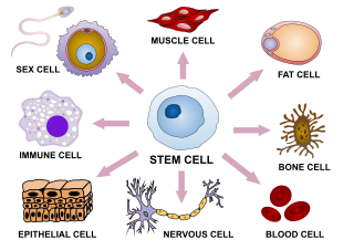
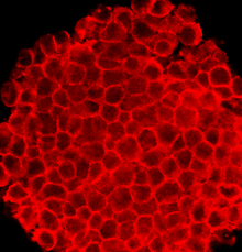
Cellular differentiation is the process in which a stem cell changes from one type to a differentiated one. Usually, the cell changes to a more specialized type. Differentiation happens multiple times during the development of a multicellular organism as it changes from a simple zygote to a complex system of tissues and cell types. Differentiation continues in adulthood as adult stem cells divide and create fully differentiated daughter cells during tissue repair and during normal cell turnover. Some differentiation occurs in response to antigen exposure. Differentiation dramatically changes a cell's size, shape, membrane potential, metabolic activity, and responsiveness to signals. These changes are largely due to highly controlled modifications in gene expression and are the study of epigenetics. With a few exceptions, cellular differentiation almost never involves a change in the DNA sequence itself. However, metabolic composition does get altered quite dramatically where stem cells are characterized by abundant metabolites with highly unsaturated structures whose levels decrease upon differentiation. Thus, different cells can have very different physical characteristics despite having the same genome.

The development of the nervous system, or neural development (neurodevelopment), refers to the processes that generate, shape, and reshape the nervous system of animals, from the earliest stages of embryonic development to adulthood. The field of neural development draws on both neuroscience and developmental biology to describe and provide insight into the cellular and molecular mechanisms by which complex nervous systems develop, from nematodes and fruit flies to mammals.

<span class="mw-page-title-main">Notch signaling pathway</span> Series of molecular signals

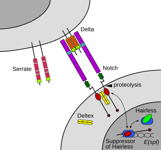
The Notch signaling pathway is a highly conserved cell signaling system present in most animals. Mammals possess four different notch receptors, referred to as NOTCH1, NOTCH2, NOTCH3, and NOTCH4. The notch receptor is a single-pass transmembrane receptor protein. It is a hetero-oligomer composed of a large extracellular portion, which associates in a calcium-dependent, non-covalent interaction with a smaller piece of the notch protein composed of a short extracellular region, a single transmembrane-pass, and a small intracellular region.

<span class="mw-page-title-main">MyoD</span> Mammalian protein found in Homo sapiens

MyoD, also known as myoblast determination protein 1, is a protein in animals that plays a major role in regulating muscle differentiation. MyoD, which was discovered in the laboratory of Harold M. Weintraub, belongs to a family of proteins known as myogenic regulatory factors (MRFs). These bHLH transcription factors act sequentially in myogenic differentiation. Vertebrate MRF family members include MyoD1, Myf5, myogenin, and MRF4 (Myf6). In non-vertebrate animals, a single MyoD protein is typically found.

<span class="mw-page-title-main">Myogenesis</span> Formation of muscular tissue, particularly during embryonic development

Myogenesis is the formation of skeletal muscular tissue, particularly during embryonic development.

mir-133 microRNA precursor family

mir-133 is a type of non-coding RNA called a microRNA that was first experimentally characterised in mice. Homologues have since been discovered in several other species including invertebrates such as the fruitfly Drosophila melanogaster. Each species often encodes multiple microRNAs with identical or similar mature sequence. For example, in the human genome there are three known miR-133 genes: miR-133a-1, miR-133a-2 and miR-133b found on chromosomes 18, 20 and 6 respectively. The mature sequence is excised from the 3' arm of the hairpin. miR-133 is expressed in muscle tissue and appears to repress the expression of non-muscle genes.

<span class="mw-page-title-main">Retinoic acid receptor alpha</span> Protein found in humans

Retinoic acid receptor alpha (RAR-α), also known as NR1B1, is a nuclear receptor that in humans is encoded by the RARA gene.

<span class="mw-page-title-main">Retinoic acid receptor beta</span> Protein-coding gene in the species Homo sapiens

Retinoic acid receptor beta (RAR-beta), also known as NR1B2 is a nuclear receptor that in humans is encoded by the RARB gene.

<span class="mw-page-title-main">GPRC5A</span> Protein-coding gene in the species Homo sapiens

Retinoic acid-induced protein 3 is a protein that in humans is encoded by the GPRC5A gene. This gene and its encoded mRNA was first identified as a phorbol ester-induced gene, and named Phorbol Ester Induced Gen 1 (PEIG-1); two years later it was rediscovered as a retinoic acid-inducible gene, and named Retinoic Acid-Inducible Gene 1 (RAIG1). Its encoded protein was later named Retinoic acid-induced protein 3.

<span class="mw-page-title-main">CRABP2</span> Protein-coding gene in the species Homo sapiens

Cellular retinoic acid-binding protein 2 is a cytoplasmic binding protein that in humans is encoded by the CRABP2 gene.

<span class="mw-page-title-main">CYP26A1</span> Protein-coding gene in the species Homo sapiens

Cytochrome P450 26A1 is a protein that in humans is encoded by the CYP26A1 gene.

<span class="mw-page-title-main">CRABP1</span> Protein-coding gene in the species Homo sapiens

Cellular retinoic acid-binding protein 1 is a protein that in humans is encoded by the CRABP1 gene.

<span class="mw-page-title-main">ALDH1A2</span> Protein-coding gene in the species Homo sapiens

Aldehyde dehydrogenase 1 family, member A2, also known as ALDH1A2 or retinaldehyde dehydrogenase 2 (RALDH2), is an enzyme that in humans is encoded by the ALDH1A2 gene.

<span class="mw-page-title-main">LIN28</span>

Lin-28 homolog A is a protein that in humans is encoded by the LIN28 gene.

<span class="mw-page-title-main">Rex1</span> Known marker of pluripotency, and is usually found in undifferentiated embryonic stem cells

Rex1 (Zfp-42) is a known marker of pluripotency, and is usually found in undifferentiated embryonic stem cells. In addition to being a marker for pluripotency, its regulation is also critical in maintaining a pluripotent state. As the cells begin to differentiate, Rex1 is severely and abruptly downregulated.

<span class="mw-page-title-main">NTERA-2</span>

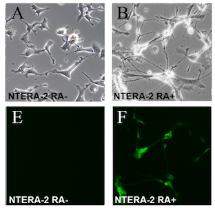
The NTERA-2 cell line is a clonally derived, pluripotent human embryonal carcinoma cell line.

Directed differentiation is a bioengineering methodology at the interface of stem cell biology, developmental biology and tissue engineering. It is essentially harnessing the potential of stem cells by constraining their differentiation in vitro toward a specific cell type or tissue of interest. Stem cells are by definition pluripotent, able to differentiate into several cell types such as neurons, cardiomyocytes, hepatocytes, etc. Efficient directed differentiation requires a detailed understanding of the lineage and cell fate decision, often provided by developmental biology.

Cardiomyocyte proliferation refers to the ability of cardiac muscle cells to progress through the cell cycle and continue to divide. Traditionally, cardiomyocytes were believed to have little to no ability to proliferate and regenerate after birth. Although other types of cells, such as gastrointestinal epithelial cells, can proliferate and differentiate throughout life, cardiac tissue contains little intrinsic ability to proliferate, as adult human cells arrest in the cell cycle. However, a recent paradigm shift has occurred. Recent research has demonstrated that human cardiomyocytes do proliferate to a small extent for the first two decades of life. Also, cardiomyocyte proliferation and regeneration has been demonstrated to occur in various neonatal mammals in response to injury in the first week of life. Current research aims to further understand the biological mechanism underlying cardiomyocyte proliferation in hopes to turn this capability back on in adults in order to combat heart disease.

References

  1. 1 2 3 McBurney, MW; Rogers, BJ (Feb 1982). "Isolation of male embryonal carcinoma cells and their chromosome replication patterns". Developmental Biology. 89 (2): 503–8. doi:10.1016/0012-1606(82)90338-4. PMID   7056443.
  2. McBurney, MW (1993). "P19 embryonal carcinoma cells". Int J Dev Biol. 37 (1): 135–140. PMID   8507558.
  3. 1 2 Rossant, J; McBurney, MW (Aug 1982). "The developmental potential of a euploid male teratocarcinoma cell line after blastocyst injection". Journal of Embryology and Experimental Morphology. 70: 99–112. PMID   7142904.
  4. 1 2 Fahnestock, M; Koshland DE, Jr (Feb 1979). "Control of the receptor for galactose taxis in Salmonella typhimurium". Journal of Bacteriology. 137 (2): 758–63. doi:10.1128/jb.137.2.758-763.1979. PMC   218354 . PMID   370099.
  5. Craine, BL; Rupert, CS (Feb 1979). "Deoxyribonucleic acid-membrane interactions near the origin of replication and initiation of deoxyribonucleic acid synthesis in Escherichia coli". Journal of Bacteriology. 137 (2): 740–5. doi:10.1128/jb.137.2.740-745.1979. PMC   218351 . PMID   370098.
  6. 1 2 3 van der Heyden, MA; Defize, LH (2003-05-01). "Twenty one years of P19 cells: what an embryonal carcinoma cell line taught us about cardiomyocyte differentiation". Cardiovascular Research. 58 (2): 292–302. doi: 10.1016/S0008-6363(02)00771-X . PMID   12757864.
  7. McBurney, MW (Nov 1976). "Clonal lines of teratocarcinoma cells in vitro: differentiation and cytogenetic characteristics". Journal of Cellular Physiology. 89 (3): 441–55. doi:10.1002/jcp.1040890310. PMID   988033.
  8. Martin, GR; Evans MJ (1975). "Multiple differentiation of clonal teratocarcinoma stem cells following embryoid body formation in vitro". Cell. 6 (4): 467–74. doi:10.1016/0092-8674(75)90035-5.
  9. Edwards, MK; Harris, JF; McBurney, MW (Dec 1983). "Induced muscle differentiation in an embryonal carcinoma cell line". Molecular and Cellular Biology. 3 (12): 2280–6. doi:10.1128/mcb.3.12.2280. PMC   370099 . PMID   6656767.
  10. 1 2 3 Jones-Villeneuve, EM; Rudnicki, MA; Harris, JF; McBurney, MW (Dec 1983). "Retinoic acid-induced neural differentiation of embryonal carcinoma cells". Molecular and Cellular Biology. 3 (12): 2271–9. doi:10.1128/mcb.3.12.2271. PMC   370098 . PMID   6656766.
  11. McBurney, MW; Reuhl, KR; Ally, AI; Nasipuri, S; Bell, JC; Craig, J (Mar 1988). "Differentiation and maturation of embryonal carcinoma-derived neurons in cell culture". The Journal of Neuroscience. 8 (3): 1063–73. doi: 10.1523/JNEUROSCI.08-03-01063.1988 . PMC   6569242 . PMID   2894413.
  12. Staines, WA; Craig, J; Reuhl, K; McBurney, MW (Apr 1996). "Retinoic acid treated P19 embryonal carcinoma cells differentiate into oligodendrocytes capable of myelination". Neuroscience. 71 (3): 845–53. doi:10.1016/0306-4522(95)00494-7. PMID   8867053.
  13. Pratt, MA; Kralova, J; McBurney, MW (Dec 1990). "A dominant negative mutation of the alpha retinoic acid receptor gene in a retinoic acid-nonresponsive embryonal carcinoma cell". Molecular and Cellular Biology. 10 (12): 6445–53. doi:10.1128/mcb.10.12.6445. PMC   362921 . PMID   2174108.
  14. Chen, Y; Reese, DH (Oct 2011). "The retinol signaling pathway in mouse pluripotent P19 cells". Journal of Cellular Biochemistry. 112 (10): 2865–72. doi:10.1002/jcb.23200. PMID   21618588.
  15. Nye, JS; Kopan, R; Axel, R (Sep 1994). "An activated Notch suppresses neurogenesis and myogenesis but not gliogenesis in mammalian cells". Development. 120 (9): 2421–30. doi:10.1242/dev.120.9.2421. PMID   7956822.
  16. Hamada-Kanazawa, M; Ishikawa, K; Nomoto, K; Uozumi, T; Kawai, Y; Narahara, M; Miyake, M (2004-02-27). "Sox6 overexpression causes cellular aggregation and the neuronal differentiation of P19 embryonic carcinoma cells in the absence of retinoic acid". FEBS Letters. 560 (1–3): 192–8. Bibcode:2004FEBSL.560..192H. doi: 10.1016/S0014-5793(04)00086-9 . PMID   14988021.
  17. Tan, Y; Xie, Z; Ding, M; Wang, Z; Yu, Q; Meng, L; Zhu, H; Huang, X; Yu, L; Meng, X; Chen, Y (Sep 2010). "Increased levels of FoxA1 transcription factor in pluripotent P19 embryonal carcinoma cells stimulate neural differentiation". Stem Cells and Development. 19 (9): 1365–74. doi:10.1089/scd.2009.0386. PMID   19916800.
  18. Lako, M; Lindsay, S; Lincoln, J; Cairns, PM; Armstrong, L; Hole, N (2001). "Characterisation of Wnt gene expression during the differentiation of murine embryonic stem cells in vitro: Role of Wnt3 in enhancing haematopoietic differentiation". Mechanisms of Development. 103 (1–2): 49–59. doi: 10.1016/S0925-4773(01)00331-8 . PMID   11335111.
  19. Tralka, TS; Rabson, AS (Dec 1976). "Cilia formation in cultures of human lung cancer cells treated with dimethyl sulfoxide". Journal of the National Cancer Institute. 57 (6): 1383–8. doi:10.1093/jnci/57.6.1383. PMID   1003564.
  20. Littauer, UZ; Palfrey, C; Kimhi, Y; Spector, I (May 1978). "Induction of differentiation in mouse neuroblastoma cells". National Cancer Institute Monograph (48): 333–7. PMID   748753.
  21. McBurney, MW; Jones-Villeneuve, EM; Edwards, MK; Anderson, PJ (1982-09-09). "Control of muscle and neuronal differentiation in a cultured embryonal carcinoma cell line". Nature. 299 (5879): 165–7. Bibcode:1982Natur.299..165M. doi:10.1038/299165a0. PMID   7110336. After 2 days of exposure, the endoderm—like cells appeared and resembled primitive extraembryonic endoderm. After 6 days of exposure, the cardiac muscle appeared in the interior of the aggregates. The content of cardiac muscle cells were 25% of the cells. After 10 days of exposure, skeletal muscle cells appeared around the embryo body.
  22. Skerjanc, IS; Petropoulos, H; Ridgeway, AG; Wilton, S (1998-12-25). "Myocyte enhancer factor 2C and Nkx2-5 up-regulate each other's expression and initiate cardiomyogenesis in P19 cells". The Journal of Biological Chemistry. 273 (52): 34904–10. doi: 10.1074/jbc.273.52.34904 . PMID   9857019.
  23. Grépin, C; Nemer, G; Nemer, M (Jun 1997). "Enhanced cardiogenesis in embryonic stem cells overexpressing the GATA-4 transcription factor". Development. 124 (12): 2387–95. doi:10.1242/dev.124.12.2387. PMID   9199365.
  24. Monzen, K; Shiojima, I; Hiroi, Y; Kudoh, S; Oka, T; Takimoto, E; Hayashi, D; Hosoda, T; Habara-Ohkubo, A; Nakaoka, T; Fujita, T; Yazaki, Y; Komuro, I (Oct 1999). "Bone morphogenetic proteins induce cardiomyocyte differentiation through the mitogen-activated protein kinase kinase kinase TAK1 and cardiac transcription factors Csx/Nkx-2.5 and GATA-4". Molecular and Cellular Biology. 19 (10): 7096–105. doi:10.1128/mcb.19.10.7096. PMC   84704 . PMID   10490646.