Related Research Articles

Blaschko's lines, also called the lines of Blaschko, are lines of normal cell development in the skin. These lines are only visible in those with a mosaic skin condition or in chimeras where different cell lines contain different genes. These lines may express different amounts of melanin, or become visible due to a differing susceptibility to disease. In such individuals, they can become apparent as whorls, patches, streaks or lines in a linear or segmental distribution over the skin. They follow a V shape over the back, S-shaped whirls over the chest and sides, and wavy shapes on the head. Not all mosaic skin conditions follow Blaschko's lines.

Nevus of Ota is a hyperpigmentation that occurs on the face, most often appearing on the white of the eye. It also occurs on the forehead, nose, cheek, periorbital region, and temple.
Eccrine angiomatous hamartoma (EAH), first described by Lotzbeck in 1859, is a rare benign vascular hamartoma characterized histologically by a proliferation of eccrine and vascular components. EAH exists on a spectrum of cutaneous tumors that include eccrine nevus, mucinous eccrine nevus and EAH. Each diagnostic subtype is characterized by an increase in the number as well as size of mature eccrine glands or ducts, with EAH being distinguished by the added vascular component.

Acrokeratoelastoidosis of Costa or Acrokeratoelastoidosis is a hereditary form of marginal keratoderma, and can be defined as a palmoplantar keratoderma. It is distinguished by tiny, firm pearly or warty papules on the sides of the hands and, occasionally, the feet. It is less common than the hereditary type of marginal keratoderma, keratoelastoidosis marginalis.

Nevus lipomatosus superficialis is characterized by soft, yellowish papules or cerebriform plaques, usually of the buttock or thigh, less often of the ear or scalp, with a wrinkled rather than warty surface. It is usually congenital in origin or appears within the first three decades.

Angioma serpiginosum is characterized by minute, copper-colored to bright red angiomatous puncta that have a tendency to become papular.
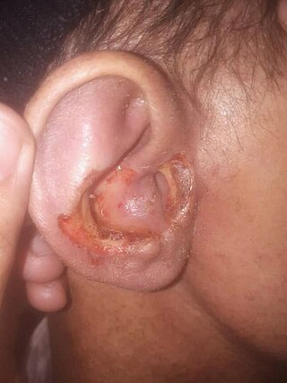
Targetoid hemosiderotic hemangioma, also known as a hobnail hemangioma is a skin condition characterized by a central brown or purplish papule that is surrounded by an ecchymotic halo. It may appear similar to melanoma. It was first described by Santa Cruz and Aronberg in 1988.
Granulosis rubra nasi is a rare familial disease of children, occurring on the nose, cheeks, and chin, characterized by diffuse redness, persistent excessive sweating, and small dark red papules that disappear on diascopic pressure.
Disseminate and recurrent infundibulofolliculitis, also called disseminate and recurrent infundibular folliculitis or Hitch and Lund disease, is a rare follicular skin condition that presents with irregularly shaped papules pierced by hair, is mildly itchy at times, and is chronic with recurrent exacerbations.
Traumatic anserine folliculosis is a curious gooseflesh-like follicular hyperkeratosis that may result from persistent pressure and lateral friction of one skin surface against another. Traumatic anserine folliculosis is caused by trauma. Topical keratolytics are the treatment of choice.
Plica neuropathica, also known as felted hair, is a curling, looping, intertwisting, and felting or matting of the hair in localized areas of the scalp.
Trigeminal trophic syndrome is a rare disease caused by the interruption of peripheral or central sensory pathways of the trigeminal nerve. A slowly enlarging, uninflamed ulcer can occur in the area that has had trigeminal nerve damage; including but not limited to the cheek beside the ala nasi.
Annular elastolytic giant-cell granuloma is a cutaneous condition characterized histologically by a dermal infiltrate of macrophages.
Benign cephalic histiocytosis(BCH) is a non-Langerhan's histiocytosis that is uncommon and self-limiting, usually beginning towards the end of the first year of life. Gianotti et al. originally described it in 1971. Initially affecting the head and neck, this condition is characterized by several small eruptions of yellow to reddish-brown papules that heal on their own. Histological investigations have demonstrated that this disorder is associated with dermal proliferation of histiocytes, characterized by intracytoplasmic comma-shaped bodies, covered vesicles, and desmosome-like structure.
Generalized eruptive histiocytoma is a rare cutaneous condition characterized by widespread, erythematous, essentially symmetrical papules, particularly involving the trunk and proximal extremities.
Progressive nodular histiocytosis is a cutaneous condition clinically characterized by the development of two types of skin lesions: superficial papules and deeper larger subcutaneous nodules. Progressive nodular histiocytosis was first reported in 1978 by Taunton et al. It is a subclass of non-Langerhans cell histiocytosis and a subgroup of xanthogranuloma.
An eccrine nevus is an extremely rare cutaneous condition that, histologically, is characterized by an increase in size or number of eccrine secretory coils. Hyperhidrosis is the most common symptom. It can present as discoloured nodules, papules, or plaques. Eccrine nevus mostly affects the extremities. Eccrine nevus are diagnosed based of histology. Treatment includes surgical excision or topical medications.
Pseudoepitheliomatous keratotic and micaceous balanitis, (PKMB) is a cutaneous condition characterized by skin lesions on the glans penis that are wart-like with scaling. It can present as a cutaneous horn. PKMB is usually asymptomatic, with occasional irritation, burning sensation, fissuring, or maceration.

Cylindroma is a rare, slow-growing, benign tumour of the skin. It mostly affects the face, scalp, and neck regions.
Nevoid hypertrichosis is a cutaneous condition characterized by the growth of terminal hairs in a circumscribed area. Nevoid hypertrichosis often presents shortly after birth. The cause of nevoid hypertrichosis is unknown. The diagnosis is made based of clinical and histopathological examination.
References
- ↑ Rapini, Ronald P.; Bolognia, Jean L.; Jorizzo, Joseph L. (2007). Dermatology: 2-Volume Set. St. Louis: Mosby. ISBN 978-1-4160-2999-1.
- 1 2 Masferrer E, Vicente MA, Bassas-Vila J, Rovira C, González-Enseñat MA (Jul 2010). "Porokeratotic eccrine ostial and dermal duct naevus: report of 10 cases". J Eur Acad Dermatol Venereol. 24 (7): 847–51. doi:10.1111/j.1468-3083.2009.03498.x. PMID 19925595. S2CID 205589524.
- 1 2 Bandyopadhyay, Debabrata; Saha, Abanti; Das, Dipti; Das, Anupam (2015). "Porokeratotic eccrine ostial and dermal duct nevus". Indian Dermatology Online Journal. 6 (2). Medknow: 117–119. doi: 10.4103/2229-5178.153016 . ISSN 2229-5178. PMC 4375756 . PMID 25821735.
- ↑ Cambiaghi, Stefano; Gianotti, Raffaele; Caputo, Ruggero (2007). "Widespread Porokeratotic Eccrine Ostial and Dermal Duct Nevus Along Blaschko Lines". Pediatric Dermatology. 24 (2). Wiley: 162–167. doi:10.1111/j.1525-1470.2007.00367.x. ISSN 0736-8046. PMID 17461816.
- ↑ Zade, John; Jfri, Abdulhadi; Nabatian, Adam; Alajaji, Abdullah; Geller, Lauren; Khorasani, Hooman (2017). "Porokeratotic eccrine ostial and dermal duct nevus: a unique case treated with CO 2 laser". Clinical Case Reports. 5 (5): 675–678. doi:10.1002/ccr3.846. ISSN 2050-0904. PMC 5412763 . PMID 28469874.
- 1 2 Naraghi, Mona Masoumeh; Nikoo, Azita; Goodarzi, Azadeh (2013). "Porokeratotic Eccrine Ostial and Dermal Duct Nevus". Case Reports in Dermatological Medicine. 2013. Hindawi Limited: 1–3. doi: 10.1155/2013/953840 . ISSN 2090-6463. PMC 3834619 . PMID 24307955.
- ↑ Wong, Jillian W.; Summers, Erika M.; Taylor, Mark B.; Harris, Ronald M. (2011-09-15). "Porokeratotic eccrine ostial and dermal duct nevus treated with a combination erbium/CO2 laser: a case and brief review". Dermatology Online Journal. 17 (9): 10. doi:10.5070/D36550J2TT. ISSN 1087-2108. PMID 21971275.
- ↑ Easton, Jennifer A.; Donnelly, Steven; Kamps, Miriam A.F.; Steijlen, Peter M.; Martin, Patricia E.; Tadini, Gianluca; Janssens, René; Happle, Rudolf; van Geel, Michel; van Steensel, Maurice A.M. (2012). "Porokeratotic Eccrine Nevus May Be Caused by Somatic Connexin26 Mutations". Journal of Investigative Dermatology. 132 (9). Elsevier BV: 2184–2191. doi:10.1038/jid.2012.143. ISSN 0022-202X. PMC 3422696 . PMID 22592158.
- ↑ Pathak, Deeptara; Kubba, Raj; Kubba, Asha (2011). "Porokeratotic eccrine ostial and dermal duct nevus". Indian Journal of Dermatology, Venereology and Leprology. 77 (2). Medknow: 174–176. doi: 10.4103/0378-6323.77457 . ISSN 0378-6323. PMID 21393947.
- ↑ Kumar, Piyush; Mondal, Avijit; Das, Anupam; Debbarman, Panchami; Mandal, RajeshKumar (2015). "Porokeratotic eccrine ostial and dermal duct nevus: A noteworthy presentation". Indian Dermatology Online Journal. 6 (2). Medknow: 130–131. doi: 10.4103/2229-5178.153021 . ISSN 2229-5178. PMC 4375761 . PMID 25821740.
- ↑ Goddard, Deborah S.; Rogers, Maureen; Frieden, Ilona J.; Krol, Alfons L.; White, Clifton R.; Jayaraman, Anu G.; Robinson-Bostom, Leslie; Bruckner, Anna L.; Ruben, Beth S. (2009). "Widespread porokeratotic adnexal ostial nevus: Clinical features and proposal of a new name unifying porokeratotic eccrine ostial and dermal duct nevus and porokeratotic eccrine and hair follicle nevus". Journal of the American Academy of Dermatology. 61 (6). Elsevier BV: 1060.e1–1060.e14. doi:10.1016/j.jaad.2009.03.036. ISSN 0190-9622. PMID 19664847.