Rhododendrol

Last updated
Rhododendrol
Rhododendrol.svg
Names
IUPAC name
4-[(3R)-3-hydroxybutyl]phenol
Other names
Rhododenol, RD, 4-(4-hydroxyphenyl)-2-butanol, (-)-Betuligenol, (R)-Frambinol, 4-Hydroxy-α-methyl-benzenepropanol
Identifiers
3D model (JSmol)
ChEMBL
ChemSpider
ECHA InfoCard 100.237.232 OOjs UI icon edit-ltr-progressive.svg
EC Number
  • 809-359-5
PubChem CID
UNII
  • Key: SFUCGABQOMYVJW-MRVPVSSYSA-N
  • InChI=1S/C10H14O2/c1-8(11)2-3-9-4-6-10(12)7-5-9/h4-8,11-12H,2-3H2,1H3/t8-/m1/s1
  • CC(CCC1=CC=C(C=C1)O)O
Properties
C10H14O2
Molar mass 166.22 g/mol
AppearanceWhite solid powder
Density 1.1±0.1 g/cm3
Melting point 68-71 °C
Boiling point 315.4±17.0 °C at 760 mmHg
Hazards
Occupational safety and health (OHS/OSH):
Main hazards
cytotoxicity
GHS labelling:
GHS-pictogram-exclam.svg
Warning
H302, H319
P270, P280, P301+P312, P305+P351+P338, P330, P337+P313, P501
Flash point 153.4±15.5 °C
Except where otherwise noted, data are given for materials in their standard state (at 25 °C [77 °F], 100 kPa).

Rhododendrol (RD) also called 4-[(3R)-3-hydroxybutyl]phenol (systemic name), is an organic compound with the formula C10H14O2. It is a naturally occurring ingredient present in many plants, such as the Rhododendron. [1] The phenolic compound was first developed in 2010 as a tyrosinase inhibitor for skin-lightening cosmetics. In 2013, after rhododendrol reportedly caused skin depigmentation in consumers using RD-containing skin-brightening cosmetics, the cosmetics were withdrawn from the market. The skin condition, caused by RD, is called RD-induced leukoderma. Rhododendrol exerts melanocyte cytotoxicity via a tyrosinase-dependent mechanism. It has been shown to impair the normal proliferation of melanocytes through reactive oxygen species-dependent activation of GADD45. [2] It is now well established that rhododendrol is a potent tyrosinase inhibitor. [3] [4]

Contents

Structure and synthesis

Structure

Rhododendrol occurs as the glucoside rhododendrin in leaves of the Rhododendron (Ericacae), and it naturally occurs as a phenolic compound in plants such as Acer nikoense , Betula platyphylla, and the Chinese red birch Betula Alba. The compound can be obtained from alkylation of phenols (C6H5OH). The molecule has a para-substituted structure, and one chiral center. Also, the compound has a natural charge.

Biosynthesis

There are several ways to synthesise rhododendrol. First, the synthesis can be achieved in six steps from benzaldehyde. The key reactions in this method include aldol condensation and trichloroacetimidate glycosylation. [5] The compound can also be prepared by reducing raspberry ketone (4-(4-hydroxyphenyl)-2- butanone) with Raney nickel in EtOH. [6] In addition, Rhododendrol can be synthesised from p-coumaric acid. This pathway involves reduction of the aliphatic double bond present in p-coumaric acid.

Mechanisms of action

The mechanism of action of rhododendrol has been investigated in multiple studies which revealed that RD competes with tyrosine for hydroxylation by tyrosinase and interferes with melanin synthesis. [7] [8] [9] First, RD is catalysed by tyrosinase to produce toxic metabolites as RD-cyclic catechol. These reactive metabolites cause damage to the melanocytes. There is still uncertainty, however, how the metabolites result in melanocyte damage.

A previous report reported that the melanocyte toxicity of rhododendrol is caused by the production of cytotoxic reactive oxygen species (ROS). [2] However, another study stated that there was no ROS detected in the rhododendrol-treated melanocytes, but a tyrosinase-dependent accumulation of endoplasmic reticulum stress and activation of the apoptotic pathway. [10] [9] Even though there is still no full agreement on the exact mechanism of action, it is suggested that the mechanism of RD-induced leukoderma closely resembles the mechanism displayed in the figure below (Suggested mechanism of Rhododendrol.png).

Suggested mechanism of Rhododendrol Suggested mechanism of Rhododendrol.png
Suggested mechanism of Rhododendrol

In some individuals, a T-cell response is observed. The melanocyte cell lysates may sensitise T-cells, and the immunised cytotoxic T-lymphocytes (specific to Melan A, which is a melanocytic differentiation marker) may enhance the RD-induced leukoderma or evoke vitiligo-like lesions on the non-applied skin. [7]

Metabolism

Rhododendrol is metabolised via tyrosinase-catalysed oxidation. Therefore, the enzyme tyrosinase is necessary for the oxidation of rhododendrol. Tyrosinase regularly plays an essential role in the production of melanocytes called the melanogenesis. After oxidation of rhododendrol by the tyrosinase enzyme, several kinds of phenols and catechols are formed. These phenols and catechols together form ortho-quinones (o-quinones). [2] Presence of o-quinones can lead to cytotoxicity via the production of reactive oxygen species (ROS) or by the binding to enzymes or DNA. [3]

When rhododendrol is metabolised via the tyrosinase-catalysed oxidation RD-quinone will be formed. [1] This formation gives rise to the formation of secondary quinones. As described in the mechanisms of action, the presence of quinones could cause cytotoxicity to melanocytes by the production of ROS or by binding to DNA and enzymes.

Adverse effects

Considering the use of rhododendrol is prohibited since 2013, the knowledge about the side effects rhodendodrol causes is limited. As stated above, the main known adverse effect of rhododendrol is melanocyte toxicity. [11] Melanocytes are melanin-producing cells, primarily responsible for skin colour. Melanocyte toxicity induces apoptosis of the cell, causing the melanocytes to die. This is due to an increased expression of caspase-3 and caspase-8. [1] Caspase proteins are crucial mediators of apoptosis, with caspase-3 and caspase-8 being death proteases. [12] Considering melanocytes are responsible for skin colour, apoptosis of these cells causes the colour of the skin to vanish. [13] This disease caused by rhododendrol is called leukoderma. Leukoderma, also known as vitiligo, is a skin disease characterized by patches of the skin losing their pigment. This rhododendrol-induced depigmentation can be either long-term and short term. In most cases, repigmentation and cessation of further depigmentation occur after discontinuing the exposure to the substance. However, some patients develop vitiligo vulgaris through the spread of depigmentation into non-exposed areas. This only occurs after severe chemical damage. [14] In addition, rhododendrol not only causes melanocytes to go into apoptosis but it also inhibits melanogenesis. Meaning that the use of rhododendrol not only causes melanocytes to die, but also prevents the development of new melanocytes. [1]

Toxicity

Various studies have shown that there is more than one mechanism by which rhododendrol can have a toxic effect. This toxic effect of rhododendrol is found in the melanocytes, which gives rise to skin depigmentation.

ROS

Rhododendrol can have a toxic effect via the production of reactive oxygen species (ROS). This will cause an impairment in the further development of melanocytes in the skin. Impairment is caused by upregulation of the GADD45 gene. A study of Kim et al. showed that the production of ROS, which gives rise to more production of GADD45, is already found at low concentrations of rhododendrol. At the time that rhododendrol was used in cosmetic products, it contained concentrations of 2%. The study of Kim et al. suggests that the production of reactive oxygen species at low concentrations may have contributed to the development of leukoderma in users of these cosmetic products. [15]

Reactive metabolites

The study of Ito et al. showed that rhododendrol exerts its toxic effect in the melanocytes via tyrosinase-dependent mechanisms. This tyrosinase enzyme breaks rhododendrol down into the following reactive metabolites: RD-quinone and RD-cyclic quinone. [16] These reactive metabolites can bind to proteins which contain a thiol-group [17] or it can form radicals. These radicals are toxic to the melanocytes as it causes auto-oxidation of the cells. [16] Auto-oxidation, in turn, causes oxidative stress to cells, which will impair the natural growth and function of the melanocytes.

Rhododenol and raspberry ketone impair the regular proliferation of melanocytes through reactive oxygen species-dependent activation of GADD45. [15]

Effects on animals

The effect of rhododendrol (4-(4-hydroxyphenyl)-2-butanol) is measured in mice as well as in guinea pigs. [18] [19] These studies were performed to elucidate the aetiology of RD-induced leukoderma. The data of these studies revealed that the amount of RD applied to the skin is highly relevant considering that high doses of RD are required in order to cause cytotoxicity. This finding is contrary to the results presented in the study of Kim et al., which is performed in humans. Furthermore, the animal studies enlightened the importance of the ER-stress response. It is suggested that the activity of the ER-stress response may determine whether melanocytes survive or die. Also, the study of Abe et al. revealed that the autophagy pathway may be involved in the resistance to the cytotoxicity of RD. [18]

Since the biochemical and histological characteristics of the used mice in the animal studies (hairless hk14-SCF Tg mice) closely resembled the characteristics of the human skin, these newly generated mice could be used as experimental animal models to investigate chemical vitiligo further.

Related Research Articles

<span class="mw-page-title-main">Melanin</span> Group of natural pigments found in most organisms

Melanin is a broad term for a group of natural pigments found in most organisms. The melanin pigments are produced in a specialized group of cells known as melanocytes.

<span class="mw-page-title-main">Melanocyte</span> Melanin-producing cells of the skin

Melanocytes are melanin-producing neural crest-derived cells located in the bottom layer of the skin's epidermis, the middle layer of the eye, the inner ear, vaginal epithelium, meninges, bones, and heart. Melanin is a dark pigment primarily responsible for skin color. Once synthesized, melanin is contained in special organelles called melanosomes which can be transported to nearby keratinocytes to induce pigmentation. Thus darker skin tones have more melanosomes present than lighter skin tones. Functionally, melanin serves as protection against UV radiation. Melanocytes also have a role in the immune system.

Neurotoxicity is a form of toxicity in which a biological, chemical, or physical agent produces an adverse effect on the structure or function of the central and/or peripheral nervous system. It occurs when exposure to a substance – specifically, a neurotoxin or neurotoxicant– alters the normal activity of the nervous system in such a way as to cause permanent or reversible damage to nervous tissue. This can eventually disrupt or even kill neurons, which are cells that transmit and process signals in the brain and other parts of the nervous system. Neurotoxicity can result from organ transplants, radiation treatment, certain drug therapies, recreational drug use, exposure to heavy metals, bites from certain species of venomous snakes, pesticides, certain industrial cleaning solvents, fuels and certain naturally occurring substances. Symptoms may appear immediately after exposure or be delayed. They may include limb weakness or numbness, loss of memory, vision, and/or intellect, uncontrollable obsessive and/or compulsive behaviors, delusions, headache, cognitive and behavioral problems and sexual dysfunction. Chronic mold exposure in homes can lead to neurotoxicity which may not appear for months to years of exposure. All symptoms listed above are consistent with mold mycotoxin accumulation.

<span class="mw-page-title-main">Hypopigmentation</span> Area of skin becoming lighter than the baseline skin color

Hypopigmentation is characterized specifically as an area of skin becoming lighter than the baseline skin color, but not completely devoid of pigment. This is not to be confused with depigmentation, which is characterized as the absence of all pigment. It is caused by melanocyte or melanin depletion, or a decrease in the amino acid tyrosine, which is used by melanocytes to make melanin. Some common genetic causes include mutations in the tyrosinase gene or OCA2 gene. As melanin pigments tend to be in the skin, eye, and hair, these are the commonly affected areas in those with hypopigmentation.

<span class="mw-page-title-main">Melasma</span> Medical condition

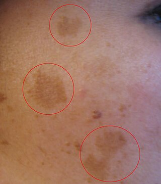
Melasma is a tan or dark skin discoloration. Melasma is thought to be caused by sun exposure, genetic predisposition, hormone changes, and skin irritation. Although it can affect anyone, it is particularly common in women, especially pregnant women and those who are taking oral or patch contraceptives or hormone replacement therapy medications.

<span class="mw-page-title-main">Tyrosinase</span> Enzyme for controlling the production of melanin

Tyrosinase is an oxidase that is the rate-limiting enzyme for controlling the production of melanin. The enzyme is mainly involved in two distinct reactions of melanin synthesis otherwise known as the Raper Mason pathway. Firstly, the hydroxylation of a monophenol and secondly, the conversion of an o-diphenol to the corresponding o-quinone. o-Quinone undergoes several reactions to eventually form melanin. Tyrosinase is a copper-containing enzyme present in plant and animal tissues that catalyzes the production of melanin and other pigments from tyrosine by oxidation. It is found inside melanosomes which are synthesized in the skin melanocytes. In humans, the tyrosinase enzyme is encoded by the TYR gene.

<span class="mw-page-title-main">Skin whitening</span> Practice of using chemical substances to lighten the skin

Skin whitening, also known as skin lightening and skin bleaching, is the practice of using chemical substances in an attempt to lighten the skin or provide an even skin color by reducing the melanin concentration in the skin. Several chemicals have been shown to be effective in skin whitening, while some have proven to be toxic or have questionable safety profiles. This includes mercury compounds which may cause neurological problems and kidney problems.

<span class="mw-page-title-main">Citrinin</span> Chemical compound

Citrinin is a mycotoxin which is often found in food. It is a secondary metabolite produced by fungi that contaminates long-stored food and it causes different toxic effects, like nephrotoxic, hepatotoxic and cytotoxic effects. Citrinin is mainly found in stored grains, but sometimes also in fruits and other plant products.

<span class="mw-page-title-main">Betulinic acid</span> Chemical compound

Betulinic acid is a naturally occurring pentacyclic triterpenoid which has antiretroviral, antimalarial, and anti-inflammatory properties, as well as a more recently discovered potential as an anticancer agent, by inhibition of topoisomerase. It is found in the bark of several species of plants, principally the white birch from which it gets its name, but also the ber tree, selfheal, the tropical carnivorous plants Triphyophyllum peltatum and Ancistrocladus heyneanus, Diospyros leucomelas, a member of the persimmon family, Tetracera boiviniana, the jambul, flowering quince, rosemary, and Pulsatilla chinensis.

Polyphenol oxidase, an enzyme involved in fruit browning, is a tetramer that contains four atoms of copper per molecule.

<i>o</i>-Toluidine Aryl amine

o-Toluidine (ortho-toluidine) is an organic compound with the chemical formula CH3C6H4NH2. It is the most important of the three isomeric toluidines. It is a colorless liquid although commercial samples are often yellowish. It is a precursor to the herbicides metolachlor and acetochlor.

<span class="mw-page-title-main">Cephaloridine</span> Chemical compound

Cephaloridine is a first-generation semisynthetic derivative of antibiotic cephalosporin C. It is a Beta lactam antibiotic, like penicillin. Its chemical structure contains 3 cephems, 4 carboxyl groups and three pyridinium methyl groups.

<span class="mw-page-title-main">Protocatechuic acid</span> Chemical compound

Protocatechuic acid (PCA) is a dihydroxybenzoic acid, a type of phenolic acid. It is a major metabolite of antioxidant polyphenols found in green tea. It has mixed effects on normal and cancer cells in in vitro and in vivo studies.

<span class="mw-page-title-main">Oxidopamine</span> Chemical compound

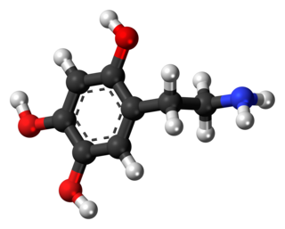
Oxidopamine, also known as 6-hydroxydopamine (6-OHDA) or 2,4,5-trihydroxyphenethylamine, is a neurotoxic synthetic organic compound used by researchers to selectively destroy dopaminergic and noradrenergic neurons in the brain.

<span class="mw-page-title-main">Dinoseb</span> Chemical compound used as a herbicide

Dinoseb is a common industry name for 6-sec-butyl-2,4-dinitrophenol, a herbicide in the dinitrophenol family. It is a crystalline orange solid which does not readily dissolve in water. Dinoseb is banned as an herbicide in the European Union (EU) and the United States because of its toxicity.

<span class="mw-page-title-main">Acute generalized exanthematous pustulosis</span> Medical condition

Acute generalized exanthematous pustulosis (AGEP) is a rare skin reaction that in 90% of cases is related to medication.

Arsenic biochemistry refers to biochemical processes that can use arsenic or its compounds, such as arsenate. Arsenic is a moderately abundant element in Earth's crust, and although many arsenic compounds are often considered highly toxic to most life, a wide variety of organoarsenic compounds are produced biologically and various organic and inorganic arsenic compounds are metabolized by numerous organisms. This pattern is general for other related elements, including selenium, which can exhibit both beneficial and deleterious effects. Arsenic biochemistry has become topical since many toxic arsenic compounds are found in some aquifers, potentially affecting many millions of people via biochemical processes.

<span class="mw-page-title-main">HU-331</span> Chemical compound

HU-331 is a quinone anticarcinogenic drug synthesized from cannabidiol, a cannabinoid in the Cannabis sativa plant. It showed a great efficacy against oncogenic human cells. HU-331 does not cause arrest in cell cycle, cell apoptosis or caspase activation. HU-331 inhibits DNA topoisomerase II even at nanomolar concentrations, but has shown a negligible effect on the action of DNA topoisomerase I. The cannabinoid quinone HU-331 is a very specific inhibitor of topoisomerase II, compared with most known anticancer quinones. One of the main objectives of these studies is the development of a new quinone derived compound that produces anti-neoplastic activity while maintaining low toxicity at therapeutic doses.

<span class="mw-page-title-main">Glyceryl octyl ascorbic acid</span> Chemical compound

Glyceryl octyl ascorbic acid (GO-VC) is an amphipathic derivative of vitamin C consisting of two ether linkages: a 1-octyl at position 2 and a glycerin at position 3. The chemical name is 2-glyceryl-3-octyl ascorbic acid. The isomer in which these two groups are swapped is also known.

The p-i concept refers to the pharmacological interaction of drugs with immune receptors. It explains a form of drug hypersensitivity, namely T cell stimulation, which can lead to various acute inflammatory manifestations such as exanthems, eosinophilia and systemic symptoms, Stevens–Johnson syndrome, toxic epidermal nercrolysis, and complications upon withdrawing the drug.

References

  1. 1 2 3 4 KUBO, MASAYOSHI; INOUE, TAKAO; NAGAI, MASAHIRO (1980). "Studies on the constituents of aceraceae plants. III. Structure of acerogenin B from Acer nikoense Maxim". Chemical & Pharmaceutical Bulletin. 28 (4): 1300–1303. doi: 10.1248/cpb.28.1300 . ISSN   0009-2363.
  2. 1 2 3 Ito, Shosuke; Ojika, Makoto; Yamashita, Toshiharu; Wakamatsu, Kazumasa (2014-06-27). "Tyrosinase-catalyzed oxidation of rhododendrol produces 2-methylchromane-6,7-dione, the putative ultimate toxic metabolite: implications for melanocyte toxicity". Pigment Cell & Melanoma Research. 27 (5): 744–753. doi: 10.1111/pcmr.12275 . ISSN   1755-1471. PMID   24903082. S2CID   31988006.
  3. 1 2 Gabe, Yu; Miyaji, Akimitsu; Kohno, Masahiro; Hachiya, Akira; Moriwaki, Shigeru; Baba, Toshihide (September 2018). "Substantial evidence for the rhododendrol-induced generation of hydroxyl radicals that causes melanocyte cytotoxicity and induces chemical leukoderma". Journal of Dermatological Science. 91 (3): 311–316. doi: 10.1016/j.jdermsci.2018.06.007 . ISSN   0923-1811. PMID   30005897.
  4. Ichiro Katayama, Lingli Yang (2015). "4-(4-Hydroroxyphenyl)-2-butanol (rhododendrol) activates the autophagy-lysosome pathway in melanocytes: Insights into the mechanisms of rhododendrol-induced leukoderma". Journal of Dermatological Science. 77 (3): 182–185. doi:10.1016/j.jdermsci.2015.01.006. ISSN   0923-1811. PMID   25680854.
  5. Iwadate, Takehiro; Kashiwakura, Yutaka; Masuoka, Noriyoshi; Yamada, Yoichi; Nihei, Ken-ichi (January 2014). "Chemical synthesis and tyrosinase inhibitory activity of rhododendrol glycosides". Bioorganic & Medicinal Chemistry Letters. 24 (1): 122–125. doi: 10.1016/j.bmcl.2013.11.063 . ISSN   0960-894X. PMID   24332496.
  6. Carruthers, W. (1978). Some modern methods of or organic synthesis. University Press. OCLC   969539863.
  7. 1 2 Tokura, Yoshiki; Fujiyama, Toshiharu; Ikeya, Shigeki; Tatsuno, Kazuki; Aoshima, Masahiro; Kasuya, Akira; Ito, Taisuke (March 2015). "Biochemical, cytological, and immunological mechanisms of rhododendrol-induced leukoderma". Journal of Dermatological Science. 77 (3): 146–149. doi:10.1016/j.jdermsci.2015.02.001. ISSN   0923-1811. PMID   25726326.
  8. Kasamatsu, Shinya; Hachiya, Akira; Nakamura, Shun; Yasuda, Yuka; Fujimori, Taketoshi; Takano, Kei; Moriwaki, Shigeru; Hase, Tadashi; Suzuki, Tamio (October 2014). "Depigmentation caused by application of the active brightening material, rhododendrol, is related to tyrosinase activity at a certain threshold". Journal of Dermatological Science. 76 (1): 16–24. doi:10.1016/j.jdermsci.2014.07.001. ISSN   0923-1811. PMID   25082450.
  9. 1 2 Sasaki, Minoru; Kondo, Masatoshi; Sato, Kohji; Umeda, Mai; Kawabata, Keigo; Takahashi, Yoshito; Suzuki, Tamio; Matsunaga, Kayoko; Inoue, Shintaro (2014-06-26). "Rhododendrol, a depigmentation-inducing phenolic compound, exerts melanocyte cytotoxicity via a tyrosinase-dependent mechanism". Pigment Cell & Melanoma Research. 27 (5): 754–763. doi: 10.1111/pcmr.12269 . ISSN   1755-1471. PMID   24890809.
  10. Yang, Lingli; Yang, Fei; Wataya-Kaneda, Mari; Tanemura, Atsuhi; Tsuruta, Daisuke; Katayama, Ichiro (March 2015). "4-(4-Hydroroxyphenyl)-2-butanol (rhododendrol) activates the autophagy-lysosome pathway in melanocytes: Insights into the mechanisms of rhododendrol-induced leukoderma". Journal of Dermatological Science. 77 (3): 182–185. doi:10.1016/j.jdermsci.2015.01.006. ISSN   0923-1811. PMID   25680854.
  11. Lee, Chang Seok; Joo, Yung Hyup; Baek, Heung Soo; Park, Miyoung; Kim, Jeong-Hwan; Shin, Hong-Ju; Park, Nok-Hyun; Lee, John Hwan; Park, Young-Ho (2016). "Different effects of five depigmentary compounds, rhododendrol, raspberry ketone, monobenzone, rucinol and AP736 on melanogenesis and viability of human epidermal melanocytes". Experimental Dermatology. 25 (1): 44–49. doi:10.1111/exd.12871. ISSN   1600-0625. PMID   26440747. S2CID   30626521.
  12. RU, Porter AG and Jänicke (1999). "Emerging roles of caspase-3 in apoptosis. - PubMed - NCBI". Cell Death and Differentiation. 6 (2): 99–104. doi: 10.1038/sj.cdd.4400476 . PMID   10200555.
  13. Ito, Shosuke; Ojika, Makoto; Yamashita, Toshiharu; Wakamatsu, Kazumasa (2014). "Tyrosinase-catalyzed oxidation of rhododendrol produces 2-methylchromane-6,7-dione, the putative ultimate toxic metabolite: implications for melanocyte toxicity". Pigment Cell & Melanoma Research. 27 (5): 744–753. doi: 10.1111/pcmr.12275 . ISSN   1755-148X. PMID   24903082. S2CID   31988006.
  14. Yoshikawa, Momoko; Sumikawa, Yasuyuki; Hida, Tokimasa; Kamiya, Takafumi; Kase, Kimi; Ishii-Osai, Yasue; Kato, Junji; Kan, Yuji; Kamiya, Shiori (2016-11-24). "Clinical and epidemiological analysis in 149 cases of rhododendrol-induced leukoderma". The Journal of Dermatology. 44 (5): 582–587. doi: 10.1111/1346-8138.13694 . ISSN   0385-2407. PMID   27882588.
  15. 1 2 Kim, Minjeong; Baek, Heung Soo; Lee, Miri; Park, Hyeonji; Shin, Song Seok; Choi, Dal Woong; Lim, Kyung-Min (2016-04-01). "Rhododenol and raspberry ketone impair the normal proliferation of melanocytes through reactive oxygen species-dependent activation of GADD45". Toxicology in Vitro. 32: 339–346. doi:10.1016/j.tiv.2016.02.003. ISSN   0887-2333. PMID   26867644.
  16. 1 2 Ito, Shosuke; Ojika, Makoto; Yamashita, Toshiharu; Wakamatsu, Kazumasa (2014). "Tyrosinase-catalyzed oxidation of rhododendrol produces 2-methylchromane-6,7-dione, the putative ultimate toxic metabolite: implications for melanocyte toxicity". Pigment Cell & Melanoma Research. 27 (5): 744–753. doi: 10.1111/pcmr.12275 . ISSN   1755-148X. PMID   24903082. S2CID   31988006.
  17. Ito, Shosuke; Okura, Masae; Nakanishi, Yukiko; Ojika, Makoto; Wakamatsu, Kazumasa; Yamashita, Toshiharu (2015). "Tyrosinase-catalyzed metabolism of rhododendrol (RD) in B16 melanoma cells: production of RD-pheomelanin and covalent binding with thiol proteins". Pigment Cell & Melanoma Research. 28 (3): 295–306. doi:10.1111/pcmr.12363. ISSN   1755-148X. PMID   25713930. S2CID   22821661.
  18. 1 2 Abe, Yuko; Okamura, Ken; Kawaguchi, Masakazu; Hozumi, Yutaka; Aoki, Hitomi; Kunisada, Takahiro; Ito, Shosuke; Wakamatsu, Kazumasa; Matsunaga, Kayoko (January 2016). "Rhododenol-induced leukoderma in a mouse model mimicking Japanese skin". Journal of Dermatological Science. 81 (1): 35–43. doi:10.1016/j.jdermsci.2015.10.011. ISSN   0923-1811. PMID   26547111.
  19. Kuroda, Yasutaka; Takahashi, Yutaka; Sakaguchi, Hitoshi; Matsunaga, Kayoko; Suzuki, Tamio (2014). "Depigmentation of the skin induced by 4-(4-hydroxyphenyl)-2-butanol is spontaneously re-pigmented in brown and black guinea pigs". The Journal of Toxicological Sciences. 39 (4): 615–623. doi: 10.2131/jts.39.615 . ISSN   0388-1350. PMID   25056786.

Category:Chemical compounds