Related Research Articles

Veins are blood vessels in the circulatory system of humans and most other animals that carry blood toward the heart. Most veins carry deoxygenated blood from the tissues back to the heart; exceptions are those of the pulmonary and fetal circulations which carry oxygenated blood to the heart. In the systemic circulation arteries carry oxygenated blood away from the heart, and veins return deoxygenated blood to the heart, in the deep veins.

The atrium is one of the two upper chambers in the heart that receives blood from the circulatory system. The blood in the atria is pumped into the heart ventricles through the atrioventricular mitral and tricuspid heart valves.

The coronary sinus is the largest vein of the heart. It drains over half of the deoxygenated blood from the heart muscle into the right atrium. It begins on the backside of the heart, in between the left atrium, and left ventricle; it begins at the junction of the great cardiac vein, and oblique vein of the left atrium. It receives multiple tributaries. It passes across the backside of the heart along a groove between left atrium and left ventricle, then drains into the right atrium at the orifice of the coronary sinus.

The occipital sinus is the smallest of the dural venous sinuses. It is usually unpaired, and is sometimes altogether absent. It is situated in the attached margin of the falx cerebelli. It commences near the foramen magnum, and ends by draining into the confluence of sinuses.

The great cardiac vein is a vein of the heart. It begins at the apex of the heart and ascends along the anterior interventricular sulcus before joining the oblique vein of the left atrium to form the coronary sinus upon the posterior surface of the heart.
The middle cardiac vein commences at the apex of the heart. It passes posteriorly along the inferior interventricular sulcus to end at the coronary sinus near the sinus' termination.

The anterior ethmoidal artery is a branch of the ophthalmic artery in the orbit. It exits the orbit through the anterior ethmoidal foramen alongside the anterior ethmoidal nerve. It contributes blood supply to the ethmoid sinuses, frontal sinuses, the dura mater, lateral nasal wall, and nasal septum. It issues a meningeal branch, and nasal branches.

The sinus venosus is a large quadrangular cavity which precedes the atrium on the venous side of the chordate heart.
The ethmoid bulla is a rounded elevation upon the lateral wall of the middle nasal meatus produced by one or more of the underlying middle ethmoidal air cells. It varies significantly based on the size of the underlying air cells.

The posterior superior alveolar artery is a branch of the maxillary artery. It is one of two or three superior alveolar arteries. It provides arterial suply to the molar and premolar teeth, maxillary sinus and adjacent bone, and the gingiva.
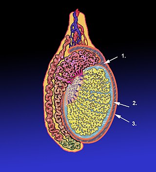
The tunica vaginalis is a pouch of serous membrane within the scrotum that lines the testis and epididymis, and the inner surface of the scrotum. It is the outermost of the three layers that constitute the capsule of the testis, with the tunica albuginea of penis situated beneath it.
The terminal sulcus is a groove on the outer surface of the right atrium of the heart marking the transition between the sinus venarum cavarum and the rest of the right atrium. The terminal sulcus corresponds to the position of the terminal crest on the inner surface of the right atium. The terminal sulcus indicate the postition of the sinoatrial node.

The crista terminalis is a vertical ridge on the posterolateral inner surface of the adult right atrium extending between the superior vena cava, and the inferior vena cava. The crista terminalis denotes where the junction of the embryologic sinus venosus and the right atrium occurred during embryonic development. It forms a boundary between the rough trabecular portion and the smooth, sinus venosus-derived portion of the internal surface of the right atrium. The sinoatrial node is located within the crista terminalis.
The tunica vasculosa testis is the inner-most of the three layers that form the capsule of the testis. It consists of a vascular plexus and loose connective tissue. It extends into the testis itself to line the surfaces of individual septa of testis.

The right marginal vein is a vein of the heart running along the inferior margin of the heart. It drains adjacent region of the right ventricle. It usually opens directly into the right atrium, but may sometimes instead empty into the anterior cardiac veins, or the coronary sinus.
The anterior cardiac veins are a variable number of small veins which drain blood from the anterior portion of the right ventricle into the right atrium.

The sinoatrial nodal artery is an artery of the heart which supplies the sinoatrial node, the natural pacemaker center of the heart. It is usually a branch of the right coronary artery. It passes between the right atrium, and the opening of the superior vena cava.

The ethmoidal infundibulum is a funnel-shaped/slit-like/curved opening/passage/space/cleft upon the anterosuperior portion of the middle nasal meatus at the hiatus semilunaris. The anterior ethmoidal air cells, and (usually) the frontonasal duct open into the ethmoidal infundibulum. The ethmoidal infundibulum extends anterosuperiorly from its opening into the nasal cavity.
The cistern of lamina terminalis is one of the a subarachnoid cisterns. It is situated rostral/anterior to the lamina terminalis and anterior commissure between the two frontal lobes of the cerebrum. It is situated rostral/anterior to the third ventricle. The cistern is an extension of interpeduncular cistern. The cistern of lamina terminalis interconnects the chiasmatic cistern and pericallosal cistern.
The heart is a muscular organ situated in the mediastinum. It consists of four chambers, four valves, two main arteries, and the conduction system. The left and right sides of the heart have different functions: the right side receives de-oxygenated blood through the superior and inferior venae cavae and pumps blood to the lungs through the pulmonary artery, and the left side receives saturated blood from the lungs.
References
- 1 2 3 "Sinus venarum". TheFreeDictionary.com. Retrieved 2023-01-05.
- 1 2 3 T. W., Sadler (2018). Langman's Medical Embryology (14th ed.). Philadelphia. p. 188. ISBN 978-1-4963-8390-7. OCLC 1042400100.
{{cite book}}: CS1 maint: location missing publisher (link) - 1 2 3 4 Morton, David A. (2019). The Big Picture: Gross Anatomy. K. Bo Foreman, Kurt H. Albertine (2nd ed.). New York. p. 56. ISBN 978-1-259-86264-9. OCLC 1044772257.
{{cite book}}: CS1 maint: location missing publisher (link) - 1 2 Waschke, Jens; Böckers, Tobias M.; Paulsen, Friedrich; Arnold, Wolfgang; Bechmann, Ingo, eds. (2018). Sobotta Anatomy Textbook: English Edition with Latin Nomenclature (1st ed.). München: Elsevier. p. 264. ISBN 978-0-7020-6760-0.