
An abscess is a collection of pus that has built up within the tissue of the body, usually caused by bacterial infection. Signs and symptoms of abscesses include redness, pain, warmth, and swelling. The swelling may feel fluid-filled when pressed. The area of redness often extends beyond the swelling. Carbuncles and boils are types of abscess that often involve hair follicles, with carbuncles being larger. A cyst is related to an abscess, but it contains a material other than pus, and a cyst has a clearly defined wall. Abscesses can also form internally on internal organs and after surgery.

Skin is the layer of usually soft, flexible outer tissue covering the body of a vertebrate animal, with three main functions: protection, regulation, and sensation.

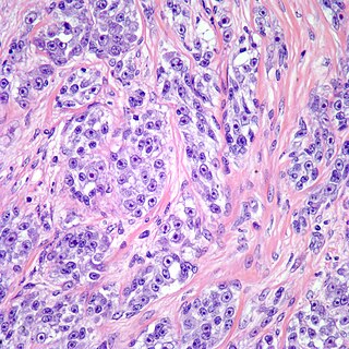
A sarcoma is a malignant tumor, a type of cancer that arises from cells of mesenchymal origin. Connective tissue is a broad term that includes bone, cartilage, muscle, fat, vascular, or other structural tissues, and sarcomas can arise in any of these types of tissues. As a result, there are many subtypes of sarcoma, which are classified based on the specific tissue and type of cell from which the tumor originates.

Dermatofibrosarcoma protuberans (DFSP) is a rare locally aggressive malignant cutaneous soft-tissue sarcoma. DFSP develops in the connective tissue cells in the middle layer of the skin (dermis). Estimates of the overall occurrence of DFSP in the United States are 0.8 to 4.5 cases per million persons per year. In the United States, DFSP accounts for between 1 and 6 percent of all soft-tissue sarcomas and 18 percent of all cutaneous soft-tissue sarcomas. In the Surveillance, Epidemiology and End Results (SEER) tumor registry from 1992 through 2004, DFSP was second only to Kaposi sarcoma.

A leiomyosarcoma (LMS) is a rare malignant (cancerous) smooth muscle tumor. The word is from leio- 'smooth' myo- 'muscle' and sarcoma 'tumor of connective tissue'. The stomach, bladder, uterus, blood vessels, and intestines are examples of hollow organs made up of smooth muscles where LMS can be located; however, the uterus and abdomen are the most common sites.

A neurofibroma is a benign nerve-sheath tumor in the peripheral nervous system. In 90% of cases, they are found as stand-alone tumors, while the remainder are found in persons with neurofibromatosis type I (NF1), an autosomal-dominant genetically inherited disease. They can result in a range of symptoms from physical disfiguration and pain to cognitive disability.

In medicine, nodules are small firm lumps, usually greater than 1 cm in diameter. If filled with fluid they are referred to as cysts. Smaller raised soft tissue bumps may be termed papules.
Malignant histiocytosis is a rare hereditary disease found in the Bernese Mountain Dog and humans, characterized by histiocytic infiltration of the lungs and lymph nodes. The liver, spleen, and central nervous system can also be affected. Histiocytes are a component of the immune system that proliferate abnormally in this disease. In addition to its importance in veterinary medicine, the condition is also important in human pathology.
A skin infection is an infection of the skin in humans and other animals, that can also affect the associated soft tissues such as loose connective tissue and mucous membranes. They comprise a category of infections termed skin and skin structure infections (SSSIs), or skin and soft tissue infections (SSTIs), and acute bacterial SSSIs (ABSSSIs). They are distinguished from dermatitis, although skin infections can result in skin inflammation.
Cutaneous asthenia is a skin disorder caused by a collagen defect. Collagen is the protein that binds the cells of the dermis together. It is also called dermatoproxy, hereditary skin fragility or cutis elastica and is found in humans, cats, dogs, mink, horses, cattle and sheep. In cattle and sheep, it is called dermatosparaxis. The skin is also abnormally fragile. The skin flaps peel or slough off very easily, often without causing bleeding. This explains why cats with the condition suddenly "molt" their wings.

Focal dermal hypoplasia is a form of ectodermal dysplasia. It is a multisystem disorder characterized primarily by skin manifestations to the atrophic and hypoplastic areas of skin which are present at birth. These defects manifest as yellow-pink bumps on the skin and pigmentation changes. The disorder is also associated with shortness of stature and some evidence suggests that it can cause epilepsy.

Juvenile hyaline fibromatosis is a very rare, autosomal recessive disease due to mutations in capillary morphogenesis protein-2. It occurs from early childhood to adulthood, and presents as slow-growing, pearly white or skin-colored dermal or subcutaneous papules or nodules on the face, scalp, and back, which may be confused clinically with neurofibromatosis. The World Health Organization in 2020 reclassified the papules and nodules that occur in juvenile hyaline fibromatosis as one of the specific benign types of tumors in the category of fibroblastic and myofibroblastic tumors.
Clear cell sarcoma is a sub-type of a rare form of cancer called a sarcoma. It is known to occur mainly in the soft tissues and dermis. Rare forms were thought to occur in the gastrointestinal tract before they were discovered to be different and redesignated as gastrointestinal neuroectodermal tumors.

Photoaging or photoageing is a term used for the characteristic changes to skin induced by chronic UVA and UVB exposure. Tretinoin is the best studied retinoid in the treatment of photoaging.

Poikiloderma vasculare atrophicans (PVA), is a cutaneous condition characterized by hypo- or hyperpigmentation, telangiectasia and skin atrophy. Other names for the condition include prereticulotic poikiloderma and atrophic parapsoriasis. The condition was first described by pioneer American pediatrician Abraham Jacobi in 1906. PVA causes areas of affected skin to appear speckled red and inflamed, yellowish and/or brown, gray or grayish-black, with scaling and a thinness that may be described as "cigarette paper". On the surface of the skin, these areas may range in size from small patches, to plaques, to neoplasms.
Histiocytic diseases in dogs are a group of diseases in dogs which may involve the skin, and which can be difficult to differentiate from granulomatous, reactive inflammatory or lymphoproliferative diseases. The clinical presentation and behaviour as well as response to therapy vary greatly among the syndromes.
Injectable filler is a special type of substance made for injections into connective tissues, such as skin, cartilage or even bone, for cosmetic or medical purposes. The most common application of injectable fillers is to change one's facial appearance, but they also are used to reduce symptoms of osteoarthritis, treat tendon or ligament injuries, support bone and gum regeneration, and for other medical applications. Injectable fillers can be in the form of hydrogel or gels made from pulverized grafts.

Congenital dermal sinus is an uncommon form of cranial or spinal dysraphism. It occurs in 1 in 2500 live births. It occurs as a dermal indentation, found along the midline of the neuraxis and often presents alongside infection and neurological deficit. Congenital dermal sinus form due to a focal failure of dysjunction between the cutaneous ectoderm and neuroectoderm during the third to eight week of gestation. Typically observed in the lumbar and lumbosacral region, congenital dermal sinus can occur from the nasion and occiput region down.
The Walleye dermal sarcoma virus (WDSV) is a retrovirus that infects walleye often causing oncogenesis. WDSV is an exogenous retrovirus belonging to the subfamily Orthoretrovirinae. This virus is related to the walleye epidermal hyperplasia viruses type 1 and type 2, all belonging to the epsilonretrovirus genus based on similarities of the gene coding for the reverse transcriptase conserved in retroviruses.
