Somatosensory evoked potential (SEP or SSEP) is the electrical activity of the brain that results from the stimulation of touch. SEP tests measure that activity and are a useful, noninvasive means of assessing somatosensory system functioning. By combining SEP recordings at different levels of the somatosensory pathways, it is possible to assess the transmission of the afferent volley from the periphery up to the cortex. SEP components include a series of positive and negative deflections that can be elicited by virtually any sensory stimuli. For example, SEPs can be obtained in response to a brief mechanical impact on the fingertip or to air puffs. [1] However, SEPs are most commonly elicited by bipolar transcutaneous electrical stimulation applied on the skin over the trajectory of peripheral nerves of the upper limb (e.g., the median nerve) or lower limb (e.g., the posterior tibial nerve), and then recorded from the scalp. [2] In general, somatosensory stimuli evoke early cortical components (N25, P60, N80), generated in the contralateral primary somatosensory cortex (S1), related to the processing of the physical stimulus attributes. About 100 ms after stimulus application, additional cortical regions are activated, such as the secondary somatosensory cortex (S2), and the posterior parietal and frontal cortices, marked by a parietal P100 and bilateral frontal N140. SEPs are routinely used in neurology today to confirm and localize sensory abnormalities, to identify silent lesions and to monitor changes during surgical procedures. [3]
The modern history of SEPs began with George Dawson's 1947 recordings of somatosensory cortical responses in patients with myoclonus, a neurological condition characterized by abrupt, involuntary, jerk-like contractions of a muscle or muscle group. Because of their relatively large amplitude and low frequency compatible with a low sampling rate of A/D conversion, the cortical SEPs were the first studied in normal subjects and patients. [2] In the 1970s and early 1980s spinal and subcortical (far-field) potentials were identified. Although the origins and mechanisms of far-field SEPs are still debated in the literature, correlations among abnormal waveforms, lesion site, and clinical observations are fairly well established. However, the most recent advances were brought about by multichannel recordings of evoked potentials coupled with source modeling and source localization in 3D images of brain volume provided by magnetic resonance imaging (MRI).
Modeling sources from the field distribution results in models of brain activation that may substantially differ from the observations of clinical correlations between the abnormal waveform and the lesion site. The approach based on clinical correlations supports the idea of a single generator for each SEP component, which is suitable for responses reflecting the sequential activation fibers and synaptic relays of the somatosensory pathways. Conversely, source modeling suggests that the evoked field distribution at a given moment may result from activities of multiple distributed sources that overlap in time. This model fits better with the parallel activation and the feedback controls that characterize the processing of somatosensory inputs at the cortical level. [2]
When recording SEPs, one usually seeks to study peripheral, spinal, brainstem, and early cortical SEPs during the same run. Electrodes placed on the scalp pick up both SEPs generated in the cortex and thalamocortical fibers (which are picked up as near-field responses located in restricted areas) and far-field positivities reflecting the evoked activity generated in peripheral, spinal and brainstem somatosensory fibers.
The literature is filled with discussions about the most appropriate site for the reference electrode to record each of the components. Considering the field distribution, the optimal recording condition is in theory that in which the reference is not influenced by the activity under study. Most of the far-field potentials are widely distributed over the scalp. Consequently, they reach their maximal amplitude when the reference electrode is non-cephalic. A non-cephalic reference common to all channels is adequate for all near-field recordings. One relevant issue is that electrical physiological (electrocardiogram, electromyogram, etc.) noise level increases with the distance between the active and reference electrodes in non-cephalic reference montages. The routine four-channel montages proposed in the International Federation of Clinical Neurophysiology (IFCN) guidelines explore the afferent peripheral volley, the segmental spinal responses at the neck and lumbar spine levels, as well as the subcortical far-field and early cortical SEPs, using scalp electrodes placed in the parietal and frontal regions for upper limb SEPs and at the vertex for lower limb SEPs. [2]
Median nerve SEP begins with the delivery of an electrical stimulus to that nerve at the wrist. A 100–300 microsecond square wave electrical pulse is delivered at intensities strong enough to cause a 1–2 cm thumb twitch. Upon delivery of such a stimulus, nerve action volleys travel up sensory fibers and motor fibers to the shoulder, producing a peak as they enter. This peak is formally known as N9. In the course of conduction, the sensory fibers then transverse the cervical roots and enter the cervical cord. The median nerve pathway then joins the posterior columns, sending off collateral branches to synapse in the midcervical cord. This midcervical cord activity gives rise to a peak known as N13. The N13 is best measured over the fifth cervical spine. Further conduction in the posterior columns passes through the synapse at the cervicomedullary junction and enters the lemniscal decussation. A scalp P14 peak is generated at this level. As conduction continues up the medial lemniscus to upper midbrain and into the thalamus, a scalp negative peak is detected, the N18. After synapsing in the thalamus and traversing the internal capsule, the N20 is recorded over the somatosensory cortex contralateral to the wrist stimulated, corresponding to arrival of the nerve impulses at the primary somatosensory region. [3]
Posterior tibial nerve stimulation at the ankle gives rise to a similar series of subsequent peaks. An N8 potential can be detected over the posterior tibial nerve at the knee. An N22 potential can be detected over the upper lumbar spine, corresponding to the collateral activity as the sensory fibers synapse in the lumbar spinal cord. More rostrally, a cervical potential can occasionally be detected over the mid- or upper cervical spine. Finally, a P37 scalp potential is seen over the midline scalp lateral to the midsagittal plane, but ipsilateral to the leg stimulated. [3]
The effects of age on SEP latencies mainly reflect conduction slowing in the peripheral nerves evidenced by the increase of the N9 component after median nerve stimulation. Shorter central conduction times (CCT, the transit time of the ascending volley in the central segments of the somatosensory pathways) have also been reported in females as compared to males, and conduction velocities are also known to be affected by changes in limb temperature. It has always been assumed that cortical SEPs peaking before 50 ms following stimulation of the upper limb are not significantly affected by cognitive processes. However, Desmedt et al. (1983) [4] identified a P40 potential in response to target stimuli in an oddball task, suggesting that attention-related processes could affect early cortical SEPs. Finally, some changes in the amplitude, waveform, and latency of the parietal N20 have been reported during natural sleep in normal subjects. [2]
Median and posterior tibial SEPs are used in a variety of clinical settings. They can detect, localize and quantify focal interruptions along the somatosensory pathways, which may be due to any number of focal neurological problems, including trauma, compression, multiple sclerosis, tumor or other focal lesions. SEPs are also sensitive to cortical attenuation due to diffuse central nervous system (CNS) disorders. This is seen in a variety of neurodegenerative disorders and metabolic problems such as vitamin B12 deficiency. When a patient suffers from sensory impairment, and when the clinical localization of the sensory impairment is unclear, SEPs can be helpful in distinguishing whether the sensory impairment is due to CNS problems as opposed to peripheral nervous system problems. Median nerve SEP is also helpful in predicting neurological sequelae following cardiac arrest: if the cortical N20 and subsequent components are completely absent 24 hours or more after the cardiac arrest, essentially all of the patients go on to die or have vegetative neurological sequelae. [3]
In the recent decade, the clinical usefulness of SEPs entered the operating room, allowing the intraoperative monitoring of the CNS and, thus, safeguarding CNS structures during high risk surgeries. Continuous SEP monitoring can warn a surgeon and prompt intervention before impairment becomes permanent. [5] Testing with median nerve SEPs is used to identify the sensory and motor cortex during craniotomies and in monitoring surgery at the midcervical or upper cervical levels. Posterior tibial nerve SEP monitoring is widely used for monitoring the spinal cord during scoliosis procedures and other surgical interventions in which the spinal cord is at risk for damage. [3] Recording of far field intracranially generated peaks can facilitate monitoring even when the primary cortical peaks are impaired due to anesthetic agents. Over time, SEP testing and monitoring in surgery have become standard techniques widely used to reduce risk of postoperative neurologic problems for the patient. Continuous SEP monitoring can warn a surgeon about potential spinal cord damage, which can prompt intervention before impairment becomes permanent. Overall, SEPs can meet a variety of specific clinical objectives, including:
Besides the clinical setting, SEPs have shown to be useful in distinct experimental paradigms. Schubert et al. (2006) [6] used SEPs to investigate the differential processing of consciously perceived versus unperceived somatosensory stimuli. The authors used an 'extinction' paradigm to examine the connection between activation of S1 and somatosensory awareness, and observed that early SEPs (P60, N80), generated in the contralateral S1, were independent of stimulus perception. In contrast, amplitude enhancements were observed for the P100 and N140 for consciously perceived stimuli. The authors concluded that early activation of S1 is not sufficient to warrant conscious stimulus perception. Conscious stimulus processing differs significantly from unconscious processing starting around 100 ms after stimulus presentation when the signal is processed in parietal and frontal cortices, brain regions crucial for stimulus access into conscious perception. In another study, Iwadate et al. (2005) looked [7] at the relationship between physical exercise and somatosensory processing using SEPs. The study compared SEPs in athletes (soccer players) and non-athletes, using two oddball tasks following separate somatosensory stimulation at the median nerve and at the tibial nerve. In the athlete group the N140 amplitudes were larger during upper- and lowerlimb tasks when compared to non-athletes. The authors concluded that plastic changes in somatosensory processing might be induced by performing physical exercises that require attention and skilled movements.
In neuroscience, an F wave is one of several motor responses which may follow the direct motor response (M) evoked by electrical stimulation of peripheral motor or mixed nerves. F-waves are the second of two late voltage changes observed after stimulation is applied to the skin surface above the distal region of a nerve, in addition to the H-reflex which is a muscle reaction in response to electrical stimulation of innervating sensory fibers. Traversal of F-waves along the entire length of peripheral nerves between the spinal cord and muscle, allows for assessment of motor nerve conduction between distal stimulation sites in the arm and leg, and related motoneurons (MN's) in the cervical and lumbosacral cord. F-waves are able to assess both afferent and efferent loops of the alpha motor neuron in its entirety. As such, various properties of F-wave motor nerve conduction are analyzed in nerve conduction studies (NCS), and often used to assess polyneuropathies, resulting from states of neuronal demyelination and loss of peripheral axonal integrity.
In physiology, nociception, also nocioception; from Latin nocere 'to harm/hurt') is the sensory nervous system's process of encoding noxious stimuli. It deals with a series of events and processes required for an organism to receive a painful stimulus, convert it to a molecular signal, and recognize and characterize the signal to trigger an appropriate defensive response.
Clinical neurophysiology is a medical specialty that studies the central and peripheral nervous systems through the recording of bioelectrical activity, whether spontaneous or stimulated. It encompasses both research regarding the pathophysiology along with clinical methods used to diagnose diseases involving both central and peripheral nervous systems. Examinations in the clinical neurophysiology field are not limited to tests conducted in a laboratory. It is thought of as an extension of a neurologic consultation. Tests that are conducted are concerned with measuring the electrical functions of the brain, spinal cord, and nerves in the limbs and muscles. It can give the precise definition of site, the type and degree of the lesion, along with revealing the abnormalities that are in question. Due to these abilities, clinical neurophysiology is used to mainly help diagnose diseases rather than treat them.
An evoked potential or evoked response is an electrical potential in a specific pattern recorded from a specific part of the nervous system, especially the brain, of a human or other animals following presentation of a stimulus such as a light flash or a pure tone. Different types of potentials result from stimuli of different modalities and types. Evoked potential is distinct from spontaneous potentials as detected by electroencephalography (EEG), electromyography (EMG), or other electrophysiologic recording method. Such potentials are useful for electrodiagnosis and monitoring that include detections of disease and drug-related sensory dysfunction and intraoperative monitoring of sensory pathway integrity.
A spinal nerve is a mixed nerve, which carries motor, sensory, and autonomic signals between the spinal cord and the body. In the human body there are 31 pairs of spinal nerves, one on each side of the vertebral column. These are grouped into the corresponding cervical, thoracic, lumbar, sacral and coccygeal regions of the spine. There are eight pairs of cervical nerves, twelve pairs of thoracic nerves, five pairs of lumbar nerves, five pairs of sacral nerves, and one pair of coccygeal nerves. The spinal nerves are part of the peripheral nervous system.
In neuroanatomy, the trigeminal nerve (lit. triplet nerve), also known as the fifth cranial nerve, cranial nerve V, or simply CN V, is a cranial nerve responsible for sensation in the face and motor functions such as biting and chewing; it is the most complex of the cranial nerves. Its name (trigeminal, from Latin tri- 'three' and -geminus 'twin') derives from each of the two nerves (one on each side of the pons) having three major branches: the ophthalmic nerve (V1), the maxillary nerve (V2), and the mandibular nerve (V3). The ophthalmic and maxillary nerves are purely sensory, whereas the mandibular nerve supplies motor as well as sensory (or "cutaneous") functions. Adding to the complexity of this nerve is that autonomic nerve fibers as well as special sensory fibers (taste) are contained within it.
The grey columns are three regions of the somewhat ridge-shaped mass of grey matter in the spinal cord. These regions present as three columns: the anterior grey column, the posterior grey column, and the lateral grey column, all of which are visible in cross-section of the spinal cord.
The pyramidal tracts include both the corticobulbar tract and the corticospinal tract. These are aggregations of efferent nerve fibers from the upper motor neurons that travel from the cerebral cortex and terminate either in the brainstem (corticobulbar) or spinal cord (corticospinal) and are involved in the control of motor functions of the body.
The spinothalamic tract is a nerve tract in the anterolateral system in the spinal cord. This tract is an ascending sensory pathway to the thalamus. From the ventral posterolateral nucleus in the thalamus, sensory information is relayed upward to the somatosensory cortex of the postcentral gyrus.
The dorsal column–medial lemniscus pathway (DCML) (also known as the posterior column-medial lemniscus pathway is the major sensory pathway of the central nervous system that conveys sensations of fine touch, vibration, two-point discrimination, and proprioception from the skin and joints. It transmits this information to the somatosensory cortex of the postcentral gyrus in the parietal lobe of the brain. The pathway receives information from sensory receptors throughout the body, and carries this in the gracile fasciculus and the cuneate fasciculus, tracts that make up the white matter dorsal columns of the spinal cord. At the level of the medulla oblongata, the fibers of the tracts decussate and are continued in the medial lemniscus, on to the thalamus and relayed from there through the internal capsule and transmitted to the somatosensory cortex. The name dorsal-column medial lemniscus comes from the two structures that carry the sensory information: the dorsal columns of the spinal cord, and the medial lemniscus in the brainstem.
Neurapraxia is a disorder of the peripheral nervous system in which there is a temporary loss of motor and sensory function due to blockage of nerve conduction, usually lasting an average of six to eight weeks before full recovery. Neurapraxia is derived from the word apraxia, meaning “loss or impairment of the ability to execute complex coordinated movements without muscular or sensory impairment”.
A nerve conduction study (NCS) is a medical diagnostic test commonly used to evaluate the function, especially the ability of electrical conduction, of the motor and sensory nerves of the human body. These tests may be performed by medical specialists such as clinical neurophysiologists, physical therapists, physiatrists, and neurologists who subspecialize in electrodiagnostic medicine. In the United States, neurologists and physiatrists receive training in electrodiagnostic medicine (performing needle electromyography as part of residency training and, in some cases, acquire additional expertise during a fellowship in clinical neurophysiology, electrodiagnostic medicine, or neuromuscular medicine. Outside the US, clinical neurophysiologists learn needle EMG and NCS testing.
The human secondary somatosensory cortex is a region of sensory cortex in the parietal operculum on the ceiling of the lateral sulcus.
Monoplegia is paralysis of a single limb, usually an arm. Common symptoms associated with monoplegic patients are weakness, numbness, and pain in the affected limb. Monoplegia is a type of paralysis that falls under hemiplegia. While hemiplegia is paralysis of half of the body, monoplegia is localized to a single limb or to a specific region of the body. Monoplegia of the upper limb is sometimes referred to as brachial monoplegia, and that of the lower limb is called crural monoplegia. Monoplegia in the lower extremities is not as common of an occurrence as in the upper extremities. Monoparesis is a similar, but less severe, condition because one limb is very weak, not paralyzed. For more information, see paresis.
The vestibulospinal tract is a nerve tract in the central nervous system. Specifically, it is a component of the extrapyramidal system and is classified as a component of the medial pathway. Like other descending motor pathways, the vestibulospinal fibers of the tract relay information from nuclei to motor neurons. The vestibular nuclei receive information through the vestibulocochlear nerve about changes in the orientation of the head. The nuclei relay motor commands through the vestibulospinal tract. The function of these motor commands is to alter muscle tone, extend, and change the position of the limbs and head with the goal of supporting posture and maintaining balance of the body and head.
The posterolateral tract is a small strand situated in relation to the tip of the posterior column close to the entrance of the posterior nerve roots. It is present throughout the spinal cord, and is most developed in the upper cervical regions.
Group C nerve fibers are one of three classes of nerve fiber in the central nervous system (CNS) and peripheral nervous system (PNS). The C group fibers are unmyelinated and have a small diameter and low conduction velocity, whereas Groups A and B are myelinated. Group C fibers include postganglionic fibers in the autonomic nervous system (ANS), and nerve fibers at the dorsal roots. These fibers carry sensory information.
Pallesthesia, or vibratory sensation, is the ability to perceive vibration. This sensation, often conducted through skin and bone, is usually generated by mechanoreceptors such as Pacinian corpuscles, Merkel disk receptors, and tactile corpuscles. All of these receptors stimulate an action potential in afferent nerves found in various layers of the skin and body. The afferent neuron travels to the spinal column and then to the brain where the information is processed. Damage to the peripheral nervous system or central nervous system can result in a decline or loss of pallesthesia.
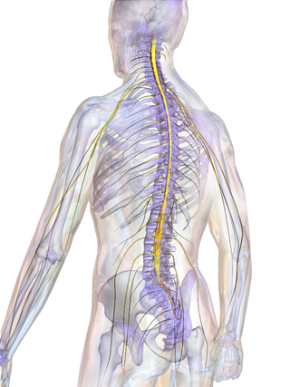
The spinal cord is a long, thin, tubular structure made up of nervous tissue that extends from the medulla oblongata in the lower brainstem to the lumbar region of the vertebral column (backbone) of vertebrate animals. The center of the spinal cord is hollow and contains a structure called the central canal, which contains cerebrospinal fluid. The spinal cord is also covered by meninges and enclosed by the neural arches. Together, the brain and spinal cord make up the central nervous system.
In neuroscience, the N100 or N1 is a large, negative-going evoked potential measured by electroencephalography ; it peaks in adults between 80 and 120 milliseconds after the onset of a stimulus, and is distributed mostly over the fronto-central region of the scalp. It is elicited by any unpredictable stimulus in the absence of task demands. It is often referred to with the following P200 evoked potential as the "N100-P200" or "N1-P2" complex. While most research focuses on auditory stimuli, the N100 also occurs for visual, olfactory, heat, pain, balance, respiration blocking, and somatosensory stimuli.