SorCS2

Last updated
SORCS2
Available structures
PDB Ortholog search: PDBe RCSB
Identifiers
Aliases SORCS2 , sortilin related VPS10 domain containing receptor 2
External IDs OMIM: 606284; MGI: 1932289; HomoloGene: 56899; GeneCards: SORCS2; OMA:SORCS2 - orthologs
Orthologs
SpeciesHumanMouse
Entrez
Ensembl
UniProt
RefSeq (mRNA)

NM_020777

NM_030889

RefSeq (protein)

NP_065828

NP_112151

Location (UCSC) Chr 4: 7.19 – 7.74 Mb Chr 5: 36.17 – 36.56 Mb
PubMed search [3] [4]
Wikidata
View/Edit Human View/Edit Mouse

The SorCS2 (sortilin-related Vps10p domain containing receptor 2) gene is found on chromosome 4 (4p16.1), and is composed of 28 exons. The N-terminal exons which encode the Vps10p domain are spaced by large introns. The functional receptor protein is largely present in the brain. It is 1109 amino acids long, largely neutral, and has a single transmembrane pass.... [5]

Contents

SorCS2 is a member of the mammalian Vps10p (vacuolar protein sorting 10 protein) domain family consisting of five transmembrane proteins with structural similarities: SorCS1, SorCS2, SorCS3, SorLA (sorting protein-related receptor with A-type repeats), and sortilin. [6] SorCS2 specifically has critical roles in neuronal viability and function. Single nucleotide polymorphisms (SNPs) in the protein has been associated with a range of diseases including attention-deficit hyperactivity disorder (ADHD), [7] bipolar disorders, [8] and schizophrenia, [9] and the receptor family has also been associated with Alzheimer's disease [10] and type 2 diabetes. [11]

Discovery

The Vps10p domain receptor family was based on the discovery of SorLA in 1996 [12] and sortilin in 1997, [13] and has since been expanded with the SorCS subfamily with SorCS2 being described in 2001 [14] [15]

SorCS2 was first found from isolated cDNA in murine floor plate samples of the central nervous system (CNS) as well as in regions of the brain. The cDNA contained the characteristic Vps10p domain enabling its classification as a SorCS protein. [14] Not long after, a corresponding partial cDNA was found in human samples, and it was possible to determine the missing N-terminal by homology to murine SorCS2. [15]

Structure

SorCS2 is composed of a small intracellular region making a single pass into the extracellular environment where the large Vps10p domain make up a beta-propeller structure consisting of 10 propeller blade-like beta sheet regions. The Vps10p domain contains at least 2 unspecific ligand binding sites. [6] The domain also contains a furin cleavage site. [16] The extracellular region of SorCS proteins also include a LR (leucine rich domain) containing imperfect LR repeats (LRRs) which are known to serve as interaction and adhesion domains [15]

Modifications in Vps10p-type receptors include glycosylations. [17] and they also contain a propeptide which is proteolytically cleaved off to make them active [6]

In the non-neuronal glia cells, SorCS2 is cleaved and a linkage forms a two-chained product distinct from that in neurons which is a single chained. The processing in glia cells have been linked to proapoptotic properties not found in neuronal SorCS2. [18] This differential processing is thought to be common in Vps10p domain proteins where it regulates receptor functionality [6]

Dimerization

Efforts have been made to elucidate the structure of SorCS2, and this has allowed determination of dimerization of SorCS2 and the other two SorCS proteins with only few monomeric structures found. This dimerization is promoted by deglycosylation at least in SorCS1. [6] Structurally, the Vps10p domains in SorCS proteins can be found next to each other, but uniquely for SorCS2 it is prevalently found in a dimer where the domains are located away from each other and connected at a two-fold rotation axis for the dimer. [6] The different types of dimers could explain correspondingly different functions of SorCS2 found in different tissues. In addition to the homodimers described, the SorCS proteins also forms heterodimers within this subfamily. [6] Crystal structures of the full extracellular portion of SorCS2 have uncovered that SorCS2 consists of six domains. [19] Five domains contribute to the dimerization of SorCS2. Despite the extensive dimerization interface, SorCS2 has substantial conformational plasticity. [19]

Localization

SorCS2 and related proteins in the Vps10p domain family are predominantly found in neurons in the brain, but are also present in other tissues. [20] In terms of brain localization SorCS2 has been found predominantly in thalamus, floor plate of the midbrain and spinal cord, ventricular zones of hippocampal and accumbens areas, meninges, and Schwann cells. The localization is distinct from the other Vps10p receptor sortilin [21]

SorCS2 has further been found in tissues that are not brain related in smaller amounts e.g. in structures of mesodermal origin such as adipose tissue, striated muscle tissues, and developing bone as well as connective tissue such as the dermis, submucosal, and submesothelial tissues in the gut, and the bronchial system. Although the presence in these tissues are largely uninvestigated, they still form the basis for further specific functions in non-brain tissue. [20]

Function

All members of the Vps10p protein family are multiligand receptors. [22] [23] They can take part in cellular trafficking and signaling through ligand binding in response to cellular conditions. [24] [25] [26] Examples of ligands are neurotrophic factors, amyloid precursor protein (APP), lipoproteins, and cytokines. [27] In addition to depending on the cellular context, the affinity for specific ligands can also be modulated by the monomer/dimer ratio. [6]

BDNF-dependent plasticity

Hippocampal N-methyl-D-aspartate (NMDA) receptor-dependent synaptic plasticity is found to be deficient at least in SorCS2 mutant mice, strongly suggesting a link between the two. SorCS2 deficient mice also show decreased long-term memory, higher tendency to take risks, and to have a more stimuli seeking behaviour than corresponding SorCS2 normal mice. [26]

The decrease in plasticity is attributed to the fact that SorCS2 forms a complex with p75NTR, a neurotrophin receptor which interacts with proBDNF (brain-derived neurotrophic factor) and TrkB (BDNF receptor tyrosine kinase) inside neurons in the hippocampal region of the brain to modulate synapse depression and potentiation respectively. Thus, SorCS2 could be the link between BDNF/proBDNF signaling and mental disorders. Deficiency in this signaling can affect the strengthening and weakening of synapses, that is, neuronal plasticity. [26]

Clinical significance

Alcohol withdrawal

When trying to stop excessive alcohol consumption alcohol withdrawal (AW) is physiological responses that in some cases can cause life-threatening seizures. SorCS2 has been associated with the severity of AW in genome analysis of European American test subjects, although no such connection could be made in African American samples [28]

A specific SorCS2 risk haplotype disrupts a transcription factor (TF) binding site in a stress hormone-modulated regulatory enhancer element with activity in human hippocampus. This region of the brain is already known for its association with AW. This increases the severity of AW in patients with alcoholism. Exposure to ethanol and glucocorticoids have been found to act as up-regulators of SorCS2, causing worsening of the problems if the risk variant of SorCS2 is present. [28]

See also

Related Research Articles

<span class="mw-page-title-main">Axon</span> Long projection on a neuron that conducts signals to other neurons

An axon or nerve fiber is a long, slender projection of a nerve cell, or neuron, in vertebrates, that typically conducts electrical impulses known as action potentials away from the nerve cell body. The function of the axon is to transmit information to different neurons, muscles, and glands. In certain sensory neurons, such as those for touch and warmth, the axons are called afferent nerve fibers and the electrical impulse travels along these from the periphery to the cell body and from the cell body to the spinal cord along another branch of the same axon. Axon dysfunction can be the cause of many inherited and acquired neurological disorders that affect both the peripheral and central neurons. Nerve fibers are classed into three types – group A nerve fibers, group B nerve fibers, and group C nerve fibers. Groups A and B are myelinated, and group C are unmyelinated. These groups include both sensory fibers and motor fibers. Another classification groups only the sensory fibers as Type I, Type II, Type III, and Type IV.

The development of the nervous system, or neural development (neurodevelopment), refers to the processes that generate, shape, and reshape the nervous system of animals, from the earliest stages of embryonic development to adulthood. The field of neural development draws on both neuroscience and developmental biology to describe and provide insight into the cellular and molecular mechanisms by which complex nervous systems develop, from nematodes and fruit flies to mammals.

<span class="mw-page-title-main">Brain-derived neurotrophic factor</span> Protein found in humans

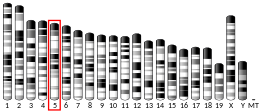
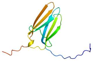
Brain-derived neurotrophic factor (BDNF), or abrineurin, is a protein that, in humans, is encoded by the BDNF gene. BDNF is a member of the neurotrophin family of growth factors, which are related to the canonical nerve growth factor (NGF), a family which also includes NT-3 and NT-4/NT-5. Neurotrophic factors are found in the brain and the periphery. BDNF was first isolated from a pig brain in 1982 by Yves-Alain Barde and Hans Thoenen.

<span class="mw-page-title-main">Neurotrophin</span> Family of proteins

Neurotrophins are a family of proteins that induce the survival, development, and function of neurons.

<span class="mw-page-title-main">Tropomyosin receptor kinase A</span> Protein-coding gene in the species Homo sapiens

Tropomyosin receptor kinase A (TrkA), also known as high affinity nerve growth factor receptor, neurotrophic tyrosine kinase receptor type 1, or TRK1-transforming tyrosine kinase protein is a protein that in humans is encoded by the NTRK1 gene.

<span class="mw-page-title-main">Tropomyosin receptor kinase B</span> Protein and coding gene in humans

Tropomyosin receptor kinase B (TrkB), also known as tyrosine receptor kinase B, or BDNF/NT-3 growth factors receptor or neurotrophic tyrosine kinase, receptor, type 2 is a protein that in humans is encoded by the NTRK2 gene. TrkB is a receptor for brain-derived neurotrophic factor (BDNF). The standard pronunciation for this protein is "track bee".

<span class="mw-page-title-main">Low-affinity nerve growth factor receptor</span> Human protein-coding gene

The p75 neurotrophin receptor (p75NTR) was first identified in 1973 as the low-affinity nerve growth factor receptor (LNGFR) before discovery that p75NTR bound other neurotrophins equally well as nerve growth factor. p75NTR is a neurotrophic factor receptor. Neurotrophic factor receptors bind Neurotrophins including Nerve growth factor, Neurotrophin-3, Brain-derived neurotrophic factor, and Neurotrophin-4. All neurotrophins bind to p75NTR. This also includes the immature pro-neurotrophin forms. Neurotrophic factor receptors, including p75NTR, are responsible for ensuring a proper density to target ratio of developing neurons, refining broader maps in development into precise connections. p75NTR is involved in pathways that promote neuronal survival and neuronal death.

<span class="mw-page-title-main">Tropomyosin receptor kinase C</span> Protein-coding gene in the species Homo sapiens

Tropomyosin receptor kinase C (TrkC), also known as NT-3 growth factor receptor, neurotrophic tyrosine kinase receptor type 3, or TrkC tyrosine kinase is a protein that in humans is encoded by the NTRK3 gene.

Neurotrophic factors (NTFs) are a family of biomolecules – nearly all of which are peptides or small proteins – that support the growth, survival, and differentiation of both developing and mature neurons. Most NTFs exert their trophic effects on neurons by signaling through tyrosine kinases, usually a receptor tyrosine kinase. In the mature nervous system, they promote neuronal survival, induce synaptic plasticity, and modulate the formation of long-term memories. Neurotrophic factors also promote the initial growth and development of neurons in the central nervous system and peripheral nervous system, and they are capable of regrowing damaged neurons in test tubes and animal models. Some neurotrophic factors are also released by the target tissue in order to guide the growth of developing axons. Most neurotrophic factors belong to one of three families: (1) neurotrophins, (2) glial cell-line derived neurotrophic factor family ligands (GFLs), and (3) neuropoietic cytokines. Each family has its own distinct cell signaling mechanisms, although the cellular responses elicited often do overlap.

<span class="mw-page-title-main">Neurotrophin-3</span> Protein-coding gene in the species Homo sapiens

Neurotrophin-3 is a protein that in humans is encoded by the NTF3 gene.

Neurturin (NRTN) is a protein that is encoded in humans by the NRTN gene. Neurturin belongs to the glial cell line-derived neurotrophic factor (GDNF) family of neurotrophic factors, which regulate the survival and function of neurons. Neurturin’s role as a growth factor places it in the transforming growth factor beta (TGF-beta) subfamily along with its homologs persephin, artemin, and GDNF. It shares a 42% similarity in amino acid sequence with mature GDNF. It is also considered a trophic factor and critical in the development and growth of neurons in the brain. Neurotrophic factors like neurturin have been tested in several clinical trial settings for the potential treatment of neurodegenerative diseases, specifically Parkinson's disease.

<span class="mw-page-title-main">Artemin</span> Protein-coding gene in the species Homo sapiens

Artemin, also known as enovin or neublastin, is a protein that in humans is encoded by the ARTN gene.

<span class="mw-page-title-main">Sortilin 1</span> Protein-coding gene in the species Homo sapiens

Sortilin (SORT1) is a protein that in humans is encoded by the SORT1 gene on chromosome 1. This protein is a type I membrane glycoprotein in the vacuolar protein sorting 10 protein (Vps10p) family of sorting receptors. While it is ubiquitously expressed in many tissues, sortilin is most abundant in the central nervous system. At the cellular level, sortilin functions in protein transport between the Golgi apparatus, endosome, lysosome, and plasma membrane, leading to its involvement in multiple biological processes such as glucose and lipid metabolism as well as neural development and cell death. Moreover, the function and role of sortilin is now emerging in several major human diseases such as hypertension, atherosclerosis, coronary artery disease, Alzheimer’s disease, and cancer. The SORT1 gene also contains one of 27 loci associated with increased risk of coronary artery disease.

Trk receptors are a family of tyrosine kinases that regulates synaptic strength and plasticity in the mammalian nervous system. Trk receptors affect neuronal survival and differentiation through several signaling cascades. However, the activation of these receptors also has significant effects on functional properties of neurons.

The glutamate hypothesis of schizophrenia models the subset of pathologic mechanisms of schizophrenia linked to glutamatergic signaling. The hypothesis was initially based on a set of clinical, neuropathological, and, later, genetic findings pointing at a hypofunction of glutamatergic signaling via NMDA receptors. While thought to be more proximal to the root causes of schizophrenia, it does not negate the dopamine hypothesis, and the two may be ultimately brought together by circuit-based models. The development of the hypothesis allowed for the integration of the GABAergic and oscillatory abnormalities into the converging disease model and made it possible to discover the causes of some disruptions.

Tobias Bonhoeffer is a German-American neurobiologist. He is director of the department Synapses – Circuits – Plasticity and current managing director at the Max Planck Institute for Biological Intelligence in Martinsried near Munich.

<span class="mw-page-title-main">Nonsynaptic plasticity</span> Form of neuroplasticity

Nonsynaptic plasticity is a form of neuroplasticity that involves modification of ion channel function in the axon, dendrites, and cell body that results in specific changes in the integration of excitatory postsynaptic potentials and inhibitory postsynaptic potentials. Nonsynaptic plasticity is a modification of the intrinsic excitability of the neuron. It interacts with synaptic plasticity, but it is considered a separate entity from synaptic plasticity. Intrinsic modification of the electrical properties of neurons plays a role in many aspects of plasticity from homeostatic plasticity to learning and memory itself. Nonsynaptic plasticity affects synaptic integration, subthreshold propagation, spike generation, and other fundamental mechanisms of neurons at the cellular level. These individual neuronal alterations can result in changes in higher brain function, especially learning and memory. However, as an emerging field in neuroscience, much of the knowledge about nonsynaptic plasticity is uncertain and still requires further investigation to better define its role in brain function and behavior.

Epigenetics of depression is the study of how epigenetics contribute to depression.

TLQP-62 (amino acid 556-617) is a VGF-derived C-terminal peptide that was first discovered by Trani et al. TLQP-62 is derived from VGF precursor protein via proteolytic cleavage by prohormone convertases PC1/3 at the RPR555 site. TLQP-62 is named after its first four N-terminal amino acids and its peptide length.

The neurotrophic hypothesis of depression proposes that major depressive disorder (MDD) is caused, at least partly, by impaired neurotrophic support. Neurotrophic factors are a family of closely related proteins which regulate the survival, development, and function of neurons in both the central and peripheral nervous systems. 

References

  1. 1 2 3 GRCh38: Ensembl release 89: ENSG00000184985 Ensembl, May 2017
  2. 1 2 3 GRCm38: Ensembl release 89: ENSMUSG00000029093 Ensembl, May 2017
  3. "Human PubMed Reference:". National Center for Biotechnology Information, U.S. National Library of Medicine.
  4. "Mouse PubMed Reference:". National Center for Biotechnology Information, U.S. National Library of Medicine.
  5. "SORCS2 sortilin related VPS10 domain containing receptor 2 [Homo sapiens (human)] - Gene - NCBI". www.ncbi.nlm.nih.gov. Retrieved 2018-11-07.
  6. 1 2 3 4 5 6 7 8 Januliene, Dovile; Manavalan, Arulmani; Ovesen, Peter Lund; Pedersen, Karen-Marie; Thirup, Søren; Nykjær, Anders; Moeller, Arne (September 2017). "Hidden Twins: SorCS Neuroreceptors Form Stable Dimers". Journal of Molecular Biology. 429 (19): 2907–2917. doi:10.1016/j.jmb.2017.08.006. ISSN   0022-2836. PMID   28827148.
  7. Alemany, Silvia; Ribasés, Marta; Vilor-Tejedor, Natàlia; Bustamante, Mariona; Sánchez-Mora, Cristina; Bosch, Rosa; Richarte, Vanesa; Cormand, Bru; Casas, Miguel (2015-07-14). "New suggestive genetic loci and biological pathways for attention function in adult attention-deficit/hyperactivity disorder". American Journal of Medical Genetics Part B: Neuropsychiatric Genetics. 168 (6): 459–470. doi: 10.1002/ajmg.b.32341 . ISSN   1552-4841. PMID   26174813. S2CID   38488226.
  8. Baum, A E; Akula, N; Cabanero, M; Cardona, I; Corona, W; Klemens, B; Schulze, T G; Cichon, S; Rietschel, M (2007-05-08). "A genome-wide association study implicates diacylglycerol kinase eta (DGKH) and several other genes in the etiology of bipolar disorder". Molecular Psychiatry. 13 (2): 197–207. doi:10.1038/sj.mp.4002012. ISSN   1359-4184. PMC   2527618 . PMID   17486107.
  9. Christoforou, A; McGhee, K A; Morris, S W; Thomson, P A; Anderson, S; McLean, A; Torrance, H S; Le Hellard, S; Pickard, B S (2010-03-30). "Convergence of linkage, association and GWAS findings for a candidate region for bipolar disorder and schizophrenia on chromosome 4p". Molecular Psychiatry. 16 (3): 240–242. doi:10.1038/mp.2010.25. ISSN   1359-4184. PMID   20351716. S2CID   10244703.
  10. Reitz, Christiane; Tokuhiro, Shinya; Clark, Lorraine N.; Conrad, Christopher; Vonsattel, Jean-Paul; Hazrati, Lili-Naz; Palotás, András; Lantigua, Raphael; Medrano, Martin (January 2011). "SORCS1 alters amyloid precursor protein processing and variants may increase Alzheimer's disease risk". Annals of Neurology. 69 (1): 47–64. doi:10.1002/ana.22308. ISSN   0364-5134. PMC   3086759 . PMID   21280075.
  11. Goodarzi, Mark O.; Lehman, Donna M.; Taylor, Kent D.; Guo, Xiuqing; Cui, Jinrui; Quiñones, Manuel J.; Clee, Susanne M.; Yandell, Brian S.; Blangero, John (2007-07-01). "SORCS1: A Novel Human Type 2 Diabetes Susceptibility Gene Suggested by the Mouse". Diabetes. 56 (7): 1922–1929. doi: 10.2337/db06-1677 . ISSN   0012-1797. PMID   17426289.
  12. Jacobsen, L.; Madsen, P.; Moestrup, S. K.; Lund, A. H.; Tommerup, N.; Nykjaer, A.; Sottrup-Jensen, L.; Gliemann, J.; Petersen, C. M. (1996-12-06). "Molecular characterization of a novel human hybrid-type receptor that binds the alpha2-macroglobulin receptor-associated protein". The Journal of Biological Chemistry. 271 (49): 31379–31383. doi: 10.1074/jbc.271.49.31379 . ISSN   0021-9258. PMID   8940146.
  13. Petersen, C. M.; Nielsen, M. S.; Nykjaer, A.; Jacobsen, L.; Tommerup, N.; Rasmussen, H. H.; Roigaard, H.; Gliemann, J.; Madsen, P. (1997-02-07). "Molecular identification of a novel candidate sorting receptor purified from human brain by receptor-associated protein affinity chromatography". The Journal of Biological Chemistry. 272 (6): 3599–3605. doi: 10.1074/jbc.272.6.3599 . ISSN   0021-9258. PMID   9013611.
  14. 1 2 Rezgaoui, M.; Hermey, G.; Riedel, I. B.; Hampe, W.; Schaller, H. C.; Hermans-Borgmeyer, I. (February 2001). "Identification of SorCS2, a novel member of the VPS10 domain containing receptor family, prominently expressed in the developing mouse brain". Mechanisms of Development. 100 (2): 335–338. doi:10.1016/S0925-4773(00)00523-2. ISSN   0925-4773. PMID   11165493. S2CID   18829884.
  15. 1 2 3 Hampe, W.; Rezgaoui, M.; Hermans-Borgmeyer, I.; Schaller, H. C. (June 2001). "The genes for the human VPS10 domain-containing receptors are large and contain many small exons". Human Genetics. 108 (6): 529–536. doi:10.1007/s004390100504. ISSN   0340-6717. PMID   11499680. S2CID   23375354.
  16. Hosaka, M.; Nagahama, M.; Kim, W. S.; Watanabe, T.; Hatsuzawa, K.; Ikemizu, J.; Murakami, K.; Nakayama, K. (1991-07-05). "Arg-X-Lys/Arg-Arg motif as a signal for precursor cleavage catalyzed by furin within the constitutive secretory pathway". The Journal of Biological Chemistry. 266 (19): 12127–12130. doi: 10.1016/S0021-9258(18)98867-8 . ISSN   0021-9258. PMID   1905715.
  17. Hermey, Guido; Riedel, I.Björn; Hampe, Wolfgang; Schaller, H.Chica; Hermans-Borgmeyer, Irm (1999-12-20). "Identification and Characterization of SorCS, a Third Member of a Novel Receptor Family". Biochemical and Biophysical Research Communications. 266 (2): 347–351. doi:10.1006/bbrc.1999.1822. ISSN   0006-291X. PMID   10600506.
  18. Glerup, Simon; Olsen, Ditte; Vaegter, Christian B.; Gustafsen, Camilla; Sjoegaard, Susanne S.; Hermey, Guido; Kjolby, Mads; Molgaard, Simon; Ulrichsen, Maj (June 2014). "SorCS2 Regulates Dopaminergic Wiring and Is Processed into an Apoptotic Two-Chain Receptor in Peripheral Glia". Neuron. 82 (5): 1074–1087. doi: 10.1016/j.neuron.2014.04.022 . hdl: 11858/00-001M-0000-0027-792B-3 . ISSN   0896-6273. PMID   24908487.
  19. 1 2 Leloup, Nadia; Chataigner, Lucas M. P.; Janssen, Bert J. C. (2018-07-30). "Structural insights into SorCS2–Nerve Growth Factor complex formation". Nature Communications. 9 (1): 2979. Bibcode:2018NatCo...9.2979L. doi:10.1038/s41467-018-05405-z. ISSN   2041-1723. PMC   6065357 . PMID   30061605.
  20. 1 2 Boggild, Simon; Molgaard, Simon; Glerup, Simon; Nyengaard, Jens Randel (2016-03-10). "Spatiotemporal patterns of sortilin and SorCS2 localization during organ development". BMC Cell Biology. 17 (1): 8. doi: 10.1186/s12860-016-0085-9 . ISSN   1471-2121. PMC   4785631 . PMID   26964886.
  21. Boggild, Simon; Molgaard, Simon; Glerup, Simon; Nyengaard, Jens Randel (2018-02-20). "Highly segregated localization of the functionally related vps10p receptors sortilin and SorCS2 during neurodevelopment". Journal of Comparative Neurology. 526 (8): 1267–1286. doi:10.1002/cne.24403. ISSN   0021-9967. PMID   29405286. S2CID   46869016.
  22. Willnow, Thomas E.; Petersen, Claus M.; Nykjaer, Anders (2008-11-12). "VPS10P-domain receptors — regulators of neuronal viability and function". Nature Reviews Neuroscience. 9 (12): 899–909. doi:10.1038/nrn2516. ISSN   1471-003X. PMID   19002190. S2CID   25776764.
  23. Willnow, Thomas E; Kjølby, Mads; Nykjaer, Anders (April 2011). "Sortilins: new players in lipoprotein metabolism". Current Opinion in Lipidology. 22 (2): 79–85. doi:10.1097/mol.0b013e3283416f2b. ISSN   0957-9672. PMID   21124217. S2CID   205829395.
  24. Nykjaer, Anders; Lee, Ramee; Teng, Kenneth K.; Jansen, Pernille; Madsen, Peder; Nielsen, Morten S.; Jacobsen, Christian; Kliemannel, Marco; Schwarz, Elisabeth (2004-02-26). "Sortilin is essential for proNGF-induced neuronal cell death". Nature. 427 (6977): 843–848. Bibcode:2004Natur.427..843N. doi:10.1038/nature02319. ISSN   0028-0836. PMID   14985763. S2CID   4343450.
  25. Larsen, Jakob Vejby; Hansen, Maria; Møller, Bjarne; Madsen, Peder; Scheller, Jürgen; Nielsen, Morten; Petersen, Claus Munck (2010-09-01). "Sortilin Facilitates Signaling of Ciliary Neurotrophic Factor and Related Helical Type 1 Cytokines Targeting the gp130/Leukemia Inhibitory Factor Receptor β Heterodimer". Molecular and Cellular Biology. 30 (17): 4175–4187. doi:10.1128/MCB.00274-10. ISSN   0270-7306. PMC   2937557 . PMID   20584990.
  26. 1 2 3 Glerup, S; Bolcho, U; Mølgaard, S; Bøggild, S; Vaegter, C B; Smith, A H; Nieto-Gonzalez, J L; Ovesen, P L; Pedersen, L F (2016-07-26). "SorCS2 is required for BDNF-dependent plasticity in the hippocampus". Molecular Psychiatry. 21 (12): 1740–1751. doi: 10.1038/mp.2016.108 . ISSN   1359-4184. PMID   27457814. S2CID   21763820.
  27. Glerup, S.; Nykjaer, A.; Vaegter, C. B. (2014), "Sortilins in Neurotrophic Factor Signaling", Neurotrophic Factors, Handbook of Experimental Pharmacology, vol. 220, Springer Berlin Heidelberg, pp. 165–189, doi:10.1007/978-3-642-45106-5_7, ISBN   9783642451058, PMID   24668473
  28. 1 2 Smith, Andrew H.; Ovesen, Peter L.; Skeldal, Sune; Yeo, Seungeun; Jensen, Kevin P.; Olsen, Ditte; Diazgranados, Nancy; Zhao, Hongyu; Farrer, Lindsay A. (2018-10-25). "Risk Locus Identification Ties Alcohol Withdrawal Symptoms to SORCS2". Alcoholism: Clinical and Experimental Research. 42 (12): 2337–2348. doi:10.1111/acer.13890. ISSN   0145-6008. PMC   6317871 . PMID   30252935.