Susan McConnell

Last updated
Susan McConnell
Susan K McConnell.jpg
Born1958
NationalityAmerican
Alma mater Harvard University
Known for Neural development
Scientific career
Fields Neurobiology
Institutions Stanford University
Doctoral advisor Simon LeVay

Susan McConnell is a neurobiologist who studies the development of neural circuits in the mammalian cerebral cortex. She is a professor in the Department of Biology at Stanford University, where she is the Susan B. Ford Professor of Humanities and Sciences, a Bass University Fellow, and a Howard Hughes Medical Institute Professor. She is an elected member of the National Academy of Sciences and the American Academy of Arts and Sciences.

Contents

Education

McConnell graduated with a joint A.B. degree from Harvard College and Radcliffe College in 1980. She did her doctoral work in the research group of Simon LeVay and received her PhD in neurobiology from Harvard University in 1987. She was a postdoctoral fellow in the laboratory of Carla J. Shatz at Stanford University.

Research

McConnell's research focuses on understanding how neurons in the developing cerebral cortex are produced, differentiated, and connected to form functional circuits.

McConnell's research showed that progression through the cell cycle plays a key role in determining the final differentiated state of a neural progenitor cell. [1] McConnell also confirmed the hypothesis that asymmetric cell division, as determined by the orientation of the dividing progenitor's cleavage plane, regulates cortical development. Her work elucidated the first molecular mechanism for this process, showing that asymmetrically inherited Notch proteins determine whether a new daughter cell will differentiate into a neuron or remain a neural progenitor. [2]

Her work also showed that developing cortical neurons use a variety of different migratory paths as they move from their birthplace to their final destination in the cortex. [3] This work stood in contrast to a prevailing theory at the time, that all neuronal migration in the cortex was dependent upon radial glia.

McConnell's recent work has continued to outline the molecular mechanisms underlying neural differentiation, [4] [5] [6] [7] neuronal migration [8] [9] and axon guidance. [10] [11]

Teaching

Stanford University has recognized McConnell with its two highest teaching honors, the Hoagland Prize for Undergraduate Teaching and the Walter J. Gores Award for Excellence in Teaching. She has taught undergraduate courses on neural development since joining Stanford's faculty in 1989.

From 2010 to 2012, McConnell co-chaired a university-wide commission that evaluated undergraduate education at Stanford. [12] The commission's recommendations encouraged students and teachers "to reconsider what they do, how they do it, and why it matters", [13] and urged reforms to the university's general education programs.

In addition, Dr. McConnell and author Andrew Todhunter were the principal force behind the creation of Stanford's Senior Reflection in Biology, a capstone course for senior undergraduates where life-science students undertake creative projects synthesizing the arts and sciences. [14] [15]

Conservation photography

In addition to her career in research and teaching, McConnell is an accomplished wildlife photographer. After photographing animals during a trip to the Svalbard archipelago in Norway, she developed an interest in using photography to tell stories about animal behavior. [16] [17] She teaches undergraduate classes on conservation photography at Stanford. Her photos have been featured in various publications including Smithsonian [18] and National Geographic . [19]

McConnell was the first non-art Stanford faculty member to have a show in the Stanford Art Gallery. [20] Her show was called On the Shoulders of Giants and focused on elephants, their poaching, and the crisis of the ivory trade. [21]

Personal life

McConnell is married to Richard Scheller, former chief scientific officer and head of therapeutics at 23andMe and the former executive vice president of research and early development at Genentech.

Awards and honors

McConnell is a member of the National Academy of Sciences and the American Academy of Arts and Sciences, and is a Howard Hughes Medical Institute Professor. She has received many other awards and honors, including:

Related Research Articles

<span class="mw-page-title-main">Brain</span> Organ central to the nervous system

The brain is an organ that serves as the center of the nervous system in all vertebrate and most invertebrate animals. It consists of nervous tissue and is typically located in the head (cephalization), usually near organs for special senses such as vision, hearing and olfaction. Being the most specialized organ, it is responsible for receiving information from the sensory nervous system, processing those information and the coordination of motor control.

<span class="mw-page-title-main">Central nervous system</span> Brain and spinal cord

The central nervous system (CNS) is the part of the nervous system consisting primarily of the brain and spinal cord. The CNS is so named because the brain integrates the received information and coordinates and influences the activity of all parts of the bodies of bilaterally symmetric and triploblastic animals—that is, all multicellular animals except sponges and diploblasts. It is a structure composed of nervous tissue positioned along the rostral to caudal axis of the body and may have an enlarged section at the rostral end which is a brain. Only arthropods, cephalopods and vertebrates have a true brain, though precursor structures exist in onychophorans, gastropods and lancelets.

<span class="mw-page-title-main">Cerebral cortex</span> Outer layer of the cerebrum of the mammalian brain

The cerebral cortex, also known as the cerebral mantle, is the outer layer of neural tissue of the cerebrum of the brain in humans and other mammals. It is the largest site of neural integration in the central nervous system, and plays a key role in attention, perception, awareness, thought, memory, language, and consciousness. The cerebral cortex is the part of the brain responsible for cognition.

The development of the nervous system, or neural development (neurodevelopment), refers to the processes that generate, shape, and reshape the nervous system of animals, from the earliest stages of embryonic development to adulthood. The field of neural development draws on both neuroscience and developmental biology to describe and provide insight into the cellular and molecular mechanisms by which complex nervous systems develop, from nematodes and fruit flies to mammals.

<span class="mw-page-title-main">Pyramidal cell</span> Projection neurons in the cerebral cortex and hippocampus

Pyramidal cells, or pyramidal neurons, are a type of multipolar neuron found in areas of the brain including the cerebral cortex, the hippocampus, and the amygdala. Pyramidal cells are the primary excitation units of the mammalian prefrontal cortex and the corticospinal tract. One of the main structural features of the pyramidal neuron is the conic shaped soma, or cell body, after which the neuron is named. Other key structural features of the pyramidal cell are a single axon, a large apical dendrite, multiple basal dendrites, and the presence of dendritic spines.

Synaptogenesis is the formation of synapses between neurons in the nervous system. Although it occurs throughout a healthy person's lifespan, an explosion of synapse formation occurs during early brain development, known as exuberant synaptogenesis. Synaptogenesis is particularly important during an individual's critical period, during which there is a certain degree of synaptic pruning due to competition for neural growth factors by neurons and synapses. Processes that are not used, or inhibited during their critical period will fail to develop normally later on in life.

<span class="mw-page-title-main">Reticular formation</span> Spinal trigeminal nucleus

The reticular formation is a set of interconnected nuclei that are located in the brainstem, hypothalamus, and other regions. It is not anatomically well defined, because it includes neurons located in different parts of the brain. The neurons of the reticular formation make up a complex set of networks in the core of the brainstem that extend from the upper part of the midbrain to the lower part of the medulla oblongata. The reticular formation includes ascending pathways to the cortex in the ascending reticular activating system (ARAS) and descending pathways to the spinal cord via the reticulospinal tracts.

In developmental psychology and developmental biology, a critical period is a maturational stage in the lifespan of an organism during which the nervous system is especially sensitive to certain environmental stimuli. If, for some reason, the organism does not receive the appropriate stimulus during this "critical period" to learn a given skill or trait, it may be difficult, ultimately less successful, or even impossible, to develop certain associated functions later in life. Functions that are indispensable to an organism's survival, such as vision, are particularly likely to develop during critical periods. "Critical period" also relates to the ability to acquire one's first language. Researchers found that people who passed the "critical period" would not acquire their first language fluently.

<span class="mw-page-title-main">Basket cell</span>

Basket cells are inhibitory GABAergic interneurons of the brain, found throughout different regions of the cortex and cerebellum.

<span class="mw-page-title-main">Thalamocortical radiations</span> Neural pathways between the thalamus and cerebral cortex

In neuroanatomy, thalamocortical radiations, also known as thalamocortical fibers, are the efferent fibers that project from the thalamus to distinct areas of the cerebral cortex. They form fiber bundles that emerge from the lateral surface of the thalamus.

<span class="mw-page-title-main">Retinotopy</span> Mapping of visual input from the retina to neurons

Retinotopy is the mapping of visual input from the retina to neurons, particularly those neurons within the visual stream. For clarity, 'retinotopy' can be replaced with 'retinal mapping', and 'retinotopic' with 'retinally mapped'.

<span class="mw-page-title-main">Subplate</span>

The subplate, also called the subplate zone, together with the marginal zone and the cortical plate, in the fetus represents the developmental anlage of the mammalian cerebral cortex. It was first described, as a separate transient fetal zone by Ivica Kostović and Mark E. Molliver in 1974.

<span class="mw-page-title-main">Radial glial cell</span> Bipolar-shaped progenitor cells of all neurons in the cerebral cortex and some glia

Radial glial cells, or radial glial progenitor cells (RGPs), are bipolar-shaped progenitor cells that are responsible for producing all of the neurons in the cerebral cortex. RGPs also produce certain lineages of glia, including astrocytes and oligodendrocytes. Their cell bodies (somata) reside in the embryonic ventricular zone, which lies next to the developing ventricular system.

The development of the nervous system in humans, or neural development, or neurodevelopment involves the studies of embryology, developmental biology, and neuroscience. These describe the cellular and molecular mechanisms by which the complex nervous system forms in humans, develops during prenatal development, and continues to develop postnatally.

<span class="mw-page-title-main">EMX1</span> Protein-coding gene in the species Homo sapiens

Homeobox protein EMX1 is a protein that in humans is encoded by the EMX1 gene. The transcribed EMX1 gene is a member of the EMX family of transcription factors. The EMX1 gene, along with its family members, are expressed in the developing cerebrum. EMX1 plays a role in specification of positional identity, the proliferation of neural stem cells, differentiation of layer-specific neuronal phenotypes and commitment to a neuronal or glial cell fate.

<span class="mw-page-title-main">TBR1</span> Protein-coding gene in Homo sapiens

T-box, brain, 1 is a transcription factor protein important in vertebrate embryo development. It is encoded by the TBR1 gene. This gene is also known by several other names: T-Brain 1, TBR-1, TES-56, and MGC141978. TBR1 is a member of the TBR1 subfamily of T-box family transcription factors, which share a common DNA-binding domain. Other members of the TBR1 subfamily include EOMES and TBX21. TBR1 is involved in the differentiation and migration of neurons and is required for normal brain development. TBR1 interacts with various genes and proteins in order to regulate cortical development, specifically within layer VI of the developing six-layered human cortex. Studies show that TBR1 may play a role in major neurological diseases such as Alzheimer's disease (AD), Parkinson's disease (PD) and autism spectrum disorder (ASD).
<span class="mw-page-title-main">Primary motor cortex</span> Brain region

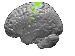
The primary motor cortex is a brain region that in humans is located in the dorsal portion of the frontal lobe. It is the primary region of the motor system and works in association with other motor areas including premotor cortex, the supplementary motor area, posterior parietal cortex, and several subcortical brain regions, to plan and execute voluntary movements. Primary motor cortex is defined anatomically as the region of cortex that contains large neurons known as Betz cells, which, along with other cortical neurons, send long axons down the spinal cord to synapse onto the interneuron circuitry of the spinal cord and also directly onto the alpha motor neurons in the spinal cord which connect to the muscles.

<span class="mw-page-title-main">Eomesodermin</span> Protein-coding gene in the species Homo sapiens

Eomesodermin also known as T-box brain protein 2 (Tbr2) is a protein that in humans is encoded by the EOMES gene.

The development of the cerebral cortex, known as corticogenesis is the process during which the cerebral cortex of the brain is formed as part of the development of the nervous system of mammals including its development in humans. The cortex is the outer layer of the brain and is composed of up to six layers. Neurons formed in the ventricular zone migrate to their final locations in one of the six layers of the cortex. The process occurs from embryonic day 10 to 17 in mice and between gestational weeks seven to 18 in humans.

An axo-axonic synapse is a type of synapse, formed by one neuron projecting its axon terminals onto another neuron's axon.

References

  1. McConnell, SK; Kaznowski, CE (11 October 1991). "Cell cycle dependence of laminar determination in developing neocortex". Science . 254 (5029): 282–285. Bibcode:1991Sci...254..282M. doi:10.1126/science.1925583. PMID   1925583.
  2. Chenn, A; McConnell, SK (25 August 1995). "Cleavage orientation and the asymmetric inheritance of Notch1 immunoreactivity in mammalian neurogenesis". Cell . 82 (4): 631–641. doi: 10.1016/0092-8674(95)90035-7 . PMID   7664342. S2CID   3200167.
  3. O'Rourke, NA; Dailey, ME; Smith, SJ; McConnell, SK (9 October 1992). "Diverse migratory pathways in the developing cerebral cortex". Science . 258 (5080): 299–302. Bibcode:1992Sci...258..299O. doi:10.1126/science.1411527. PMID   1411527.
  4. Hébert, JM; Mishina, Y; McConnell, SK (12 September 2002). "BMP signaling is required locally to pattern the dorsal telencephalic midline". Neuron . 35 (6): 1029–1041. doi: 10.1016/s0896-6273(02)00900-5 . PMID   12354394. S2CID   17084431.
  5. Alcamo, EA; Chirivella, L; Dautzenberg, M; Dobreva, G; Fariñas, I; Grosschedl, R; McConnell, SK (7 February 2008). "Satb2 regulates callosal projection neuron identity in the developing cerebral cortex". Neuron. 57 (3): 364–377. doi: 10.1016/j.neuron.2007.12.012 . PMID   18255030. S2CID   6716396.
  6. Chen, B; Wang, SS; Hattox, AM; Rayburn, H; Nelson, SB; McConnell, SK (12 August 2008). "The Fezf2-Ctip2 genetic pathway regulates the fate choice of subcortical projection neurons in the developing cerebral cortex". Proceedings of the National Academy of Sciences of the United States of America . 105 (32): 11382–11387. Bibcode:2008PNAS..10511382C. doi: 10.1073/pnas.0804918105 . PMC   2495013 . PMID   18678899.
  7. Srinivasan, K; Leone, DP; Bateson, RK; Dobreva, G; Kohwi, Y; Kohwi-Shigematsu, T; Grosschedl, R; McConnell, SK (20 November 2012). "A network of genetic repression and derepression specifies projection fates in the developing neocortex". Proceedings of the National Academy of Sciences of the United States of America. 109 (47): 19071–19078. doi: 10.1073/pnas.1216793109 . PMC   3511157 . PMID   23144223.
  8. Schaar, BT; Kinoshita, K; McConnell, SK (22 January 2004). "Doublecortin microtubule affinity is regulated by a balance of kinase and phosphatase activity at the leading edge of migrating neurons". Neuron. 41 (2): 203–213. doi: 10.1016/s0896-6273(03)00843-2 . PMID   14741102. S2CID   14896246.
  9. Schaar, BT; McConnell, SK (20 September 2005). "Cytoskeletal coordination during neuronal migration". Proceedings of the National Academy of Sciences of the United States of America. 102 (38): 13652–71365. Bibcode:2005PNAS..10213652S. doi: 10.1073/pnas.0506008102 . PMC   1199551 . PMID   16174753.
  10. Okada, A; Charron, F; Morin, S; Shin, DS; Wong, K; Fabre, PJ; Tessier-Lavigne, M; McConnell, SK (16 November 2006). "Boc is a receptor for sonic hedgehog in the guidance of commissural axons". Nature . 444 (7117): 369–673. Bibcode:2006Natur.444..369O. doi:10.1038/nature05246. PMID   17086203. S2CID   4412154.
  11. Chen, B; Schaevitz, LR; McConnell, SK (22 November 2005). "Fezl regulates the differentiation and axon targeting of layer 5 subcortical projection neurons in cerebral cortex". Proceedings of the National Academy of Sciences of the United States of America. 102 (47): 17184–17189. Bibcode:2005PNAS..10217184C. doi: 10.1073/pnas.0508732102 . PMC   1282569 . PMID   16284245.
  12. "The Study of Undergraduate Education at Stanford" . Retrieved February 28, 2015.
  13. "The Study of Undergraduate Education at Stanford University" (PDF). Retrieved February 28, 2015.
  14. "Susan K. McConnell, PhD". Howard Hughes Medical Institute . Retrieved February 27, 2015.
  15. Peterson, Erin. "Bold Experiments". HHMI Bulletin. Retrieved February 27, 2015.
  16. Keim, Brandon. "Second Calling". HHMI Bulletin. Retrieved February 27, 2015.
  17. McConnell, Susan. "In a Last Wild Place". Stanford Alumni Magazine. Retrieved February 27, 2015.
  18. O'Connell-Rodwell, Caitlin. "How Male Elephants Bond". Smithsonian.com. Smithsonian. Retrieved February 27, 2015.
  19. Marsh, Laura (2013). Meerkats . National Geographic Children's Books. p.  15. ISBN   978-1426313424.
  20. University, Stanford (2019-01-30). "Science meets art". Stanford News. Retrieved 2020-07-12.
  21. "On the Shoulders of Giants - photographs by Susan McConnell | Department of Art & Art History". art.stanford.edu. Retrieved 2020-07-12.