
A bone is a rigid organ that constitutes part of the skeleton in most vertebrate animals. Bones protect the various other organs of the body, produce red and white blood cells, store minerals, provide structure and support for the body, and enable mobility. Bones come in a variety of shapes and sizes and have complex internal and external structures. They are lightweight yet strong and hard and serve multiple functions.

Bone healing, or fracture healing, is a proliferative physiological process in which the body facilitates the repair of a bone fracture.
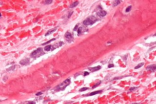
Osteoblasts are cells with a single nucleus that synthesize bone. However, in the process of bone formation, osteoblasts function in groups of connected cells. Individual cells cannot make bone. A group of organized osteoblasts together with the bone made by a unit of cells is usually called the osteon.

The internal carotid artery is an artery in the neck which supplies the anterior circulation of the brain.

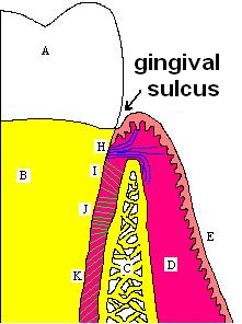
The periosteum is a membrane that covers the outer surface of all bones, except at the articular surfaces of long bones. Endosteum lines the inner surface of the medullary cavity of all long bones.

A hematoma, also spelled haematoma, or blood suffusion is a localized bleeding outside of blood vessels, due to either disease or trauma including injury or surgery and may involve blood continuing to seep from broken capillaries. A hematoma is benign and is initially in liquid form spread among the tissues including in sacs between tissues where it may coagulate and solidify before blood is reabsorbed into blood vessels. An ecchymosis is a hematoma of the skin larger than 10 mm.

Volkmann's contracture is a permanent flexion contracture of the hand at the wrist, resulting in a claw-like deformity of the hand and fingers. Passive extension of fingers is restricted and painful.

A cephalohaematoma is a hemorrhage of blood between the skull and the periosteum at any age, including a newborn baby secondary to rupture of blood vessels crossing the periosteum. Because the swelling is subperiosteal, its boundaries are limited by the individual bones, in contrast to a caput succedaneum.
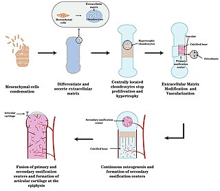
Endochondral ossification is one of the two essential pathways by which bone tissue is produced during fetal development of the mammalian skeletal system, the other pathway being intramembranous ossification. Both endochondral and intramembranous processes initiate from a precursor mesenchymal tissue, but their transformations into bone are different. In intramembranous ossification, mesenchymal tissue is directly converted into bone. On the other hand, endochondral ossification starts with mesenchymal tissue turning into an intermediate cartilage stage, which is eventually substituted by bone.

Intramembranous ossification is one of the two essential processes during fetal development of the gnathostome skeletal system by which rudimentary bone tissue is created. Intramembranous ossification is also an essential process during the natural healing of bone fractures and the rudimentary formation of bones of the head.

In osteology, the osteon or haversian system is the fundamental functional unit of much compact bone. Osteons are roughly cylindrical structures that are typically between 0.25 mm and 0.35 mm in diameter. Their length is often hard to define, but estimates vary from several millimeters to around 1 centimeter. They are present in many bones of most mammals and some bird, reptile, and amphibian species.
Haversian canals are a series of microscopic tubes in the outermost region of bone called cortical bone. They allow blood vessels and nerves to travel through them to supply the osteocytes.

A sequestrum is a piece of dead bone that has become separated during the process of necrosis from normal or sound bone.
In anatomy, a canaliculus is a small passageway.

The bony labyrinth is the rigid, bony outer wall of the inner ear in the temporal bone. It consists of three parts: the vestibule, semicircular canals, and cochlea. These are cavities hollowed out of the substance of the bone, and lined by periosteum. They contain a clear fluid, the perilymph, in which the membranous labyrinth is situated.
Sharpey's fibres are a matrix of connective tissue consisting of bundles of strong predominantly type I collagen fibres connecting periosteum to bone. They are part of the outer fibrous layer of periosteum, entering into the outer circumferential and interstitial lamellae of bone tissue.
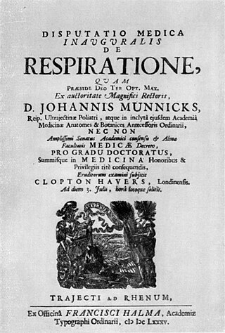
Clopton Havers was an English physician who did pioneering research on the microstructure of bone. He is believed to have been the first person to observe and almost certainly the first to describe what are now called Haversian canals and Sharpey's fibres.
Lamella means a small plate or flake in Latin, and in English may refer to:
Perforator flap surgery is a technique used in reconstructive surgery where skin and/or subcutaneous fat are removed from a distant or adjacent part of the body to reconstruct the excised part. The vessels that supply blood to the flap are isolated perforator(s) derived from a deep vascular system through the underlying muscle or intermuscular septa. Some perforators can have a mixed septal and intramuscular course before reaching the skin. The name of the particular flap is retrieved from its perforator and not from the underlying muscle. If there is a potential to harvest multiple perforator flaps from one vessel, the name of each flap is based on its anatomical region or muscle. For example, a perforator that only traverses through the septum to supply the underlying skin is called a septal perforator. Whereas a flap that is vascularised by a perforator traversing only through muscle to supply the underlying skin is called a muscle perforator. According to the distinct origin of their vascular supply, perforators can be classified into direct and indirect perforators. Direct perforators only pierce the deep fascia, they don't traverse any other structural tissue. Indirect perforators first run through other structures before piercing the deep fascia.

Orthopedic surgery is the branch of surgery concerned with conditions involving the musculoskeletal system. Orthopedic surgeons use both surgical and nonsurgical means to treat musculoskeletal injuries, sports injuries, degenerative diseases, infections, bone tumours, and congenital limb deformities. Trauma surgery and traumatology is a sub-specialty dealing with the operative management of fractures, major trauma and the multiply-injured patient.


