Wobbler disease

Last updated

Wobbler disease is a catchall term referring to several possible malformations of the cervical vertebrae that cause an unsteady (wobbly) gait and weakness in dogs and horses. A number of different conditions of the cervical (neck) spinal column cause similar clinical signs. These conditions may include malformation of the vertebrae, intervertebral disc protrusion, and disease of the interspinal ligaments, ligamenta flava, and articular facets of the vertebrae. [1] Wobbler disease is also known as cervical vertebral instability (CVI), cervical spondylomyelopathy (CSM), and cervical vertebral malformation (CVM). In dogs, the disease is most common in large breeds, especially Great Danes and Doberman Pinschers. In horses, it is not linked to a particular breed, though it is most often seen in tall, race-bred horses of Thoroughbred or Standardbred ancestry. It is most likely inherited to at least some extent in dogs and horses.[ citation needed ]

Contents

Wobbler disease in dogs

X-ray of wobbler disease in a dog DogAxisMalformation.jpg
X-ray of wobbler disease in a dog
Doberman C6-C7 and C5-C6 traction responsive myelopathy A.jpg
Doberman C6-C7 and C5-C6 traction responsive myelopathy A
Doberman C6-C7 and C5-C6 traction responsive myelopathy B.jpg
Doberman C6-C7 and C5-C6 traction responsive myelopathy B
T2 weighted MRI in neutral (A) and linear traction (B) of a seven-year-old Doberman with a two-year history of cervical pain treated with NSAIDs and presented acutely tetraplegic: A C6-C7 and C5-C6 traction responsive myelopathy are evident on MRI. The spinal-cord hyperintensity seen at the C5-C6 is suggestive of chronic lesion and most likely responsible for the chronic history of cervical pain, while the C5-C6 lesion was most likely responsible for the acute tetraplegia.
Doberman post surgery following double implant A.jpg
Doberman post surgery following double implant A
Doberman post surgery following double implant B.jpg
Doberman post surgery following double implant B
Same dog (A) treated with double implant (B) three days after surgery: The dog became ambulatory three days after surgery. Four weeks after surgery, it had ataxia without conscious proprioceptive deficits, and three months after surgery, the dog was neurologically normal. The owner reported it had been two years since the dog was able to hold its neck in an elevated position.

Wobbler disease is probably inherited in the Borzoi, Great Dane, Doberman, and Basset Hound. [2] Instability of the vertebrae of the neck (usually the caudal neck) causes spinal cord compression. In younger dogs such as Great Danes less than two years of age, wobbler disease is caused by stenosis (narrowing) of the vertebral canal [3] related to degeneration of the dorsal articular facets and subsequent thickening of the associated joint capsules and ligaments. [1] A high-protein diet may contribute to its development. [4] In middle-aged and older dogs such as Dobermans, intervertebral disc disease leads to bulging of the disc or herniation of the disc contents, and the spinal cord is compressed. [3] In Great Danes, the C4 to C6 vertebrae are most commonly affected; in Dobermans, the C5 to C7 vertebrae are affected. [5]


The disease tends to be gradually progressive. Symptoms such as weakness, ataxia, and dragging of the toes start in the rear legs. Dogs often have a crouching stance with a downward flexed neck. The disease progresses to the front legs, but the symptoms are less severe. Neck pain is sometimes seen. Symptoms are usually gradual in onset, but may progress rapidly following trauma. [6] X-rays may show misaligned vertebrae and narrow disk spaces, but it is not as effective as a myelogram, which reveals stenosis of the vertebral canal. Magnetic resonance imaging has been shown to be more effective at showing the location, nature, and severity of spinal cord compression than a myelogram. [7] Treatment is either medical to control the symptoms, usually with corticosteroids and cage rest, or surgical to correct the spinal cord compression. The prognosis is guarded in either case. Surgery may fully correct the problem, but it is technically difficult and relapses may occur. Types of surgery include ventral decompression of the spinal cord (ventral slot technique), dorsal decompression, and vertebral stabilization. [8] One study showed no significant advantage to any of the common spinal cord decompression procedures. [9] Another study showed that electroacupuncture may be a successful treatment for Wobbler disease. [10] A new surgical treatment using a proprietary medical device has been developed for dogs with disc-associated wobbler disease. It implants an artificial disc (cervical arthroplasty) in place of the affected disc space. [11]

Commonly affected dog breeds

Wobbler disease in horses

Wobbler disease or wobbler's syndrome is a broad category of cervical disorders in the horse, including the conditions listed above, as well as equine wobbles anemia and cervical vertebral myelopathy, spinal cord compression (sometimes referred to colloquially among horse owners as "cervical arthritis" due to the arthritis that accumulates in facets). Within the horse community, any neurological problem that causes limb proprioception issues or decreased performance in the horse that is suspected to be rooted in the neck is considered a type of wobbler disease. Most forms of wobbler disease have a root cause in cervical vertebral myelopathy (CVM), which causes inflammation and arthritis in the joint and is painful to affected horses. At this time it is believed to possibly be a congenital condition. Mounting evidence is growing that points to CVM and other cervical and vertebral disorders (like Kissing Spine) as hereditary and found in populations of specific families. [12]

Other forms, such as cervical arthritis and equine wobbles anemia, are concentrated in certain breeds and demonstrate possible hereditary factors. Horses with wobbler disease often exhibit ataxia (implying dysfunction of parts of the nervous system), show weakness in the hindquarters, or may knuckle over in their fetlocks, particularly in the rear. They also may show overall stiffness especially in the neck and back and may not move fluidly. A common ataxic symptom is the horse will list to one side behind. They also may demonstrate uneven strides with one leg intermittently shorter in step. Horses with wobbler disease or cervical anomalies can demonstrate difficult to diagnose lamenesses that come and go, and often are not resolved by veterinary blocks to the limbs. With advanced stages of the disease, affected horses are prone to falling due to extreme listing behind and often demonstrate difficulty getting up from lying down, or reluctance to lie down at all. While some cases are successfully treated with nutritional and medical management, surgery is also used. One method is the use of titanium baskets, placed to fuse the vertebrae, thereby preventing compression of the spinal cord. Some horses are able to return to work, with a few able to reach competitive levels. No complete cure for the condition is known. Famous horses known to have completed basket surgery include Seattle Slew. Seattle Slew underwent two basket surgeries and has sired multiple offspring that have cervical arthritis. He is one of many Thoroughbred stallions to have undergone basket surgery and still be used for stud.

Because wobbler disease is the best known of the neurological conditions that affect horses, other, unrelated conditions, such as kissing spine, equine protozoal myeloencephalitis and cerebellar abiotrophy, are sometimes misdiagnosed as wobbler disease, though the causes and symptoms differ.

Clinical Presentation

Including the symptoms listed above, clinical presentations of "wobblers" can also include neck pain and stiffness, difficulty tracking up or gait abnormalities, decreased performance, intermittent or swapping lameness particularly in diagonal pairs, forelimb lameness, abnormal head and neck posture, defensiveness or change in behavior, and abnormal sweat patterns.

Diagnosis

Wobbler disease is definitively diagnosed by x-ray, nuclear scintigraphy or bone scan. X-rays will show channel widening or filling the easiest and are often most cost-effective to horse owners. X-rays will also show any structural anomaly, arthritis, facet remodeling, or bone spurs present. Preliminary diagnosis can be made by ultrasound but x-rays are needed to measure the true depth of facet involvement. For extent of damage to associated structures, veterinarians may opt to have the horse undergo a bone scan or nuclear scintigraphy.

Commonly affected horse breeds

Related Research Articles

<span class="mw-page-title-main">Cervical spine disorder</span> Medical condition

Cervical spine disorders are illnesses that affect the cervical spine, which is made up of the upper first seven vertebrae, encasing and shielding the spinal cord. This fragment of the spine starts from the region above the shoulder blades and ends by supporting and connecting the skull.

<span class="mw-page-title-main">Spondylosis</span> Degeneration of the vertebral column

Spondylosis is the degeneration of the vertebral column from any cause. In the more narrow sense it refers to spinal osteoarthritis, the age-related degeneration of the spinal column, which is the most common cause of spondylosis. The degenerative process in osteoarthritis chiefly affects the vertebral bodies, the neural foramina and the facet joints. If severe, it may cause pressure on the spinal cord or nerve roots with subsequent sensory or motor disturbances, such as pain, paresthesia, imbalance, and muscle weakness in the limbs.

<span class="mw-page-title-main">Degenerative disc disease</span> Medical condition

Degenerative disc disease (DDD) is a medical condition typically brought on by the normal aging process in which there are anatomic changes and possibly a loss of function of one or more intervertebral discs of the spine. DDD can take place with or without symptoms, but is typically identified once symptoms arise. The root cause is thought to be loss of soluble proteins within the fluid contained in the disc with resultant reduction of the oncotic pressure, which in turn causes loss of fluid volume. Normal downward forces cause the affected disc to lose height, and the distance between vertebrae is reduced. The anulus fibrosus, the tough outer layers of a disc, also weakens. This loss of height causes laxity of the longitudinal ligaments, which may allow anterior, posterior, or lateral shifting of the vertebral bodies, causing facet joint malalignment and arthritis; scoliosis; cervical hyperlordosis; thoracic hyperkyphosis; lumbar hyperlordosis; narrowing of the space available for the spinal tract within the vertebra ; or narrowing of the space through which a spinal nerve exits with resultant inflammation and impingement of a spinal nerve, causing a radiculopathy.

<span class="mw-page-title-main">Klippel–Feil syndrome</span> Congenital condition characterised by fusion of two or more vertebrae in the neck

Klippel–Feil syndrome (KFS), also known as cervical vertebral fusion syndrome, is a rare congenital condition characterized by the abnormal fusion of any two of the seven bones in the neck. It results in a limited ability to move the neck and shortness of the neck, resulting in the appearance of a low hairline.

Middle back pain, also known as thoracic back pain, is back pain that is felt in the region of the thoracic vertebrae, which are between the bottom of the neck and top of the lumbar spine. It has a number of potential causes, ranging from muscle strain to collapse of a vertebra or rare serious diseases. The upper spine is very strong and stable to support the weight of the upper body, as well as to anchor the rib cage which provides a cavity to allow the heart and lungs to function and protect them.

Congenital vertebral anomalies are a collection of malformations of the spine. Most, around 85%, are not clinically significant, but they can cause compression of the spinal cord by deforming the vertebral canal or causing instability. This condition occurs in the womb. Congenital vertebral anomalies include alterations of the shape and number of vertebrae.

Polyneuropathy in dogs and cats is a collection of peripheral nerve disorders that often are breed-related in these animals. Polyneuropathy indicates that multiple nerves are involved, unlike mononeuropathy. Polyneuropathy usually involves motor nerve dysfunction, also known as lower motor neuron disease. Symptoms include decreased or absent reflexes and muscle tone, weakness, or paralysis. It often occurs in the rear legs and is bilateral. Most are chronic problems with a slow onset of symptoms, but some occur suddenly.

<span class="mw-page-title-main">Spinal cord compression</span> Medical condition

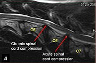
Spinal cord compression is a form of myelopathy in which the spinal cord is compressed. Causes can be bone fragments from a vertebral fracture, a tumor, abscess, ruptured intervertebral disc or other lesion.

Copenhagen disease, sometimes known as Copenhagen syndrome or progressive non-infectious anterior vertebral fusion (PAVF), is a unique spinal disorder with distinctive radiological features. This is a rare childhood disease of unknown cause, affecting females slightly more than males (60%). Prevalence is unknown, but there have been approximately 80–100 individuals with Copenhagen disease reported since 1949. However, there is still little known research due to the rarity of the disease. The disease is so rare that the National Organization for Rare Diseases does not even mention Copenhagen disease in their database.

<span class="mw-page-title-main">Spinal disc herniation</span> Injury to the connective tissue between spinal vertebrae

A spinal disc herniation is an injury to the cushioning and connective tissue between vertebrae, usually caused by excessive strain or trauma to the spine. It may result in back pain, pain or sensation in different parts of the body, and physical disability. The most conclusive diagnostic tool for disc herniation is MRI, and treatment may range from painkillers to surgery. Protection from disc herniation is best provided by core strength and an awareness of body mechanics including posture.

<span class="mw-page-title-main">Laminotomy</span> Surgical procedure

A laminotomy is an orthopaedic neurosurgical procedure that removes part of the lamina of a vertebral arch in order to relieve pressure in the vertebral canal. A laminotomy is less invasive than conventional vertebral column surgery techniques, such as laminectomy because it leaves more ligaments and muscles attached to the spinous process intact and it requires removing less bone from the vertebra. As a result, laminotomies typically have a faster recovery time and result in fewer postoperative complications. Nevertheless, possible risks can occur during or after the procedure like infection, hematomas, and dural tears. Laminotomies are commonly performed as treatment for lumbar spinal stenosis and herniated disks. MRI and CT scans are often used pre- and post surgery to determine if the procedure was successful.

<span class="mw-page-title-main">Laminoplasty</span>

Laminoplasty is an orthopaedic/neurosurgical surgical procedure for treating spinal stenosis by relieving pressure on the spinal cord. The main purpose of this procedure is to provide relief to patients who may have symptoms of numbness, pain, or weakness in arm movement. The procedure involves cutting the lamina on both sides of the affected vertebrae and then "swinging" the freed flap of bone open thus relieving the pressure on the spinal cord. The spinous process may be removed to allow the lamina bone flap to be swung open. The bone flap is then propped open using small wedges or pieces of bone such that the enlarged spinal canal will remain in place.

<span class="mw-page-title-main">Spinal disease</span> Diseases involving the vertebral column

Spinal disease refers to a condition impairing the backbone. These include various diseases of the back or spine ("dorso-"), such as kyphosis. Dorsalgia refers to back pain. Some other spinal diseases include spinal muscular atrophy, ankylosing spondylitis, lumbar spinal stenosis, spina bifida, spinal tumors, osteoporosis and cauda equina syndrome.

<span class="mw-page-title-main">Spinal stenosis</span> Disease of the bony spine that results in narrowing of the spinal canal

Spinal stenosis is an abnormal narrowing of the spinal canal or neural foramen that results in pressure on the spinal cord or nerve roots. Symptoms may include pain, numbness, or weakness in the arms or legs. Symptoms are typically gradual in onset and improve with leaning forward. Severe symptoms may include loss of bladder control, loss of bowel control, or sexual dysfunction.

<span class="mw-page-title-main">Vertebral column</span> Bony structure found in vertebrates

The vertebral column, also known as the backbone or spine, is the core part of the axial skeleton in vertebrate animals. The vertebral column is the defining characteristic of vertebrate endoskeleton in which the notochord found in all chordates has been replaced by a segmented series of mineralized irregular bones called vertebrae, separated by fibrocartilaginous intervertebral discs. The dorsal portion of the vertebral column houses the spinal canal, a cavity formed by alignment of the neural arches that encloses and protects the spinal cord.

The ventral slot technique is a procedure that allows the surgeon to reach and decompress the spinal cord and associated nerve roots from a ventral route in veterinary medicine. There are also alternative ways to open the spinal canal from dorsal by performing a hemilaminectomy, but this often gives only limited access. Even when the main pathological changes evolve from the midline, it is necessary to choose a ventral approach.

<span class="mw-page-title-main">Vertebra</span> Bone in the vertebral column

Each vertebra is an irregular bone with a complex structure composed of bone and some hyaline cartilage, that make up the vertebral column or spine, of vertebrates. The proportions of the vertebrae differ according to their spinal segment and the particular species.

<span class="mw-page-title-main">Cervicocranial syndrome</span> Medical condition

Cervicocranial syndrome or is a neurological illness. It is a combination of symptoms that are caused by an abnormality in the neck. The bones of the neck that are affected are cervical vertebrae. This syndrome can be identified by confirming cervical bone shifts, collapsed cervical bones or misalignment of the cervical bone leading to improper functioning of cervical spinal nerves.Greenberg Regenerative Medicine | Bryn Mawr, Pennsylvania Cervicocranial syndrome is either congenital or acquired. Some examples of diseases that could result in cervicocranial syndrome are Chiari disease, Klippel-Feil malformation osteoarthritis, and trauma. Treatment options include neck braces, pain medication and surgery. The quality of life for individuals suffering from CCJ syndrome can improve through surgery.

Craniocervical instability (CCI) is a medical condition where there is excessive movement of the vertebrae at the atlanto-occipital joint and the atlanto-axial joint, that is, between the skull and the top two vertebrae. This can cause neuronal injury and compression of nearby structures including the spinal cord, brain stem, vertebral artery or vagus nerve, causing a constellation of symptoms. It is frequently co-morbid with atlanto-axial instability, Chiari malformation and tethered cord syndrome.

Cervical spondylotic myelopathy (CSM) is a disorder characterised by the age-related deterioration of the cervical spinal cord. Also called spondylotic radiculomyelopathy (SRM), it is a neurological disorder related to the spinal cord and nerve roots. The severity of CSM is most commonly associated with factors including age, location and extent of spinal cord compression.

References

  1. 1 2 Bagley, Rodney S. (2006). "Acute Spinal Disease" (PDF). Proceedings of the North American Veterinary Conference. Retrieved 2007-02-19.
  2. "Spinal Cord Disorders: Small Animals". The Merck Veterinary Manual. 2006. Retrieved 2007-02-19.
  3. 1 2 Chrisman, Cheryl; Clemmons, Roger; Mariani, Christopher; Platt, Simon (2003). Neurology for the Small Animal Practitioner (1st ed.). Teton New Media. ISBN   1-893441-82-2.
  4. 1 2 Ettinger, Stephen J.; Feldman, Edward C. (1995). Textbook of Veterinary Internal Medicine (4th ed.). W.B. Saunders Company. ISBN   0-7216-6795-3.
  5. Danourdis, Anastassios M. (2004). "The Diagnostic and Therapeutic Approach to Cervical Spondylomyelopathy". Proceedings of the 29th World Congress of the World Small Animal Veterinary Association. Retrieved 2007-02-19.
  6. Braund, K.G. (2003). "Degenerative and Compressive Structural Disorders". Braund's Clinical Neurology in Small Animals: Localization, Diagnosis and Treatment. Retrieved 2007-02-19.
  7. da Costa R, Parent J, Dobson H, Holmberg D, Partlow G (2006). "Comparison of magnetic resonance imaging and myelography in 18 Doberman pinscher dogs with cervical spondylomyelopathy". Vet Radiol Ultrasound. 47 (6): 523–31. doi:10.1111/j.1740-8261.2006.00180.x. PMID   17153059.
  8. Wheeler, Simon J. (2004). "Update on Spinal Surgery I: Cervical Spine". Proceedings of the 29th World Congress of the World Small Animal Veterinary Association. Retrieved 2007-02-19.
  9. Jeffery N, McKee W (2001). "Surgery for disc-associated wobbler syndrome in the dog--an examination of the controversy". J Small Anim Pract. 42 (12): 574–81. doi: 10.1111/j.1748-5827.2001.tb06032.x . PMID   11791771.
  10. Sumano H, Bermudez E, Obregon K (2000). "Treatment of wobbler syndrome in dogs with electroacupuncture". Dtsch Tierarztl Wochenschr. 107 (6): 231–5. PMID   10916938.
  11. Adamo PF (2011). "Cervical arthroplasty in two dogs with disk-associated cervical spondylomyelopathy". J Am Vet Med Assoc. 239 (6): 808–17. doi:10.2460/javma.239.6.808. PMID   21916764.
  12. UC Davis Center for Equine Health [ dead link ]
  13. Cervical disorders [ dead link ]