Inversion (evolutionary biology)

Last updated

In evolutionary developmental biology, inversion refers to the hypothesis that during the course of animal evolution, the structures along the dorsoventral (DV) axis have taken on an orientation opposite that of the ancestral form.

Contents

Inversion was first noted in 1822 by the French zoologist Étienne Geoffroy Saint-Hilaire, when he dissected a crayfish (an arthropod) and compared it with the vertebrate body plan. The idea was heavily criticised, but periodically resurfaced, and is now supported by some molecular embryologists.

History

As early as 1822, the French zoologist Étienne Geoffroy Saint-Hilaire noted that the organization of dorsal and ventral structures in arthropods is opposite that of mammals. Five decades later, in light of Darwin's theory of "descent with modification", German zoologist Anton Dohrn proposed that these groups arose from a common ancestor which possessed a body plan similar to that of modern annelids with a ventral nerve cord and dorsal heart. [1] Whereas this arrangement is retained in arthropods and other protostomes, in chordate deuterostomes, the nerve cord is located dorsally and the heart ventrally. The inversion hypothesis was met with scornful criticism each time it was proposed, and has periodically resurfaced and been rejected. [1] However, some modern molecular embryologists suggest that recent findings support the idea of inversion.

Evidence for inversion

Generalized protostome and chordate body plans illustrating the opposite orientation of dorsoventral structures. Concomitant with the change in orientation of these structures is the inversion of domains of expression of TGF-b signals (dpp/BMPs) and their inhibitors (sog/Chordin). DV-axis-inversion.png
Generalized protostome and chordate body plans illustrating the opposite orientation of dorsoventral structures. Concomitant with the change in orientation of these structures is the inversion of domains of expression of TGF-β signals (dpp/BMPs) and their inhibitors (sog/Chordin).
Balanoglossus, an example of a Hemichordata, represents an "evolutionary link" between invertebrates and vertebrates. Ptychodera flava in New Caledonia, from Encyclopaedia Britannica (1911).png
Balanoglossus , an example of a Hemichordata, represents an "evolutionary link" between invertebrates and vertebrates.

In addition to the simple observation that the dorsoventral axes of protostomes and chordates appear to be inverted with respect to each other, molecular biology provides some support for the inversion hypothesis. The most notable piece of evidence comes from analysis of the genes involved in establishing the DV axis in these two groups. [2] In the fruit fly Drosophila melanogaster , as well as in other protostomes, the β-type transforming growth factor (TGF-β) family member decapentaplegic (dpp) is expressed dorsally and is thought to suppress neural fate. On the ventral side of the embryo, a dpp inhibitor, short gastrulation (sog), is expressed, thus allowing nervous tissue to form ventrally. In chordates, the dpp homolog BMP-4 is expressed in the prospective ventral (non-neural) part of the embryo while several sog-like BMP inhibitors (Chordin, Noggin, Follistatin) are expressed dorsally. [1]

Other patterning genes also show conserved domains of expression. The neural patterning genes vnd, ind, msh, and netrin are expressed in the Drosophila ventral nerve cells and midline mesectoderm. The chordate homologs of these genes, NK2, Gsh1/2, Msx1/3, and Netrin, are expressed in the dorsal neural tube. Furthermore, the tinman/Nkx2-5 gene is expressed very early in cells that will become the heart in both Drosophila (dorsally) and chordates (ventrally). [1]

Additional support comes from work on the development of the polychaete annelid Platynereis dumerilii , another protostome. Even more so than Drosophila, its pattern of central-nervous-system development is strikingly similar to that of vertebrates, but inverted. [3] There is also evidence from left-right asymmetry. Vertebrates have a highly conserved Nodal signaling pathway that acts on the left side of the body, determining left-right asymmetries of internal organs. Sea urchins have the same signaling pathway, but it acts on the right side of the body. [4] It was even shown that an opposing right-sided signal for regulating left-right asymmetry in vertebrates, i.e. BMP signaling pathway, is activated on the left side of the sea urchin larva, [5] suggesting an axial inversion during evolution from basal deuterostome to chordate such as amphioxus. The Nodal signaling pathway in amphioxus is on the left side of the embryo, which is the same situation as vertebrates. [6] Sea urchins, like other echinoderms, have radially-symmetric adults, but bilaterally-symmetric larvae. Since sea urchins are deuterostomes, this suggests that the ancestral deuterostome shared its orientation with protostomes, and that dorsoventral inversion originated in some ancestral chordate.

There is evidence that invertebrate chordates are also inverted. Ascidian larvae have a dorsal mouth, as one would expect from inversion. [7] The amphioxus has an odd feature: its mouth appears on the left side and migrates to the ventral side. [8] Biologist Thurston Lacalli speculates that this may be a recapitulation of the migration of the mouth from the dorsal to the ventral side in a protochordate. [9]

Hemichordates: an intermediate body plan?

Some biologists have proposed that the Hemichordates (specifically the Enteropneusta) may represent an intermediate body plan in the evolution of the "inverted" state of the chordates. [10] Though they are considered deuterostomes, the dorsoventral axis of hemichordates retains features of both protostomes and chordates. For example, enteropneusts have an ectodermally-derived dorsal nerve cord in the collar region which has been proposed to be homologous to the chordate neural tube. However, they also have a ventral nerve cord and a dorsal contractile vessel similar to protostomes. [10] Furthermore, the relative positions other "intermediate" structures in hemichordates, such as the hepatic organs and ventral pygochord, which has been proposed to be homologous to the chordate-defining notochord, [11] are retained but inverted. Nübler-Jung and Arendt argue that the principal innovation in the chordate lineage was the obliteration of the mouth on the neural side (as in hemichordates, arthropods, and annelids) and the development of a new mouth on the non-neural ventral side. [10]

Alternative hypotheses

While the idea of dorsoventral axis inversion appears to be supported by morphological and molecular data, others have proposed alternative plausible hypotheses (reviewed in Gerhart 2000). [1] One assumption of the inversion hypothesis is that the common ancestor of protostomes and chordates already possessed an organized central nervous system located at one pole of the dorsoventral axis. Alternatively, this ancestor may have possessed only a diffuse nerve net or several bundles of nervous tissue with no distinct dorsoventral localization. [1] [12] This would mean that the apparent inversion was simply a result of concentration of the central nervous system at opposite poles independently in the lineages leading to protostomes and chordates. Lacalli (1996) suggested a scenario in which the ancestor had a single opening to the digestive system, and that the neural and non-neural mouths arose independently in protostomes and chordates, respectively. [13] By this hypothesis, there is no need for inversion.

Martindale and Henry propose a ctenophore-like ancestor (biradial rather than bilateral) with a concentrated nerve cord and two anal pores on opposite sides of the animal in addition to a terminal gut opening. [1] [14] If one of these pores became the mouth in protostomes and the other became the mouth in deuterostomes, this would also preclude inversion. Another alternative, proposed by von Salvini-Plawen, states that the ancestor had a two-part nervous system - one part concentrated, the other diffuse. The nervous systems of protostome and deuterostome descendants of this ancestor may have arisen independently from these two distinct parts. [1] [15]

The Axial Twist theory proposes that not the whole body, but only the anterior region of the head is inverted. [16] [17] These theories by Kinsbourne [16] and de Lussanet & Osse [17] also explain the presence of an optic chiasm in vertebrates and the contralateral organization of the forebrain. One of these theories [17] is supported by developmental evidence and even explains the asymmetric organization of the heart and bowels. [18]

Related Research Articles

<span class="mw-page-title-main">Chordate</span> Phylum of animals having a dorsal nerve cord

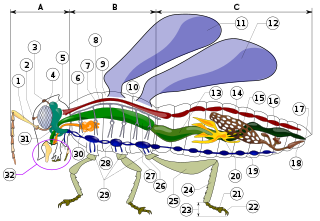
A chordate is a deuterostomic animal belonging to the phylum Chordata. All chordates possess, at some point during their larval or adult stages, five distinctive physical characteristics (synapomorphies) that distinguish them from other taxa. These five synapomorphies are a notochord, a hollow dorsal nerve cord, an endostyle or thyroid, pharyngeal slits, and a post-anal tail. The name "chordate" comes from the first of these synapomorphies, the notochord, which plays a significant role in chordate body plan structuring and movements. Chordates are also bilaterally symmetric, have a coelom, possess a closed circulatory system, and exhibit metameric segmentation.

<span class="mw-page-title-main">Nervous system</span> Part of an animal that coordinates actions and senses

In biology, the nervous system is the highly complex part of an animal that coordinates its actions and sensory information by transmitting signals to and from different parts of its body. The nervous system detects environmental changes that impact the body, then works in tandem with the endocrine system to respond to such events. Nervous tissue first arose in wormlike organisms about 550 to 600 million years ago. In vertebrates, it consists of two main parts, the central nervous system (CNS) and the peripheral nervous system (PNS). The CNS consists of the brain and spinal cord. The PNS consists mainly of nerves, which are enclosed bundles of the long fibers, or axons, that connect the CNS to every other part of the body. Nerves that transmit signals from the brain are called motor nerves or efferent nerves, while those nerves that transmit information from the body to the CNS are called sensory nerves or afferent. Spinal nerves are mixed nerves that serve both functions. The PNS is divided into three separate subsystems, the somatic, autonomic, and enteric nervous systems. Somatic nerves mediate voluntary movement. The autonomic nervous system is further subdivided into the sympathetic and the parasympathetic nervous systems. The sympathetic nervous system is activated in cases of emergencies to mobilize energy, while the parasympathetic nervous system is activated when organisms are in a relaxed state. The enteric nervous system functions to control the gastrointestinal system. Both autonomic and enteric nervous systems function involuntarily. Nerves that exit from the cranium are called cranial nerves while those exiting from the spinal cord are called spinal nerves.

<span class="mw-page-title-main">Hemichordate</span> Phylum of marine deuterostome animals

Hemichordata is a phylum which consists of triploblastic, enterocoelomate, and bilaterally symmetrical marine deuterostome animals, generally considered the sister group of the echinoderms. They appear in the Lower or Middle Cambrian and include two main classes: Enteropneusta, and Pterobranchia. A third class, Planctosphaeroidea, is known only from the larva of a single species, Planctosphaera pelagica. The class Graptolithina, formerly considered extinct, is now placed within the pterobranchs, represented by a single living genus Rhabdopleura.

<span class="mw-page-title-main">Vetulicolia</span> Extinct Cambrian taxon of deuterostomes

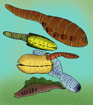
Vetulicolia is a phylum of animals encompassing several extinct species belonging to the Cambrian Period. The phylum was created by Degan Shu and his research team in 2001, and named after Vetulicola cuneata, the first species of the phylum described in 1987. The vetulicolian body comprises two parts: a voluminous anterior forebody, tipped with an anteriorly positioned mouth and lined with a row of five round to oval-shaped features on each lateral side, which have been interpreted as gills ; and a posterior section that primitively comprises seven segments and functions as a tail. All vetulicolians lack preserved appendages of any kind, having no legs, feelers or even eyes. The area where the anterior and posterior parts join is constricted.

<span class="mw-page-title-main">Bilateria</span> Animals with embryonic bilateral symmetry

Bilateria is a large clade or infrakingdom of animals called bilaterians, characterized by bilateral symmetry during embryonic development. This means their body plans are laid around a longitudinal axis with a front and a rear end, as well as a left–right–symmetrical belly (ventral) and back (dorsal) surface. Nearly all bilaterians maintain a bilaterally symmetrical body as adults; the most notable exception is the echinoderms, which achieve secondary pentaradial symmetry as adults, but are bilaterally symmetrical as an embryo. Cephalization is also a characteristic feature among most bilaterians, where the special sense organs and central nerve ganglia become concentrated at the front/rostral end.

<span class="mw-page-title-main">Neural tube</span> Developmental precursor to the central nervous system

In the developing chordate, the neural tube is the embryonic precursor to the central nervous system, which is made up of the brain and spinal cord. The neural groove gradually deepens as the neural fold become elevated, and ultimately the folds meet and coalesce in the middle line and convert the groove into the closed neural tube. In humans, neural tube closure usually occurs by the fourth week of pregnancy.

<span class="mw-page-title-main">Gastrulation</span> Stage in embryonic development in which germ layers form

Gastrulation is the stage in the early embryonic development of most animals, during which the blastula, or in mammals the blastocyst is reorganized into a two-layered or three-layered embryo known as the gastrula. Before gastrulation, the embryo is a continuous epithelial sheet of cells; by the end of gastrulation, the embryo has begun differentiation to establish distinct cell lineages, set up the basic axes of the body, and internalized one or more cell types including the prospective gut.

<span class="mw-page-title-main">Notochord</span> Flexible rod-shaped structure in all chordates

In zoology and developmental anatomy, the notochord is an elastic, rod-like anatomical structure found in many deuterostomal animals. A notochord is one of five synapomorphies used to define a species as a chordate.

<i>Pikaia</i> Extinct genus of primitive chordates

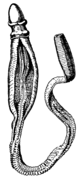
Pikaia gracilens is an extinct, primitive chordate animal known from the Middle Cambrian Burgess Shale of British Columbia. Described in 1911 by Charles Doolittle Walcott as an annelid, and in 1979 by Harry B. Whittington and Simon Conway Morris as a chordate, it became "one of the most famous early chordate fossils," or "famously known as the earliest described Cambrian chordate". It is estimated to have lived during the latter period of the Cambrian explosion. Since its initial discovery, more than a hundred specimens have been recovered.

<span class="mw-page-title-main">Lancelet</span> Order of chordates

The lancelets, also known as amphioxi, consist of some 30 to 35 species of "fish-like" benthic filter feeding chordates in the subphylum Cephalochordata, class Leptocardii, and family Branchiostomatidae.

<span class="mw-page-title-main">Acorn worm</span> Class of hemichordate invertebrates

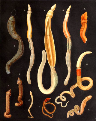
The acorn worms or Enteropneusta are a hemichordate class of invertebrates consisting of one order of the same name. The closest non-hemichordate relatives of the Enteropneusta are the echinoderms. There are 111 known species of acorn worm in the world, the main species for research being Saccoglossus kowalevskii. Two families—Harrimaniidae and Ptychoderidae—separated at least 370 million years ago.

<span class="mw-page-title-main">Pharyngeal slit</span> Repeated openings that appear along the pharynx of chordates

Pharyngeal slits are filter-feeding organs found among deuterostomes. Pharyngeal slits are repeated openings that appear along the pharynx caudal to the mouth. With this position, they allow for the movement of water in the mouth and out the pharyngeal slits. It is postulated that this is how pharyngeal slits first assisted in filter-feeding, and later, with the addition of gills along their walls, aided in respiration of aquatic chordates. These repeated segments are controlled by similar developmental mechanisms. Some hemichordate species can have as many as 200 gill slits. Pharyngeal clefts resembling gill slits are transiently present during the embryonic stages of tetrapod development. The presence of pharyngeal arches and clefts in the neck of the developing human embryo famously led Ernst Haeckel to postulate that "ontogeny recapitulates phylogeny"; this hypothesis, while false, contains elements of truth, as explored by Stephen Jay Gould in Ontogeny and Phylogeny. However, it is now accepted that it is the vertebrate pharyngeal pouches and not the neck slits that are homologous to the pharyngeal slits of invertebrate chordates. Pharyngeal arches, pouches, and clefts are, at some stage of life, found in all chordates. One theory of their origin is the fusion of nephridia which opened both on the outside and the gut, creating openings between the gut and the environment.

The dorsal nerve cord is an anatomical feature found in chordate animals, mainly in the subphylum Vertebrata. It is one of the five embryonic features unique to all chordates, the other four being a notochord, a post-anal tail, an endostyle, and pharyngeal slits.

<span class="mw-page-title-main">Ventral nerve cord</span>

The ventral nerve cord is a major structure of the invertebrate central nervous system. It is the functional equivalent of the vertebrate spinal cord. The ventral nerve cord coordinates neural signaling from the brain to the body and vice versa, integrating sensory input and locomotor output. Because arthropods have an open circulatory system, decapitated insects can still walk, groom, and mate—illustrating that the circuitry of the ventral nerve cord is sufficient to perform complex motor programs without brain input.

<span class="mw-page-title-main">Cerberus (protein)</span> Protein-coding gene in the species Homo sapiens

Cerberus is a protein that in humans is encoded by the CER1 gene. Cerberus is a signaling molecule which contributes to the formation of the head, heart and left-right asymmetry of internal organs. This gene varies slightly from species to species but its overall functions seem to be similar.

<span class="mw-page-title-main">Deuterostome</span> Superphylum of bilateral animals

Deuterostomes are bilaterian animals of the superphylum Deuterostomia, typically characterized by their anus forming before the mouth during embryonic development. Deuterostomia is further divided into 4 phyla; Chordata, Echinodermata, Hemichordata, the extinct Vetulicolia known from Cambrian fossils. The extinct clade Cambroernida is also though to be a member of Deuterostomia.

The evolution of nervous systems dates back to the first development of nervous systems in animals. Neurons developed as specialized electrical signaling cells in multicellular animals, adapting the mechanism of action potentials present in motile single-celled and colonial eukaryotes. Primitive systems, like those found in protists, use chemical signalling for movement and sensitivity; data suggests these were precursors to modern neural cell types and their synapses. When some animals started living a mobile lifestyle and eating larger food particles externally, they developed ciliated epithelia, contractile muscles and coordinating & sensitive neurons for it in their outer layer.

<span class="mw-page-title-main">Contralateral brain</span> Each side of the forebrain represents the opposite side of the body

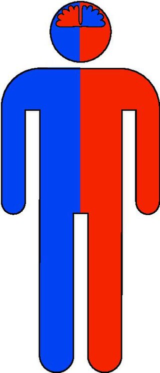
The contralateral organization of the forebrain is the property that the hemispheres of the cerebrum and the thalamus represent mainly the contralateral side of the body. Consequently, the left side of the forebrain mostly represents the right side of the body, and the right side of the brain primarily represents the left side of the body. The contralateral organization involves both executive and sensory functions. The contralateral organization is only present in vertebrates.

<span class="mw-page-title-main">Axial twist theory</span> Scientific theory in vertebrate development

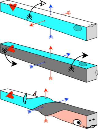
The axial twist theory is a scientific theory put forward to explain a range of unusual aspects of the body plan of vertebrates. It proposes that the rostral part of the head is "turned around" regarding the rest of the body. This end-part consists of the face as well as part of the brain. According to the theory, the vertebrate body has a left-handed chirality.

Left-right asymmetry is the process in early embryonic development that breaks the normal symmetry in the bilateral embryo. In vertebrates, left-right asymmetry is established early in development at a structure called the left-right organizer and leads to activation of different signalling pathways on the left and right of the embryo. This in turn cause several organs in adults to develop LR asymmetry, such as the tilt of the heart, the different number lung lobes on each side of the body and the position of the stomach and spleen on the right side of the body. If this process does not occur correctly in humans it can result in the syndromes heterotaxy or situs inversus.

References

  1. 1 2 3 4 5 6 7 8 Gerhart, J. (2000). "Inversion of the chordate body axis: Are there alternatives?". Proc. Natl. Acad. Sci. USA. 97 (9): 4445–8. Bibcode:2000PNAS...97.4445G. doi: 10.1073/pnas.97.9.4445 . PMC   34319 . PMID   10781041.
  2. De Robertis EM, Sasai Y (1996). "A common plan for dorsoventral patterning in Bilateria". Nature. 380 (6569): 37–40. Bibcode:1996Natur.380...37D. doi:10.1038/380037a0. PMID   8598900. S2CID   4355458.
  3. Alexandru S. Denes; Gáspár Jékely; Patrick R.H. Steinmetz; Florian Raible; Heidi Snyman; Benjamin Prud'homme; David E.K. Ferrier; Guillaume Balavoine; Detlev Arendt (2007). "Molecular Architecture of Annelid Nerve Cord Supports Common Origin of Nervous System Centralization in Bilateria". Cell. 129 (2): 277–288. doi: 10.1016/j.cell.2007.02.040 . PMID   17448990. S2CID   14904427.
  4. Véronique Duboc; Eric Röttinger; François Lapraz; Lydia Besnardeau; Thierry Lepage (2005). "Left-Right Asymmetry in the Sea Urchin Embryo Is Regulated by Nodal Signaling on the Right Side". Developmental Cell. 9 (1): 147–158. doi: 10.1016/j.devcel.2005.05.008 . PMID   15992548.
  5. Luo YJ, Su YH (2012). "Opposing Nodal and BMP Signals Regulate Left–Right Asymmetry in the Sea Urchin Larva". PLOS Biology. 10 (10): e1001402. doi: 10.1371/journal.pbio.1001402 . PMC   3467216 . PMID   23055827.
  6. Yu JK, Holland LZ, Holland ND (2002). "An amphioxus nodal gene (AmphiNodal) with early symmetrical expression in the organizer and mesoderm and later asymmetrical expression associated with left-right axis formation". Evolution & Development. 4 (6): 418–25. doi:10.1046/j.1525-142x.2002.02030.x. PMID   12492142. S2CID   44814248.
  7. Thurston C. Lacalli (2006). "Prospective protochordate homologs of vertebrate midbrain and MHB, with some thoughts on MHB origins". International Journal of Biological Sciences. 2 (3): 104–109. doi:10.7150/ijbs.2.104. PMC   1458436 . PMID   16763669.
  8. Kinya Yasui; Takao Kaji (2008). "The Lancelet and Ammocoete Mouths" (PDF). Zoological Science. 25 (10): 1012–1019. doi:10.2108/zsj.25.1012. PMID   19267637. S2CID   26726046. Archived from the original (PDF) on 2016-03-03. Retrieved 2012-06-16.
  9. Thurston C. Lacalli (2009). "The emergence of the chordate body plan: some puzzles and problems". Acta Zoologica. 91 (1): 4–10. doi:10.1111/j.1463-6395.2008.00384.x.
  10. 1 2 3 Nübler-Jung K, Arendt D (1999). "Dorsoventral axis inversion: Enteropneust anatomy links invertebrates to chordates turned upside down". J Zool Syst Evol Research. 37 (2): 93–100. doi: 10.1046/j.1439-0469.1999.372106.x .
  11. Cameron CB (2005). "A phylogeny of the hemichordates based on morphological characters". Can J Zool. 83: 196–215. doi:10.1139/Z04-190. S2CID   59467984.
  12. Martin-Duran JM, Pang K, Børve A, Semmler Lê H, Furu A, Cannon JT, Jondelius U, Hejnol A (2017). "Convergent evolution of bilaterian nerve cords". Nature. 553 (7686): 1–8. Bibcode:2018Natur.553...45M. doi:10.1038/nature25030. PMC   5756474 . PMID   29236686.
  13. Lacalli T (1996). "Dorsoventral axis inversion: a phylogenetic perspective". BioEssays. 18 (3): 251–4. doi:10.1002/bies.950180313.
  14. Martindale MQ, Henry JQ (1998). "The development of radial and biradial symmetry: The evolution of bilaterality". Am. Zool. 38 (4): 672–84. doi: 10.1093/icb/38.4.672 .
  15. von Salvini-Plawen L (1998). "The urochordate larva and archichordate organization: chordate origin and anagenesis revisited". J. Zool. Syst. Evol. Res. 36 (3): 129–45. doi: 10.1111/j.1439-0469.1998.tb00832.x .
  16. 1 2 Kinsbourne, Marcel (2013). "Somatic twist: A model for the evolution of decussation". Neuropsychology. 27 (5): 511–5. doi:10.1037/a0033662. PMID   24040928. S2CID   11646580.
  17. 1 2 3 De Lussanet, Marc H.E; Osse, Jan W.M (2012). "An ancestral axial twist explains the contralateral forebrain and the optic chiasm in vertebrates". Animal Biology. 62 (2): 193–216. arXiv: 1003.1872 . doi:10.1163/157075611X617102. S2CID   7399128.
  18. de Lussanet M. H. E., Osse J. W. M. (2015). "Decussation as an axial twist: A comment on Kinsbourne (2013)". Neuropsychology. 29 (5): 713–714. doi:10.1037/neu0000163. PMID   25528610. S2CID   6983617.