Polysomy

Last updated
Human karyogram, with annotated bands and sub-bands. It is a graphical representation of the idealized human diploid karyotype. It shows dark and white regions on G banding. Each row is vertically aligned at centromere level. It shows 22 homologous autosomal chromosome pairs, both the female (XX) and male (XY) versions of the two sex chromosomes, as well as the mitochondrial genome (at bottom left).
Further information: Karyotype Human karyotype with bands and sub-bands.png
Human karyogram, with annotated bands and sub-bands. It is a graphical representation of the idealized human diploid karyotype. It shows dark and white regions on G banding. Each row is vertically aligned at centromere level. It shows 22 homologous autosomal chromosome pairs, both the female (XX) and male (XY) versions of the two sex chromosomes, as well as the mitochondrial genome (at bottom left).
Trisomy 21 - Down syndrome, an example of a polysomy at chromosome 21 21 trisomy - Down syndrome.png
Trisomy 21 – Down syndrome, an example of a polysomy at chromosome 21

Polysomy is a condition found in many species, including fungi, plants, insects, and mammals, in which an organism has at least one more chromosome than normal, i.e., there may be three or more copies of the chromosome rather than the expected two copies. [1] Most eukaryotic species are diploid, meaning they have two sets of chromosomes, whereas prokaryotes are haploid, containing a single chromosome in each cell. Aneuploids possess chromosome numbers that are not exact multiples of the haploid number and polysomy is a type of aneuploidy. [2] A karyotype is the set of chromosomes in an organism and the suffix -somy is used to name aneuploid karyotypes. This is not to be confused with the suffix -ploidy, referring to the number of complete sets of chromosomes.

Contents

Polysomy is usually caused by non-disjunction (the failure of a pair of homologous chromosomes to separate) during meiosis, but may also be due to a translocation mutation (a chromosome abnormality caused by rearrangement of parts between nonhomologous chromosomes). Polysomy is found in many diseases, including Down syndrome in humans where affected individuals possess three copies (trisomy) of chromosome 21. [3]

Polysomic inheritance occurs during meiosis when chiasmata form between more than two homologous partners, producing multivalent chromosomes. [1] Autopolyploids may show polysomic inheritance of all the linkage groups, and their fertility may be reduced due to unbalanced chromosome numbers in the gametes. [1] In tetrasomic inheritance, four copies of a linkage group rather than two (tetrasomy) assort two-by-two. [1]

Types

Polysomy types are categorized based on the number of extra chromosomes in each set, noted as a diploid (2n) with an extra chromosome of various numbers. For example, a polysomy with three chromosomes is called a trisomy, a polysomy with four chromosomes is called tetrasomy, etc.: [4]

Number of chromosomesNameDescriptionExamples
3 trisomy Three copies of a chromosome, 2n + 1 Down syndrome (Trisomy 21), Edwards syndrome (Trisomy 18), or Patau syndrome (Trisomy 13) [3]
4 tetrasomy Four copies of a chromosome, 2n + 2 Tetrasomy 9p, Tetrasomy 18p [5]
5pentasomyFive copies of a chromosome, 2n + 3 Pentasomy X (XXXXX or 49, XXXXX) [6]
6hexasomySix copies of a chromosome, 2n + 4 Mosaic Hexasomy 21 or Partial Hexasomy 15 [7]
7heptasomySeven copies of a chromosome, 2n + 5 Heptasomy 21 in acute myeloid leukemia [8]
8octosomyEight copies of a chromosome, 2n + 6Octosomy in sturgeon fish ( Acipenser baerii , A. persicus, A. sinensis, and A. transmontanus) [9]
9nanosomyNine copies of a chromosome, 2n + 7Nanosomy in congenital skeletal polydystrophy [10]
10decasomyTen copies of a chromosome, 2n + 8decasomy 8 in histolytic carcinoma [11]

In mammals

In canines

Trisomy 13 Trisomy13.jpg
Trisomy 13

Polysomy plays a role in canine leukemia, hemangiopericytomas, and thyroid tumors. [12] Abnormalities of chromosome 13 have been observed in canine osteoid chondrosarcoma and lymphosarcoma. [13] Trisomy 13 in dogs with lymphosarcoma show a longer duration of first remission (medicine) and survival, responding well to treatments with chemotherapeutic agents. [14] Polysomy of chromosome 13 (Polysomy 13) is significant in the development of prostate cancer and is often caused by centric fusions. [12] Since canine chromosome 13 is similar to human chromosome 8q, research could provide insight to treatment for prostate cancer in humans. [15] Polysomy of chromosomes 1, 2, 4, 5, and 25 are also frequently involved in canine tumors. [16] Chromosome 1 may contain a gene responsible for tumor development and lead to changes in the karyotype, including fusion of the centromere, or centric fusions. [16] Aneuploidy due to nondisjunction is a common feature in tumor cells. [17]

In humans

Sex chromosomes

Some of the most frequent genetic disorders are abnormalities of sex chromosomes, but polysomies rarely occur. [18] 49,XXXXY chromosome polysomy occurs every 1 in 85,000 newborn males. [19] The incidence of other X polysomies (48,XXXX, 48,XXXY, 48,XXYY) is more rare than 49,XXXXY. [20] Polysomy Y (47,XYY; 48,XYYY; 48,XXYY; 49,XXYYY) occurs in 1 out of 975 males and may cause psychiatric, social, and somatic abnormalities. [21] Polysomy X may cause mental and developmental retardation and physical malformation. Klinefelter syndrome is an example of human polysomy X with the karyotype 47, XXY. X chromosome polysomies can be inherited from either a single maternal (49, X polysomies) or paternal (48, X polysomies) X chromosome. [18] Polysomy of sex chromosomes is caused by successive nondisjunctions in meiosis I and II. [6]

Karyotype of Polysomy Y (XYY) XYY diagram.gif
Karyotype of Polysomy Y (XYY)
example of Polysomy X (47,XXY, Klinefelter syndrome) Human karyotype (259 31) Karyotype Human 47,XXY (Klinefelter syndrome).jpg
example of Polysomy X (47,XXY, Klinefelter syndrome)
Effects of Polysomy X as seen in Klinefelter syndrome Klinefelter's syndrome.jpg
Effects of Polysomy X as seen in Klinefelter syndrome

Chromosome 7

CFTR gene on chromosome 7 CFTR gene on chromosome 7.svg
CFTR gene on chromosome 7

In squamous cell carcinoma, a protein from the epidermal growth factor receptor (EGFR) gene is often overexpressed in conjunction with polysomy of chromosome 7, so chromosome 7 can be used to predict the presence of EGFR in squamous cell carcinoma. [22] In colorectal cancer, EGFR expression is decreased with polysomy 7, which makes polysomy 7 easier to detect and could be used to prevent patients from having unnecessary cancer treatment. [23]

Chromosome 8

AML-M2 associated with chromosome 8 abnormality AML-M2 associated with a t(8;21) chromosome abnormality.jpg
AML-M2 associated with chromosome 8 abnormality

Tetrasomy and hexasomy 8 are rare compared to trisomy 8, which is the most common karyotypic finding in acute myeloid leukemia (AML) and myelodysplastic syndromes (MDS). [24] AML, MDS, or myeloproliferative disorder (MPD) with a high incidence of secondary diseases and a six-month survival rate are associated with a polysomy 8 syndrome. [25]

Chromosome 17

Overexpression of the HER2/neu gene on chromosome 17 and some type of polysomy has been reported in 8-68% of breast carcinomas. [26] If theHER-2/neu gene does not amplify in the case of polysomy, proteins may be overexpressed and could lead to tumerogenesis. [27] Polysomy 17 may complicate the interpretation of HER2 testing results in cancer patients. Chromosome 17 polysomy may not be present when the centromere is amplified, so it was later discovered that polysomy 17 is rare. This was discovered using array comparative genomic hybridization, a DNA-based alternative for clinical evaluation of HER2 gene copy number. [28]

Trisomy 21

Nuchal edema in Down Syndrome Dr. W. Moroder Nuchal edema in Down Syndrome Dr. W. Moroder.jpg
Nuchal edema in Down Syndrome Dr. W. Moroder

Trisomy 21 is a form of Down syndrome that occurs when there is an extra copy of chromosome 21. The result is a genetic condition in which a person has 47 chromosomes instead of the usual 46. During egg or sperm development the 21st chromosome does not separate during either the egg or sperm development. The result is a cell that has 24 chromosomes. This extra chromosome may cause problems with the manner in which the body and brain develop. [29]

Tetrasomy 9p

Tetrasomy 9p is a rare condition in which people have a small extra chromosome that contains two copies of part of chromosome 9, in addition to having two normal chromosome 9's as well. This condition may be diagnosed by analyzing a person's blood sample since 9p is found in high concentrations in the blood. Ultrasound is another tool that may be utilized to identify tetrasomy 9p in infants prior to birth. Prenatal ultrasound may reveal several common characteristics including: growth restriction, ventriculomegaly, cleft lip or palate, and renal anomalies. [30]

Tetrasomy 18p

Tetrasomy 18p occurs when the short arm of the 18th chromosome appears four times, rather than twice, in the cells of the body. It is considered to be a rare disease and usually is not inherited. The mechanism of 18p formation appears to be the result of two independent events: centromeric misdivision and nondisjunction. [31] Characteristic features of tetrasomy 18p include, but are not limited to: growth retardation, scoliosis, abnormal brain MRI, developmental delays, and strabismus. [31]

In insects

Germ line polysomy in the grasshopper

Karyotype showing chromosomes 1-22 are grouped A-G Karyotype isochromosomeX.JPG
Karyotype showing chromosomes 1–22 are grouped A-G

Germ line cells develop into eggs and sperm and the associated inherited material can be passed down to future generations. [32] As shown in the associated karyotype image, chromosomes 1–22 are grouped A-G. A population of male grasshoppers ( Chorthippus binotatus ) from the Sierra Nevada (Spain) are polysomic mosaics (coming from cells of two genetically different types) possessing an extra E group chromosome(chromosomes 16, 17 & 18) in their testicles. [33] Parents that exhibited polysomy did not pass the E chromosome abnormality to any of the offspring, so this is not something that is passed down to future generations. [33] Male grasshoppers ( Atractomorpha similis ) from Australia carry between one and ten extra copies of chromosome A9, with one being the most common in natural populations. [34] Most polysomic males produce normal sperm. However, polysomy can be transmissible through both the male and female parents through nondisjunction. [34]

Heterochromatic polysomy in the cricket

Heterochromatin contains a small number of genes and densely staining nodules in or along chromosomes. [35] The mole cricket chromosome number varies between 19 and 23 chromosomes depending on the part of the world in which they are located, including Jerusalem, Palestine, and Europe. [36] Heterochromic polysomy is seen in mole crickets with 23 chromosomes and may be a factor contributing to their evolution, specifically within the species Gryllotalpa gryllotalpa , along with various living environments and mating systems. [36] [37]

X-chromosome polysomy in the fruit fly

In the fruit fly, Drosophila , one X chromosome in the male is almost the same as two X chromosomes in the female in terms of the gene product produced. [38] Despite this, metafemales, or females having three X chromosomes, are unlikely to survive. [38] It is possible that the extra X chromosome decreases gene expression and could explain why the metafemales rarely survive this X-chromosome polysomy. [38]

In plants

A karyotype rearrangement of individual chromosomes takes place when polysomy in plants in observed. The mechanism of this type of rearrangement is "non-disjunction, mis-segregation in diploids or polyploids; mis-segregation from multivalents in interchange heterozygotes." [39] Incidences of polysomy have been identified in many species of plants, including:

Brosen flower nn1, Brassica rapa Brosen flower nn1.jpg
Brosen flower nn1, Brassica rapa

In fungi

S. cerevisiae under DIC microscopy S cerevisiae under DIC microscopy.jpg
S. cerevisiae under DIC microscopy

Few fungi have been researched so far, possibly due to the low number of chromosomes in fungi, as determined by pulsed field gel electrophoresis. [46] Polysomy of Chromosome 13 has been observed in the Flor strains of the yeast species Saccharomyces cerevisiae . Chromosome 13 contains loci, specifically the ADH2 and ADH3 loci, which encode for the isozymes of alcohol dehydrogenase. These isozymes play a primary role in the biological aging of wines via ethanol oxidative utilization. [47] Polysomy of Chromosome 13 is promoted when there is disruption of the yeast RNA1 gene with LEU2 sequences. [48]

Diagnostic tools

FISH (Fluorescent In Situ Hybridization) FISH (Fluorescent In Situ Hybridization).jpg
FISH (Fluorescent In Situ Hybridization)

Fluorescent in situ hybridization

Fluorescence in situ hybridization (FISH) is a cytogenetic technique that has proven to be useful in the diagnosis of patients with polysomy. [49] Conventional cytogenetics and fluorescence in situ hybridization (FISH) have been used to detect various polysomies, including the most common autosomies (trisomy 13, 18, 21) as well as polysomy X and Y. [50] Testing for chromosomal aneuploidy with Fluorescence in situ hybridization may increase the sensitivity of cytology and improve the accuracy of cancer diagnosis. [51] The Cervical Cancer, TERC, Fluorescence in situ hybridization test, detects amplification of the human telomerase RNA component (TERC) gene and/or polysomy of chromosome 3. [52]

Spectral karyotyping

Spectral karyotyping (SKY) looks at the entire karyotype by using fluorescent labels and assigning a particular color to each chromosome. SKY is usually performed after conventional cytogenic techniques have already detected an abnormal chromosome. FISH analysis is then used to confirm the identity of the chromosome. [50]

Giemsa banding (G-banded karyotyping)

Karyotypes are commonly analyzed using Giemsa banding (G-banded karyotyping)). Each chromosome shows unique light and dark bands after they are denatured with trypsin and polysomies can be detected by counting the stained chromosomes. Several cells have to be analysed to detect mosaicism. [53]

Microarray analysis

Submicroscopic chromosomal abnormalities that are too small to be detected via other means of karyotyping, may be identified by chromosomal microarray analysis. [54] There are several existing microarray techniques that may be utilized during the prenatal diagnosis phase, and these include SNP arrays and comparative genomic hybridization (CGH). [55] CGH is a DNA-based diagnostic tool that has been used to detect polysomy 17 in breast cancer. [27] CGH was first used in 1992 by Kallionemi at UC San Francisco. [56] When used in conjunction with ultrasound findings, microarray analysis may be instrumental in the clinical diagnosis of chromosomal abnormalities.

Prenatal diagnostic tests

Prenatal and other diagnostic techniques such as immunocytochemistry (ICC) evaluation are usually followed by FISH or Polymerase Chain Reaction to detect chromosomal aneuploidies. Maternal blood sampling for fetal cells, often used to identify risk of trisomies 18 or 21, poses less risk as compared to amniocentesis and chorionic villous sampling (CVS). [57] Chorionic villus sampling utilizes placental tissue to give information about fetal chromosome status and has been used since the 1970s. [58] In addition to CVS, amniocentesis can be used to obtain fetal karyotype by examining fetal cells in amniotic fluid. It was first performed in 1952 and became standard practice in the 1970s. [59] The odds of having a child with polysomy increases as the age of the mother increases, so pregnant women over the age of 35 are tested. [60]

Restriction fragment length polymorphism (RFLP) analysis

RFLPs can be used to determine the origin and mechanism involved with Polysomy X and other chromosome heteromorphisms or chromosomes that differ in size, shape, or staining properties. Restriction enzymes cut DNA at a specific site and the DNA fragments that are left are called restriction fragment length polymorphisms, or RFLPs. [61] RFLP also aids in the identification of the Huntingtin (HTT) gene which is predictive of an adult-onset autosomal disorder called Huntington's disease (HD). Mutations in chromosome 4 are able to be visualized when RFLP is used in conjunction with Southern blot analysis. [62]

Flow cytometry

Human lymphocyte cultures may be analyzed by flow cytometry to assess chromosomal abnormalities, such as polyploidy, hypodiploidy, and hyperdiploidy. [63] Flow cytometers have the ability to analyze thousands of cells each second and are commonly used to isolate specific cell populations.

See also

Related Research Articles

<span class="mw-page-title-main">Autosome</span> Any chromosome other than a sex chromosome

An autosome is any chromosome that is not a sex chromosome. The members of an autosome pair in a diploid cell have the same morphology, unlike those in allosomal pairs, which may have different structures. The DNA in autosomes is collectively known as atDNA or auDNA.

<span class="mw-page-title-main">Karyotype</span> Photographic display of total chromosome complement in a cell

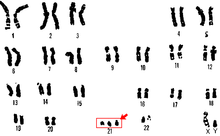
A karyotype is the general appearance of the complete set of chromosomes in the cells of a species or in an individual organism, mainly including their sizes, numbers, and shapes. Karyotyping is the process by which a karyotype is discerned by determining the chromosome complement of an individual, including the number of chromosomes and any abnormalities.

<span class="mw-page-title-main">Aneuploidy</span> Presence of an abnormal number of chromosomes in a cell

Aneuploidy is the presence of an abnormal number of chromosomes in a cell, for example a human cell having 45 or 47 chromosomes instead of the usual 46. It does not include a difference of one or more complete sets of chromosomes. A cell with any number of complete chromosome sets is called a euploid cell.

<span class="mw-page-title-main">Cytogenetics</span> Branch of genetics

Cytogenetics is essentially a branch of genetics, but is also a part of cell biology/cytology, that is concerned with how the chromosomes relate to cell behaviour, particularly to their behaviour during mitosis and meiosis. Techniques used include karyotyping, analysis of G-banded chromosomes, other cytogenetic banding techniques, as well as molecular cytogenetics such as fluorescence in situ hybridization (FISH) and comparative genomic hybridization (CGH).

<span class="mw-page-title-main">Nondisjunction</span> Failure to separate properly during cell division

Nondisjunction is the failure of homologous chromosomes or sister chromatids to separate properly during cell division (mitosis/meiosis). There are three forms of nondisjunction: failure of a pair of homologous chromosomes to separate in meiosis I, failure of sister chromatids to separate during meiosis II, and failure of sister chromatids to separate during mitosis. Nondisjunction results in daughter cells with abnormal chromosome numbers (aneuploidy).

<span class="mw-page-title-main">Chromosomal translocation</span> Phenomenon that results in unusual rearrangement of chromosomes

In genetics, chromosome translocation is a phenomenon that results in unusual rearrangement of chromosomes. This includes balanced and unbalanced translocation, with two main types: reciprocal, and Robertsonian translocation. Reciprocal translocation is a chromosome abnormality caused by exchange of parts between non-homologous chromosomes. Two detached fragments of two different chromosomes are switched. Robertsonian translocation occurs when two non-homologous chromosomes get attached, meaning that given two healthy pairs of chromosomes, one of each pair "sticks" and blends together homogeneously.

<span class="mw-page-title-main">Prenatal testing</span> Testing for diseases or conditions in a fetus

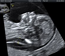
Prenatal testing is a tool that can be used to detect some birth defects at various stages prior to birth. Prenatal testing consists of prenatal screening and prenatal diagnosis, which are aspects of prenatal care that focus on detecting problems with the pregnancy as early as possible. These may be anatomic and physiologic problems with the health of the zygote, embryo, or fetus, either before gestation even starts or as early in gestation as practicable. Screening can detect problems such as neural tube defects, chromosome abnormalities, and gene mutations that would lead to genetic disorders and birth defects, such as spina bifida, cleft palate, Down syndrome, trisomy 18, Tay–Sachs disease, sickle cell anemia, thalassemia, cystic fibrosis, muscular dystrophy, and fragile X syndrome. Some tests are designed to discover problems which primarily affect the health of the mother, such as PAPP-A to detect pre-eclampsia or glucose tolerance tests to diagnose gestational diabetes. Screening can also detect anatomical defects such as hydrocephalus, anencephaly, heart defects, and amniotic band syndrome.

Comparative genomic hybridization (CGH) is a molecular cytogenetic method for analysing copy number variations (CNVs) relative to ploidy level in the DNA of a test sample compared to a reference sample, without the need for culturing cells. The aim of this technique is to quickly and efficiently compare two genomic DNA samples arising from two sources, which are most often closely related, because it is suspected that they contain differences in terms of either gains or losses of either whole chromosomes or subchromosomal regions. This technique was originally developed for the evaluation of the differences between the chromosomal complements of solid tumor and normal tissue, and has an improved resolution of 5–10 megabases compared to the more traditional cytogenetic analysis techniques of giemsa banding and fluorescence in situ hybridization (FISH) which are limited by the resolution of the microscope utilized.

<span class="mw-page-title-main">Small supernumerary marker chromosome</span> Abnormal partial or mixed chromosome

A small supernumerary marker chromosome (sSMC) is an abnormal extra chromosome. It contains copies of parts of one or more normal chromosomes and like normal chromosomes is located in the cell's nucleus, is replicated and distributed into each daughter cell during cell division, and typically has genes which may be expressed. However, it may also be active in causing birth defects and neoplasms. The sSMC's small size makes it virtually undetectable using classical cytogenetic methods: the far larger DNA and gene content of the cell's normal chromosomes obscures those of the sSMC. Newer molecular techniques such as fluorescence in situ hybridization, next generation sequencing, comparative genomic hybridization, and highly specialized cytogenetic G banding analyses are required to study it. Using these methods, the DNA sequences and genes in sSMCs are identified and help define as well as explain any effect(s) it may have on individuals.

The Pallister–Killian syndrome (PKS), also termed tetrasomy 12p mosaicism or the Pallister mosaic aneuploidy syndrome, is an extremely rare and severe genetic disorder. PKS is due to the presence of an extra and abnormal chromosome termed a small supernumerary marker chromosome (sSMC). sSMCs contain copies of genetic material from parts of virtually any other chromosome and, depending on the genetic material they carry, can cause various genetic disorders and neoplasms. The sSMC in PKS consists of multiple copies of the short arm of chromosome 12. Consequently, the multiple copies of the genetic material in the sSMC plus the two copies of this genetic material in the two normal chromosome 12's are overexpressed and thereby cause the syndrome. Due to a form of genetic mosaicism, however, individuals with PKS differ in the tissue distributions of their sSMC and therefore show different syndrome-related birth defects and disease severities. For example, individuals with the sSMC in their heart tissue are likely to have cardiac structural abnormalities while those without this sSMC localization have a structurally normal heart.

A chromosomal abnormality, chromosomal anomaly, chromosomal aberration, chromosomal mutation, or chromosomal disorder is a missing, extra, or irregular portion of chromosomal DNA. These can occur in the form of numerical abnormalities, where there is an atypical number of chromosomes, or as structural abnormalities, where one or more individual chromosomes are altered. Chromosome mutation was formerly used in a strict sense to mean a change in a chromosomal segment, involving more than one gene. Chromosome anomalies usually occur when there is an error in cell division following meiosis or mitosis. Chromosome abnormalities may be detected or confirmed by comparing an individual's karyotype, or full set of chromosomes, to a typical karyotype for the species via genetic testing.

<span class="mw-page-title-main">Molecular cytogenetics</span>

Molecular cytogenetics combines two disciplines, molecular biology and cytogenetics, and involves the analysis of chromosome structure to help distinguish normal and cancer-causing cells. Human cytogenetics began in 1956 when it was discovered that normal human cells contain 46 chromosomes. However, the first microscopic observations of chromosomes were reported by Arnold, Flemming, and Hansemann in the late 1800s. Their work was ignored for decades until the actual chromosome number in humans was discovered as 46. In 1879, Arnold examined sarcoma and carcinoma cells having very large nuclei. Today, the study of molecular cytogenetics can be useful in diagnosing and treating various malignancies such as hematological malignancies, brain tumors, and other precursors of cancer. The field is overall focused on studying the evolution of chromosomes, more specifically the number, structure, function, and origin of chromosome abnormalities. It includes a series of techniques referred to as fluorescence in situ hybridization, or FISH, in which DNA probes are labeled with different colored fluorescent tags to visualize one or more specific regions of the genome. Introduced in the 1980s, FISH uses probes with complementary base sequences to locate the presence or absence of the specific DNA regions. FISH can either be performed as a direct approach to metaphase chromosomes or interphase nuclei. Alternatively, an indirect approach can be taken in which the entire genome can be assessed for copy number changes using virtual karyotyping. Virtual karyotypes are generated from arrays made of thousands to millions of probes, and computational tools are used to recreate the genome in silico.

Confined placental mosaicism (CPM) represents a discrepancy between the chromosomal makeup of the cells in the placenta and the cells in the fetus. CPM was first described by Kalousek and Dill in 1983. CPM is diagnosed when some trisomic cells are detected on chorionic villus sampling and only normal cells are found on a subsequent prenatal test, such as amniocentesis or fetal blood sampling. In theory, CPM is when the trisomic cells are found only in the placenta. CPM is detected in approximately 1-2% of ongoing pregnancies that are studied by chorionic villus sampling (CVS) at 10 to 12 weeks of pregnancy. Chorionic villus sampling is a prenatal procedure which involves a placental biopsy. Most commonly when CPM is found it represents a trisomic cell line in the placenta and a normal diploid chromosome complement in the baby. However, the fetus is involved in about 10% of cases.

<span class="mw-page-title-main">Cat eye syndrome</span> Genetic partial duplication of chromosome 22

Not to be confused with Heterochromia, an optical condition which is commonly associated with cats

45,X/46,XY mosaicism, also known as X0/XY mosaicism and mixed gonadal dysgenesis, is a mutation of sex development in humans associated with sex chromosome aneuploidy and mosaicism of the Y chromosome. It is a fairly rare chromosomal disorder at birth, with an estimated incidence rate of about 1 in 15,000 live births. Mosaic loss of the Y chromosome in previously non-mosaic men grows increasingly common with age.

<span class="mw-page-title-main">XYYY syndrome</span> Chromosomal disorder

XYYY syndrome, also known as 48,XYYY, is a chromosomal disorder in which a male has two extra copies of the Y chromosome. The syndrome is exceptionally rare, with only twelve recorded cases. The presentation of the syndrome is heterogeneous, but appears to be more severe than its counterpart XYY syndrome. Common traits include borderline to mild intellectual disability, infertility, radioulnar synostosis, and in some cases tall stature.

<span class="mw-page-title-main">Tetrasomy X</span> Chromosomal disorder with 4 X chromosomes

Tetrasomy X, also known as 48,XXXX, is a chromosomal disorder in which a female has four, rather than two, copies of the X chromosome. It is associated with intellectual disability of varying severity, characteristic "coarse" facial features, heart defects, and skeletal anomalies such as increased height, clinodactyly, and radioulnar synostosis. Tetrasomy X is a rare condition, with few medically recognized cases; it is estimated to occur in approximately 1 in 50,000 females.

<span class="mw-page-title-main">Pentasomy X</span> Chromosomal disorder

Pentasomy X, also known as 49,XXXXX, is a chromosomal disorder in which a female has five, rather than two, copies of the X chromosome. Pentasomy X is associated with short stature, intellectual disability, characteristic facial features, heart defects, skeletal anomalies, and pubertal and reproductive abnormalities. The condition is exceptionally rare, with an estimated prevalence between 1 in 85,000 and 1 in 250,000.

<span class="mw-page-title-main">Trisomy X</span> Chromosome disorder in women

Trisomy X, also known as triple X syndrome and characterized by the karyotype 47,XXX, is a chromosome disorder in which a female has an extra copy of the X chromosome. It is relatively common and occurs in 1 in 1,000 females, but is rarely diagnosed; fewer than 10% of those with the condition know they have it.

<span class="mw-page-title-main">XXXYY syndrome</span> Chromosomal disorder

XXXYY syndrome, also known as 49,XXXYY, is a chromosomal disorder in which a male has three copies of the X chromosome and two copies of the Y chromosome. XXXYY syndrome is exceptionally rare, with only eight recorded cases. Little is known about its presentation, but associated characteristics include intellectual disability, anomalies of the external genitalia, and characteristic physical and facial features. It is not caused by characteristics of the parents, but rather occurs via nondisjunction, a random event in gamete development. The karyotype observed in the syndrome is formally known as 49,XXXYY, which represents the 49 chromosomes observed in the disorder as compared to the 46 in normal human development.

References

  1. 1 2 3 4 Rieger, R.; Michaelis, A.; Green, M.M. (1968). A glossary of genetics and cytogenetics: Classical and molecular . New York: Springer-Verlag.
  2. White, Michael James Denham (1937). The Chromosomes. London: Methuen & Co., Ltd. p. 55.
  3. 1 2 Griffiths, AJF; Miller JH; Suzuki DT; et al. (2000). An Introduction to Genetic Analysis: Aneuploidy (7th ed.). New York: W.H. Freeman & Co. Retrieved 24 March 2014.[ page needed ]
  4. Anthony J. F. Griffiths (1999). An introduction to genetic analysis (7. ed., 1. print. ed.). New York: Freeman. ISBN   978-0-7167-3520-5.[ page needed ]
  5. Calvieri F, Tozzi C, Benincori C, et al. (August 1988). "Partial tetrasomy 9 in an infant with clinical and radiological evidence of multiple joint dislocations". European Journal of Pediatrics. 147 (6): 645–8. doi:10.1007/bf00442483. PMID   3181206. S2CID   20302464.
  6. 1 2 Celik A, Eraslan S, Gökgöz N, et al. (June 1997). "Identification of the parental origin of polysomy in two 49,XXXXY cases". Clinical Genetics. 51 (6): 426–9. doi:10.1111/j.1399-0004.1997.tb02504.x. PMID   9237509. S2CID   34190928.
  7. Huang B, Bartley J (September 2003). "Partial hexasomy of chromosome 15". American Journal of Medical Genetics Part A. 121A (3): 277–80. doi:10.1002/ajmg.a.20182. PMID   12923871. S2CID   41001200.
  8. Fabarius A, Li R, Yerganian G, Hehlmann R, Duesberg P (January 2008). "Specific clones of spontaneously evolving karyotypes generate individuality of cancers". Cancer Genetics and Cytogenetics. 180 (2): 89–99. doi:10.1016/j.cancergencyto.2007.10.006. PMID   18206533.
  9. Ludwig A, Belfiore NM, Pitra C, Svirsky V, Jenneckens I (July 2001). "Genome duplication events and functional reduction of ploidy levels in sturgeon (Acipenser, Huso and Scaphirhynchus)". Genetics. 158 (3): 1203–15. doi:10.1093/genetics/158.3.1203. PMC   1461728 . PMID   11454768.
  10. Schachter M (1949). "[Nanosomy by congenital skeletal polydystrophy – correlation with a maternal gestational deficiency]". Athena (in Italian). 15 (6): 141–3. PMID   15409638.
  11. Mori M, Matsushita A, Takiuchi Y, et al. (July 2010). "Histiocytic sarcoma and underlying chronic myelomonocytic leukemia: a proposal for the developmental classification of histiocytic sarcoma". International Journal of Hematology. 92 (1): 168–73. doi:10.1007/s12185-010-0603-z. PMID   20535595. S2CID   25682085.
  12. 1 2 Reimann-Berg N, Willenbrock S, Murua Escobar H, et al. (2011). "Two new cases of polysomy 13 in canine prostate cancer". Cytogenetic and Genome Research. 132 (1–2): 16–21. doi:10.1159/000317077. PMID   20668368. S2CID   24726737.
  13. Winkler S, Murua Escobar H, Reimann-Berg N, Bullerdiek J, Nolte I (2005). "Cytogenetic investigations in four canine lymphomas". Anticancer Research. 25 (6B): 3995–8. PMID   16309190.
  14. Hahn, KA; Richardson, RC; Hahn, EA; Chrisman, CL (Sep 1994). "Diagnostic and prognostic importance of chromosomal aberrations identified in 61 dogs with lymphosarcoma". Veterinary Pathology. 31 (5): 528–40. doi: 10.1177/030098589403100504 . PMID   7801430.
  15. Yang F, Graphodatsky AS, O'Brien PC, et al. (2000). "Reciprocal chromosome painting illuminates the history of genome evolution of the domestic cat, dog and human". Chromosome Research. 8 (5): 393–404. doi:10.1023/A:1009210803123. PMID   10997780. S2CID   19318062.
  16. 1 2 Winkler S, Reimann-Berg N, Murua Escobar H, et al. (September 2006). "Polysomy 13 in a canine prostate carcinoma underlining its significance in the development of prostate cancer". Cancer Genetics and Cytogenetics. 169 (2): 154–8. doi:10.1016/j.cancergencyto.2006.03.015. PMID   16938574.
  17. Winkler S, Murua Escobar H, Eberle N, Reimann-Berg N, Nolte I, Bullerdiek J (2005). "Establishment of a cell line derived from a canine prostate carcinoma with a highly rearranged karyotype". The Journal of Heredity. 96 (7): 782–5. doi: 10.1093/jhered/esi085 . PMID   15994418.
  18. 1 2 Leal CA, Belmont JW, Nachtman R, Cantu JM, Medina C (October 1994). "Parental origin of the extra chromosomes in polysomy X". Human Genetics. 94 (4): 423–6. doi:10.1007/bf00201605. PMID   7927341. S2CID   23275179.
  19. Kleczkowska A, Fryns JP, Van den Berghe H (September 1988). "X-chromosome polysomy in the male. The Leuven experience 1966–1987". Human Genetics. 80 (1): 16–22. doi:10.1007/BF00451449. PMID   3417301. S2CID   7308546.
  20. de Grouchy J, Turleau C (October 1986). "Microcytogenetics 1984". Experientia. 42 (10): 1090–7. doi:10.1007/BF01941282. PMID   3533601. S2CID   32070898.
  21. Elias, Sherman; Shulman, Lee P. (2009). "Males with Polysomy Y and Females with Polysomy X". The Global Library of Women's Medicine. doi:10.3843/GLOWM.10358 . Retrieved 21 April 2014.
  22. Couceiro P, Sousa V, Alarcão A, Silva M, Carvalho L (2010). "Polysomy and amplification of chromosome 7 defined for EGFR gene in squamous cell carcinoma of the lung together with exons 19 and 21 wild type". Revista Portuguesa de Pneumologia. 16 (3): 453–62. doi:10.1016/s2173-5115(10)70049-x. hdl: 10316/102760 . PMID   20635059.
  23. Li YH, Wang F, Shen L, et al. (January 2011). "EGFR fluorescence in situ hybridization pattern of chromosome 7 disomy predicts resistance to cetuximab in KRAS wild-type metastatic colorectal cancer patients". Clinical Cancer Research. 17 (2): 382–90. doi: 10.1158/1078-0432.CCR-10-0208 . PMID   20884623.
  24. Paulsson K, Johansson B (February 2007). "Trisomy 8 as the sole chromosomal aberration in acute myeloid leukemia and myelodysplastic syndromes". Pathologie-biologie. 55 (1): 37–48. doi:10.1016/j.patbio.2006.04.007. PMID   16697122.
  25. Beyer V, Mühlematter D, Parlier V, et al. (July 2005). "Polysomy 8 defines a clinico-cytogenetic entity representing a subset of myeloid hematologic malignancies associated with a poor prognosis: report on a cohort of 12 patients and review of 105 published cases". Cancer Genetics and Cytogenetics. 160 (2): 97–119. doi:10.1016/j.cancergencyto.2004.12.003. PMID   15993266.
  26. Schiaovon, BN; Vassallo J; Rocha RM (2011). "Is polysomy 17 an important phenomenon to predict treatment with trastuzumab in breast cancer?". Applied Cancer Research. 31 (4): 138–142. Archived from the original on 23 April 2014. Retrieved 21 April 2014.
  27. 1 2 Yang, Liu; et al. (Dec 15, 2013). "Impact of polysomy 17 on HER2 testing of invasive breast cancer patients". International Journal of Clinical and Experimental Pathology. 7 (1): 163–173. PMC   3885470 . PMID   24427336.
  28. Yeh IT, Martin MA, Robetorye RS, et al. (September 2009). "Clinical validation of an array CGH test for HER2 status in breast cancer reveals that polysomy 17 is a rare event". Modern Pathology. 22 (9): 1169–75. doi: 10.1038/modpathol.2009.78 . PMID   19448591.
  29. "Down Syndrome". Medline Plus. Retrieved 22 April 2014.
  30. Dhandha, S; Hogge, WA; Surti, U; McPherson, E (Dec 15, 2002). "Three cases of tetrasomy 9p". American Journal of Medical Genetics. 113 (4): 375–80. doi:10.1002/ajmg.b.10826. PMID   12457411.
  31. 1 2 Sebold C, Roeder E, Zimmerman M, Soileau B, Heard P, Carter E, Schatz M, White WA, Perry B, Reinker K, O'Donnell L, Lancaster J, Li J, Hasi M, Hill A, Pankratz L, Hale DE, Cody JD (Sep 2010). "Tetrasomy 18p: report of the molecular and clinical findings of 43 individuals". American Journal of Medical Genetics Part A. 152A (9): 2164–72. doi:10.1002/ajmg.a.33597. PMID   20803640. S2CID   19758003.
  32. Merriam-Webster. "Germ Line". Merriam-Webster, Incorporated. Retrieved 7 April 2014.
  33. 1 2 Talavera, M.; López-Leon, M. D.; Cabrero, J.; Camacho, J. P. M. (June 1990). "Male germ line polysomy in the grasshopper Chorthippus binotatus: extra chromosomes are not transmitted". Genome. 33 (3): 384–388. doi:10.1139/g90-058.
  34. 1 2 Peters, G. B. (January 1981). "Germ line polysomy in the grasshopper Atractomorpha similis". Chromosoma. 81 (4): 593–617. doi:10.1007/BF00285852. S2CID   41211400.
  35. Merriam-Webster. "Heterochromatin". Merriam-Webster, Incorporated. Retrieved 7 April 2014.
  36. 1 2 Kushnir, Tuviah (February 1952). "Heterochromatic polysomy in Gryllotalpa gryllotalpa L". Journal of Genetics. 50 (3): 361–383. doi:10.1007/BF02986834. S2CID   1793646.
  37. Nevo E, Beiles A, Korol AB, Robin YI, Pavlicek T, Hamilton W (April 2000). "Extraordinary multilocus genetic organization in mole crickets, Gryllotalpidae". Evolution. 54 (2): 586–605. doi:10.1111/j.0014-3820.2000.tb00061.x. PMID   10937235. S2CID   198153947.
  38. 1 2 3 Birchler JA, Hiebert JC, Krietzman M (August 1989). "Gene expression in adult metafemales of Drosophila melanogaster". Genetics. 122 (4): 869–79. doi:10.1093/genetics/122.4.869. PMC   1203761 . PMID   2503426.
  39. Gupta, P. K.; T. Tsuchiya. (1991). Chromosome Engineering in Plants: Genetics, Breeding, Evolution. Amsterdam: Eselvier.
  40. Ruiz Rejón, C.; R. Lozano; M. Ruiz Rejón (1987). "Polysomy and supernumerary chromosomes in Ornithogalum umbellatum L. (Liliaceae)". Genome. 29 (1): 19–25. doi:10.1139/g87-004.
  41. Ahuja, MR; Neale DB (2002). "Origins of Polyploidy in Coast Redwood (Sequoia sempervirens (D. DON) ENDL.) and Relationship of Coast Redwood to other Genera of Taxodiaceae". Silvae Genetica. 51: 2–3.
  42. D'Hont, A; Grivet, L; Feldmann, P; Rao, S; Berding, N; Glaszmann, JC (Mar 7, 1996). "Characterisation of the double genome structure of modern sugarcane cultivars (Saccharum spp.) by molecular cytogenetics". Molecular & General Genetics. 250 (4): 405–13. doi:10.1007/bf02174028. PMID   8602157. S2CID   43107532.
  43. Mun, JH; et al. (2010). "Sequence and structure of Brassica rapa chromosome A3". Genome Biology. 11 (9): R94. doi: 10.1186/gb-2010-11-9-r94 . PMC   2965386 . PMID   20875114.
  44. Barker, W.R.; M. Kiehn; E. Vitek (1988). "Chromosome numbers in AustralianEuphrasia (Scrophulariaceae)". Plant Systematics and Evolution. 158 (2–4): 161–164. doi:10.1007/bf00936342. S2CID   1881882.
  45. Zhu, J.M.; LJ Davies; D Cohen; RE Rowland (1994). "Variation in chromosome number in the seedling progeny of a somaclone of Paspalum dilatatum". Cell Research. 4: 65–78. doi:10.1038/cr.1994.7. S2CID   36302051.
  46. P.M. Kirk; et al. (2008). Ainsworth & Bisby's dictionary of the fungi (10th ed.). Wallingford, Oxon, UK: CABI. ISBN   978-0-85199-826-8.
  47. Arora, Dilip K., ed. (2004). Handbook of fungal biotechnology (2. ed., rev. and expanded ed.). New York, NY [u.a.]: Marcel Dekker. ISBN   978-0-8247-4018-4.
  48. Atkinson, NS; Hopper, AK (Jul 1987). "Chromosome specificity of polysomy promotion by disruptions of the Saccharomyces cerevisiae RNA1 gene". Genetics. 116 (3): 371–5. doi:10.1093/genetics/116.3.371. PMC   1203148 . PMID   3301528.
  49. Bangarulingam, SY; Bjornsson, E; Enders, F; Barr Fritcher, EG; Gores, G; Halling, KC; Lindor, KD (Jan 2010). "Long-term outcomes of positive fluorescence in situ hybridization tests in primary sclerosing cholangitis". Hepatology. 51 (1): 174–80. doi: 10.1002/hep.23277 . PMID   19877179. S2CID   11858431.
  50. 1 2 Binns, Victoria; Nancy Hsu (20 Jun 2001). "Prenatal Diagnosis". Encyclopedia of Life Sciences. Jon Wiley & Sons. doi:10.1038/npg.els.0002291. ISBN   978-0470016176.
  51. Gonda, TA; Glick, MP; Sethi, A; Poneros, JM; Palmas, W; Iqbal, S; Gonzalez, S; Nandula, SV; Emond, JC; Brown, RS; Murty, VV; Stevens, PD (Jan 2012). "Polysomy and p16 deletion by fluorescence in situ hybridization in the diagnosis of indeterminate biliary strictures". Gastrointestinal Endoscopy. 75 (1): 74–9. doi:10.1016/j.gie.2011.08.022. PMID   22100297. S2CID   2012265.
  52. Heselmeyer-Haddad, K; Sommerfeld, K; White, NM; Chaudhri, N; Morrison, LE; Palanisamy, N; Wang, ZY; Auer, G; Steinberg, W; Ried, T (Apr 2005). "Genomic amplification of the human telomerase gene (TERC) in pap smears predicts the development of cervical cancer". The American Journal of Pathology. 166 (4): 1229–38. doi:10.1016/S0002-9440(10)62341-3. PMC   1602397 . PMID   15793301.
  53. Yunis JJ, Sanchez O (1973). "G-banding and chromosome structure". Chromosoma. 44 (1): 15–23. doi:10.1007/BF00372570. PMID   4130183. S2CID   8711896.
  54. "The Use of Chromosomal Microarray Analysis in Prenatal Diagnosis". American College of Obstetricians and Gynecologists. Retrieved 5 May 2014.
  55. Shaffer, LG; Rosenfeld, JA; Dabell, MP; Coppinger, J; Bandholz, AM; Ellison, JW; Ravnan, JB; Torchia, BS; Ballif, BC; Fisher, AJ (Oct 2012). "Detection rates of clinically significant genomic alterations by microarray analysis for specific anomalies detected by ultrasound". Prenatal Diagnosis. 32 (10): 986–95. doi:10.1002/pd.3943. PMC   3509216 . PMID   22847778.
  56. Crocker, ed. by David Burnett; John (2005). The science of laboratory diagnosis (2. ed.). Chichester [u.a.]: Wiley. p. 523. ISBN   978-0470859124.{{cite book}}: |first= has generic name (help)CS1 maint: multiple names: authors list (link)
  57. Calabrese, G; Baldi, M; Fantasia, D; Sessa, MT; Kalantar, M; Holzhauer, C; Alunni-Fabbroni, M; Palka, G; Sitar, G (Aug 2012). "Detection of chromosomal aneuploidies in fetal cells isolated from maternal blood using single-chromosome dual-probe FISH analysis". Clinical Genetics. 82 (2): 131–9. doi:10.1111/j.1399-0004.2011.01775.x. PMID   21895636. S2CID   34089887.
  58. Ross, Helen L.; Elias, Sherman (1997). "Maternal Serum Screening for Fetal Genetic Disorders". Obstetrics and Gynecology Clinics of North America. 24 (1): 33–47. doi:10.1016/S0889-8545(05)70288-6. PMID   9086517.
  59. Sherman, Elias (2013). "Amniocentesis". Genetic Disorders and the Fetus. Springer. pp. 31–52. doi:10.1007/978-1-4684-5155-9_2. ISBN   978-1-4684-5157-3.
  60. Simpson, Joe Leigh (1990). "Incidence and timing of pregnancy losses: Relevance to evaluating safety of early prenatal diagnosis". American Journal of Medical Genetics. 35 (2): 165–173. doi:10.1002/ajmg.1320350205. PMID   2178414.
  61. Deng, Han-Xiang; Abe, Kyohko; Kondo, Ikuko; Tsukahara, Masato; Inagaki, Haruyo; Hamada, Isamu; Fukushima, Yoshimitsu; Niikawa, Norio (1991). "Parental origin and mechanism of formation of polysomy X: an XXXXX case and four XXXXY cases determined with RFLPs". Human Genetics. 86 (6): 541–4. doi:10.1007/BF00201538. PMID   1673956. S2CID   11111874.
  62. Chial, Heidi. "Huntington's disease: The discovery of the Huntingtin gene". Nature Education. Retrieved 5 May 2014.
  63. Muehlbauer PA, Schuler MJ (August 2005). "Detection of numerical chromosomal aberrations by flow cytometry: a novel process for identifying aneugenic agents". Mutation Research. 585 (1–2): 156–69. doi:10.1016/j.mrgentox.2005.05.002. PMID   15996509.

Further reading

  1. Gardner, R. J. M., Grant R. Sutherland, and Lisa G. Shaffer. Chromosome Abnormalities and Genetic Counseling. 4th ed. Oxford: Oxford UP, 2012.
  2. Miller, Orlando J., and Eeva Therman. Human Chromosomes. New York: Springer, 2001.
  3. Schmid, M., and Indrajit Nanda. Chromosomes Today, Volume 14. Dordrecht: Kluwer Academic, 2004.
  4. Nussbaum, Robert L., Roderick R. McInnes, Huntington F. Willard, Ada Hamosh, and Margaret W. Thompson. Thompson & Thompson Genetics in Medicine. 7th ed. Philadelphia: Saunders/Elsevier, 2007.