Autosomal dominant cerebellar ataxia

Last updated
Autosomal dominant cerebellar ataxia
Other namesAutosomal dominant spinocerebellar ataxia [1]
Autosomal dominant - en.svg
Autosomal dominant is the manner in which this condition is inherited
Symptoms Multi system involvement [2]
TypesADCS type1, ADCA type 2, ADCA type 3 [2]
Diagnostic method MRI, CT scan [3]
TreatmentAnticonvulsants may be used [2]

Autosomal dominant cerebellar ataxia (ADCA) is a form of spinocerebellar ataxia inherited in an autosomal dominant manner. ADCA is a genetically inherited condition that causes deterioration of the nervous system leading to disorder and a decrease or loss of function to regions of the body. [2]

Contents

Degeneration occurs at the cellular level and in certain subtypes results in cellular death. Cellular death or dysfunction causes a break or faulty signal in the line of communication from the central nervous system to target muscles in the body. When there is impaired communication or a lack of communication entirely, the muscles in the body do not function correctly. Muscle control complications can be observed in multiple balance, speech, and motor or movement impairment symptoms. ADCA is divided into three types and further subdivided into subtypes known as SCAs (spinocerebellar ataxias). [4]

Types

Currently there are 27 subtypes have been identified: SCA1-SCA4, SCA8, SCA10, SCA12-SCA14, SCA15/SCA16, SCA17-SCA23, SCA25, SCA27, SCA28, SCA32, SCA34-SCA37, autosomal dominant cerebellar ataxia and dentatorubral pallidoluysian atrophy. [3]

Type 1

Type I ADCA is characterized by different symptoms of ataxia as well as other conditions that are dependent on the subtype. Type 1 ADCA is divided into 3 subclasses based on pathogenesis of the subtypes each contain. [4] [3] [5]

L-Glutamin L-Glutamin - L-Glutamine.svg
L-Glutamin

Type 2/3

Type II ADCA is composed of SCA7 and syndromes associated with pigmentary maculopathies. [4] SCA7 is a disease that specifically displays retinal degeneration, along with the common degeneration of the cerebellum. Moving further into SCA7's pathology, a similar genetic process is described, while the function of ATXN7 (an ataxin gene) is much like a component of the SAGA complex. The SAGA complex uses two histone-modifying techniques to regulate transcription. These activities are the Gcn5 histone acetyltransferase and the Usp22 deubiquitinase. Mutant ATXN7 in HAT activity causes an increase in activity, which was reported from an in-vivo analysis in the retina. There are also studies that show a loss in activity when human ATXN7 in yeast was used. The SCA7 autosomal-dominant inheritance pattern is similar to a mutant ATXN5-induced gain in Gcn5 HAT. [6] Spinocerebellar ataxia type 15 has been classified as an ADCA Type III as it has been noted to have postural and action tremor in addition to cerebellar ataxia. [4] Additionally, spinocerebellar ataxia type 20 (SCA20) is organized in ADCA III that often exhibits disease-like symptoms at an earlier age, sometime starting at fourteen years old. [4] [7] [8]

Symptoms and signs

Cerebellum Cerebellum animation small.gif
Cerebellum

Symptoms typically are onset in the adult years, although, childhood cases have also been observed. Common symptoms include a loss of coordination which is often seen in walking, and slurred speech. ADCA primarily affects the cerebellum, as well as, the spinal cord. [9] Some signs and symptoms are: [2]

Genetics

In terms of the genetics of autosomal dominant cerebellar ataxia 11 of 18 known genes are caused by repeated expansions in corresponding proteins, sharing the same mutational mechanism. SCAs can be caused by conventional mutations or large rearrangements in genes that make glutamate and calcium signaling, channel function, tau regulation and mitochondrial activity or RNA alteration. [10]

The mechanism of Type I is not completely known, however, Whaley, et al. suggest the polyglutamine product is toxic to the cell at a protein level, this effect may be done by transcriptional dysregulation and disruption of calcium homeostasis which causes apoptosis to occur earlier. [4]

Diagnosis

In diagnosing autosomal dominant cerebellar ataxia the individuals clinical history or their past health examinations, a current physical examination to check for any physical abnormalities, and a genetic screening of the patients genes and the genealogy of the family are done. [11] The large category of cerebellar ataxia is caused by a deterioration of neurons in the cerebellum, therefore magnetic resonance imaging (MRI) is used to detect any structural abnormality such as lesions which are the primary cause of the ataxia. Computed tomography (CT) scans can also be used to view neuronal deterioration, but the MRI provides a more accurate and detailed picture. [12]

Treatments

In terms of a cure there is currently none available, however for the disease to manifest itself, it requires mutant gene expression. [6] Manipulating the use of protein homeostasis regulators can be therapeutic agents, or a treatment to try and correct an altered function that makes up the pathology is one current idea put forth by Bushart, et al. [6] [13] There is some evidence that for SCA1 and two other polyQ disorders that the pathology can be reversed after the disease is underway. [6] There is no effective treatments that could alter the progression of this disease, therefore care is given, like occupational and physical therapy for gait dysfunction and speech therapy.[ medical citation needed ]

Epidemiology

In terms of frequency, it is estimated at 2 per 100,000, it has identified in different regions of the world. Some clusters of certain types of autosomal dominant cerebellar ataxia reach a prevalence of 5 per 100,000. [2]

Related Research Articles

Ataxia is a neurological sign consisting of lack of voluntary coordination of muscle movements that can include gait abnormality, speech changes, and abnormalities in eye movements, that indicates dysfunction of parts of the nervous system that coordinate movement, such as the cerebellum.

<span class="mw-page-title-main">Spinocerebellar ataxia</span> Medical condition

Spinocerebellar ataxia (SCA) is a progressive, degenerative, genetic disease with multiple types, each of which could be considered a neurological condition in its own right. An estimated 150,000 people in the United States have a diagnosis of spinocerebellar ataxia at any given time. SCA is hereditary, progressive, degenerative, and often fatal. There is no known effective treatment or cure. SCA can affect anyone of any age. The disease is caused by either a recessive or dominant gene. In many cases people are not aware that they carry a relevant gene until they have children who begin to show signs of having the disorder.

<span class="mw-page-title-main">Machado–Joseph disease</span> Genetic neurodegenerative disease

Machado–Joseph disease (MJD), also known as Machado–Joseph Azorean disease, Machado's disease, Joseph's disease or spinocerebellar ataxia type 3 (SCA3), is a rare autosomal dominantly inherited neurodegenerative disease that causes progressive cerebellar ataxia, which results in a lack of muscle control and coordination of the upper and lower extremities. The symptoms are caused by a genetic mutation that results in an expansion of abnormal "CAG" trinucleotide repeats in the ATXN3 gene that results in an abnormal form of the protein ataxin which causes degeneration of cells in the hindbrain. Some symptoms, such as clumsiness and rigidity, make MJD commonly mistaken for drunkenness or Parkinson's disease.

<span class="mw-page-title-main">Purkinje cell</span> Specialized neuron in the cerebellum

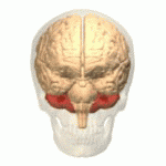
Purkinje cells or Purkinje neurons, named for Czech physiologist Jan Evangelista Purkyně who identified them in 1837, are a unique type of prominent large neurons located in the cerebellar cortex of the brain. With their flask-shaped cell bodies, many branching dendrites, and a single long axon, these cells are essential for controlling motor activity. Purkinje cells mainly release GABA neurotransmitter, which inhibits some neurons to reduce nerve impulse transmission. Purkinje cells efficiently control and coordinate the body's motor motions through these inhibitory actions.

Cerebellar ataxia is a form of ataxia originating in the cerebellum. Non-progressive congenital ataxia (NPCA) is a classical presentation of cerebral ataxias.

<span class="mw-page-title-main">Ataxin 1</span> Protein-coding gene in the species Homo sapiens

Ataxin-1 is a DNA-binding protein which in humans is encoded by the ATXN1 gene.

Ataxin 7 (ATXN7) is a protein of the SCA7 gene, which contains 892 amino acids with an expandable poly(Q) region close to the N-terminus. The expandable poly(Q) motif region in the protein contributes crucially to spinocerebellar ataxia (SCA) pathogenesis by the induction of intranuclear inclusion bodies. ATXN7 is associated with both olivopontocerebellar atrophy type 3 (OPCA3) and spinocerebellar ataxia type 7 (SCA7).

<span class="mw-page-title-main">Hemiplegic migraine</span> Medical condition

Hemiplegic migraine is a type of migraine headache characterized by motor weakness affecting only one side of the body, accompanied by aura. There is often an impairment in vision, speech, or sensation. It can run in the family, called familial hemiplegic migraine, or in a single individual, called sporadic hemiplegic migraine. The symptoms can be similar to a stroke, and may be precipitated by minor head trauma. People with FHM are advised to avoid activities that may trigger their attacks.

<span class="mw-page-title-main">Spinocerebellar ataxia type 6</span> Medical condition

Spinocerebellar ataxia type 6 (SCA6) is a rare, late-onset, autosomal dominant disorder, which, like other types of SCA, is characterized by dysarthria, oculomotor disorders, peripheral neuropathy, and ataxia of the gait, stance, and limbs due to cerebellar dysfunction. Unlike other types, SCA 6 is not fatal. This cerebellar function is permanent and progressive, differentiating it from episodic ataxia type 2 (EA2) where said dysfunction is episodic. In some SCA6 families, some members show these classic signs of SCA6 while others show signs more similar to EA2, suggesting that there is some phenotypic overlap between the two disorders. SCA6 is caused by mutations in CACNA1A, a gene encoding a calcium channel α subunit. These mutations tend to be trinucleotide repeats of CAG, leading to the production of mutant proteins containing stretches of 20 or more consecutive glutamine residues; these proteins have an increased tendency to form intracellular agglomerations. Unlike many other polyglutamine expansion disorders expansion length is not a determining factor for the age that symptoms present.

Episodic ataxia (EA) is an autosomal dominant disorder characterized by sporadic bouts of ataxia with or without myokymia. There are seven types recognized but the majority are due to two recognized entities. Ataxia can be provoked by psychological stress or startle, or heavy exertion, including exercise. Symptoms can first appear in infancy. There are at least six loci for EA, of which 4 are known genes. Some patients with EA also have migraine or progressive cerebellar degenerative disorders, symptomatic of either familial hemiplegic migraine or spinocerebellar ataxia. Some patients respond to acetazolamide though others do not.

<span class="mw-page-title-main">Spinocerebellar ataxia type-13</span> Medical condition

Spinocerebellar ataxia type 13 (SCA13) is a rare autosomal dominant disorder, which, like other types of SCA, is characterized by dysarthria, nystagmus, and ataxia of gait, stance and the limbs due to cerebellar dysfunction. Patients with SCA13 also tend to present with epilepsy, an inability to run, and increased reflexes. This cerebellar dysfunction is permanent and progressive. SCA13 is caused by mutations in KCNC3, a gene encoding a voltage-gated potassium channel KV3.3. There are two known mutations in this gene causative for SCA13. Unlike many other types of SCA, these are not polyglutamine expansions but, rather, point mutations resulting in channels with no current or altered kinetics.

<span class="mw-page-title-main">Ataxin-2</span> Mammalian protein found in Homo sapiens

Ataxin-2 is a protein that in humans is encoded by the ATXN2 gene. Mutations in ATXN2 cause spinocerebellar ataxia type 2 (SCA2).

Ca<sub>v</sub>2.1 Protein-coding gene in the species Homo sapiens

Cav2.1, also called the P/Q voltage-dependent calcium channel, is a calcium channel found mainly in the brain. Specifically, it is found on the presynaptic terminals of neurons in the brain and cerebellum. Cav2.1 plays an important role in controlling the release of neurotransmitters between neurons. It is composed of multiple subunits, including alpha-1, beta, alpha-2/delta, and gamma subunits. The alpha-1 subunit is the pore-forming subunit, meaning that the calcium ions flow through it. Different kinds of calcium channels have different isoforms (versions) of the alpha-1 subunit. Cav2.1 has the alpha-1A subunit, which is encoded by the CACNA1A gene. Mutations in CACNA1A have been associated with various neurologic disorders, including familial hemiplegic migraine, episodic ataxia type 2, and spinocerebellar ataxia type 6.

<span class="mw-page-title-main">Ataxin 10</span> Protein-coding gene in the species Homo sapiens

Ataxin-10 is a protein that in humans is encoded by the ATXN10 gene.

<span class="mw-page-title-main">PLEKHG4</span> Protein-coding gene in the species Homo sapiens

Puratrophin-1 is a protein that in humans is encoded by the PLEKHG4 gene.

<span class="mw-page-title-main">Autosomal recessive cerebellar ataxia type 1</span> Hereditary ataxia that has material basis in autosomal recessive inheritance

Autosomal recessive cerebellar ataxia type 1 (ARCA1) is a condition characterized by progressive problems with movement. Signs and symptoms of the disorder first appear in early to mid-adulthood. People with this condition initially experience impaired speech (dysarthria), problems with coordination and balance (ataxia), or both. They may also have difficulty with movements that involve judging distance or scale (dysmetria). Other features of ARCA1 include abnormal eye movements (nystagmus) and problems following the movements of objects with their eyes. The movement problems are slowly progressive, often resulting in the need for a cane, walker, or wheelchair.

<span class="mw-page-title-main">Corneal-cerebellar syndrome</span> Medical condition

Corneal-cerebellar syndrome is an autosomally recessive disease that was first described in 1985. Three cases are known: all are sisters in the same family.

<span class="mw-page-title-main">Spinocerebellar ataxia type 1</span> Rare neurodegenerative disorder

Spinocerebellar ataxia type 1 (SCA1) is a rare autosomal dominant disorder, which, like other spinocerebellar ataxias, is characterized by neurological symptoms including dysarthria, hypermetric saccades, and ataxia of gait and stance. This cerebellar dysfunction is progressive and permanent. First onset of symptoms is normally between 30 and 40 years of age, though juvenile onset can occur. Death typically occurs within 10 to 30 years from onset.

<span class="mw-page-title-main">Cerebellar degeneration</span> Medical condition

Cerebellar degeneration is a condition in which cerebellar cells, otherwise known as neurons, become damaged and progressively weaken in the cerebellum. There are two types of cerebellar degeneration; paraneoplastic cerebellar degeneration, and alcoholic or nutritional cerebellar degeneration. As the cerebellum contributes to the coordination and regulation of motor activities, as well as controlling equilibrium of the human body, any degeneration to this part of the organ can be life-threatening. Cerebellar degeneration can result in disorders in fine movement, posture, and motor learning in humans, due to a disturbance of the vestibular system. This condition may not only cause cerebellar damage on a temporary or permanent basis, but can also affect other tissues of the central nervous system, those including the cerebral cortex, spinal cord and the brainstem.

Autosomal dominant cerebellar ataxia, deafness, and narcolepsy (ADCADN) is a rare progressive genetic disorder that primarily affects the nervous system and is characterized by sensorineural hearing loss, narcolepsy with cataplexy, and dementia later in life. People with this disorder usually start showing symptoms when they are in their early-mid adulthoods. It is a type of autosomal dominant cerebellar ataxia.

References

  1. RESERVED, INSERM US14-- ALL RIGHTS. "Orphanet: Autosomal dominant cerebellar ataxia". www.orpha.net. Archived from the original on 31 August 2019. Retrieved 8 August 2019.{{cite web}}: CS1 maint: numeric names: authors list (link)
  2. 1 2 3 4 5 6 "Autosomal Dominant Cerebellar Ataxia information page. Patient | Patient". Patient. Archived from the original on 2021-08-13. Retrieved 2016-03-25.
  3. 1 2 3 RESERVED, INSERM US14 -- ALL RIGHTS. "Orphanet: Autosomal dominant cerebellar ataxia type 1". www.orpha.net. Archived from the original on 2020-11-12. Retrieved 2016-03-25.{{cite web}}: CS1 maint: numeric names: authors list (link)
  4. 1 2 3 4 5 6 7 8 9 Whaley, Nathaniel; Fujioka, Shinsuke; Wszolek, Zbigniew K (1 January 2011). "Autosomal dominant cerebellar ataxia type I: A review of the phenotypic and genotypic characteristics". Orphanet Journal of Rare Diseases. 6 (1): 33. doi: 10.1186/1750-1172-6-33 . PMC   3123548 . PMID   21619691.
  5. "SCA1". Genetics Home Reference. 2016-03-21. Archived from the original on 2021-04-17. Retrieved 2016-03-25.
  6. 1 2 3 4 Orr, H. T. (16 April 2012). "The cell biology of disease: Cell biology of spinocerebellar ataxia". The Journal of Cell Biology. 197 (2): 167–177. doi:10.1083/jcb.201105092. PMC   3328388 . PMID   22508507.
  7. Pulst, Stefan M. (1993-01-01). Pagon, Roberta A.; Adam, Margaret P.; Ardinger, Holly H.; Wallace, Stephanie E.; Amemiya, Anne; Bean, Lora J.H.; Bird, Thomas D.; Fong, Chin-To; Mefford, Heather C. (eds.). Spinocerebellar Ataxia Type 2. Seattle (WA): University of Washington, Seattle. PMID   20301452. Archived from the original on 2017-01-18. Retrieved 2017-08-30.Revised 2015
  8. Fujioka, Shinsuke; Sundal, Christina; Wszolek, Zbigniew K (2013-01-18). "Autosomal dominant cerebellar ataxia type III: a review of the phenotypic and genotypic characteristics". Orphanet Journal of Rare Diseases. 8 (1): 14. doi: 10.1186/1750-1172-8-14 . PMC   3558377 . PMID   23331413.
  9. Bird, Thomas D. (1993-01-01). Pagon, Roberta A.; Adam, Margaret P.; Ardinger, Holly H.; Wallace, Stephanie E.; Amemiya, Anne; Bean, Lora J.H.; Bird, Thomas D.; Fong, Chin-To; Mefford, Heather C. (eds.). Hereditary Ataxia Overview. Seattle (WA): University of Washington, Seattle. PMID   20301317. Archived from the original on 2021-10-17. Retrieved 2017-08-30.Last Revision: March 3, 2016.
  10. Autosomal dominant cerebellar ataxias: polyglutamine expansions and beyond. Durr A. doi : 10.1016/S1474-4422(10)70183-6   via ScienceDirect  (Subscription may be required or content may be available in libraries.)
  11. Brusse E, Maat-Kievit JA, van Swieten JC (2007). "Diagnosis and management of early- and late-onset cerebellar ataxia". Clin. Genet. 71 (1): 12–24. doi:10.1111/j.1399-0004.2006.00722.x. PMID   17204042. S2CID   25779423.
  12. Ludger, Schols (2003). "Autosomal Dominant Cerebellar Ataxia" (PDF). Orphanet. Archived from the original (PDF) on 30 August 2017. Retrieved 25 March 2016.
  13. Bushart, David D.; Murphy, Geoffrey G.; Shakkottai, Vikram G. (2016-01-01). "Precision medicine in spinocerebellar ataxias: treatment based on common mechanisms of disease". Annals of Translational Medicine. 4 (2): 25. doi:10.3978/j.issn.2305-5839.2016.01.06. ISSN   2305-5839. PMC   4731605 . PMID   26889478.

Further reading