Fatty liver disease

Last updated
Fatty liver
Other namesHepatic steatosis
Non-alcoholic fatty liver disease1.jpg
Micrograph showing a fatty liver (macrovesicular steatosis), as seen in non-alcoholic fatty liver disease. Trichrome stain.
Specialty Gastroenterology
Symptoms None, tiredness, pain in the upper right side of the abdomen [1] [2]
Complications Cirrhosis, liver cancer, esophageal varices [1] [3]
Types Non-alcoholic fatty liver disease (NAFLD), alcoholic liver disease [1]
Causes Alcohol, diabetes, obesity [3] [1]
Diagnostic method Based on the medical history supported by blood tests, medical imaging, liver biopsy [1]
Differential diagnosis Viral hepatitis, Wilson disease, primary sclerosing cholangitis [3]
TreatmentAvoiding alcohol, weight loss [3] [1]
Prognosis Good if treated early [3]
FrequencyNAFLD: 30% (Western countries) [2]
ALD: >90% of heavy drinkers [4]

Fatty liver disease (FLD), also known as hepatic steatosis and steatotic liver disease (SLD), is a condition where excess fat builds up in the liver. [1] Often there are no or few symptoms. [1] [2] Occasionally there may be tiredness or pain in the upper right side of the abdomen. [1] Complications may include cirrhosis, liver cancer, and esophageal varices. [1] [3]

Contents

The main subtypes of fatty liver disease are metabolic dysfunction–associated steatotic liver disease (MASLD, formerly "non-alcoholic fatty liver disease" (NAFLD)) and alcohol-associated liver disease (ALD), with the category "metabolic and alcohol associated liver disease" (metALD) describing an overlap of the two. [5]

The primary risks include alcohol, type 2 diabetes, and obesity. [1] [3] Other risk factors include certain medications such as glucocorticoids, and hepatitis C. [1] It is unclear why some people with NAFLD develop simple fatty liver and others develop nonalcoholic steatohepatitis (NASH), which is associated with poorer outcomes. [1] Diagnosis is based on the medical history supported by blood tests, medical imaging, and occasionally liver biopsy. [1]

Treatment of NAFLD is generally by dietary changes and exercise to bring about weight loss. [1] In those who are severely affected, liver transplantation may be an option. [1] More than 90% of heavy drinkers develop fatty liver while about 25% develop the more severe alcoholic hepatitis. [4] NAFLD affects about 30% of people in Western countries and 10% of people in Asia. [2] NAFLD affects about 10% of children in the United States. [1] It occurs more often in older people and males. [3] [6]

Classification

Fatty liver disease was classified into:

In 2023, a new nomenclature was chosen, [5] [7] with the classifications including:

Signs and symptoms

Often there are no or few symptoms. [1] Occasionally there may be tiredness or pain in the upper right side of the abdomen. [1]

Complications

Fatty liver can develop into hepatic fibrosis, cirrhosis or liver cancer. [8] For people affected by NAFLD, the 10-year survival rate was about 80%. The rate of progression of fibrosis is estimated to be one per 7 years in NASH and one per 14 years in NAFLD, with an increasing speed. [9] [10] There is a strong relationship between these pathologies and metabolic illnesses (diabetes type II, metabolic syndrome). These pathologies can also affect non-obese people, who are then at a higher risk. [8]

Less than 10% of people with cirrhotic alcoholic FLD will develop hepatocellular carcinoma, [11] the most common type of primary liver cancer in adults, but up to 45% people with NASH without cirrhosis can develop hepatocellular carcinoma. [12]

The condition is also associated with other diseases that influence fat metabolism. [13]

Causes

Different stages of liver damage Stage of liver damage.JPG
Different stages of liver damage

Fatty liver (FL) is commonly associated with metabolic syndrome (diabetes, hypertension, obesity, and dyslipidemia), but can also be due to any one of many causes: [14] [15]

Alcohol
Alcohol use disorder is one of the causes of fatty liver due to production of toxic metabolites like aldehydes during metabolism of alcohol in the liver. This phenomenon most commonly occurs with chronic alcohol use disorder.
Metabolic
abetalipoproteinemia, glycogen storage diseases, Weber–Christian disease, acute fatty liver of pregnancy, lipodystrophy
Nutritional
obesity, malnutrition, total parenteral nutrition, severe weight loss, refeeding syndrome, jejunoileal bypass, gastric bypass, jejunal diverticulosis with bacterial overgrowth
Drugs and toxins
amiodarone, methotrexate, diltiazem, expired tetracycline, highly active antiretroviral therapy, glucocorticoids, tamoxifen, [16] environmental hepatotoxins (e.g., phosphorus, mushroom poisoning)
Other
celiac disease, [17] inflammatory bowel disease, HIV, hepatitis C (especially genotype 3), and alpha 1-antitrypsin deficiency [18]

Pathology

Micrograph of periportal hepatic steatosis, as may be seen due to steroid use, trichrome stain Periportal hepatosteatosis intermed mag.jpg
Micrograph of periportal hepatic steatosis, as may be seen due to steroid use, trichrome stain

The fatty change represents the intracytoplasmatic accumulation of triglycerides (neutral fats). At the beginning, the hepatocytes present small fat vacuoles (liposomes) around the nucleus (microvesicular fatty change). In this stage, liver cells are filled with multiple fat droplets that do not displace the centrally located nucleus. In the late stages, the size of the vacuoles increases, pushing the nucleus to the periphery of the cell, giving a characteristic signet ring appearance (macrovesicular fatty change). These vesicles are well-delineated and optically "empty" because fats dissolve during tissue processing. Large vacuoles may coalesce and produce fatty cysts, which are irreversible lesions. Macrovesicular steatosis is the most common form and is typically associated with alcohol, diabetes, obesity, and corticosteroids. Acute fatty liver of pregnancy and Reye's syndrome are examples of severe liver disease caused by microvesicular fatty change. [19] The diagnosis of steatosis is made when fat in the liver exceeds 5–10% by weight. [13] [20] [21]

Mechanism leading to hepatic steatosis Steatosis.jpg
Mechanism leading to hepatic steatosis

Defects in fatty acid metabolism are responsible for pathogenesis of FLD, which may be due to imbalance in energy consumption and its combustion, resulting in lipid storage, or can be a consequence of peripheral resistance to insulin, whereby the transport of fatty acids from adipose tissue to the liver is increased. [13] [22] Impairment or inhibition of receptor molecules (PPAR-α, PPAR-γ and SREBP1) that control the enzymes responsible for the oxidation and synthesis of fatty acids appears to contribute to fat accumulation. In addition, alcohol use disorder is known to damage mitochondria and other cellular structures, further impairing cellular energy mechanism. On the other hand, non-alcoholic FLD may begin as excess of unmetabolised energy in liver cells. Hepatic steatosis is considered reversible and to some extent nonprogressive if the underlying cause is reduced or removed.

Micrograph of inflamed fatty liver (steatohepatitis) Steatohepatitis high mag.jpg
Micrograph of inflamed fatty liver (steatohepatitis)

Severe fatty liver is sometimes accompanied by inflammation, a situation referred to as steatohepatitis. Progression to alcoholic steatohepatitis (ASH) or non-alcoholic steatohepatitis (NASH) depends on the persistence or severity of the inciting cause. Pathological lesions in both conditions are similar. However, the extent of inflammatory response varies widely and does not always correlate with degree of fat accumulation. Steatosis (retention of lipid) and onset of steatohepatitis may represent successive stages in FLD progression. [23]

Liver disease with extensive inflammation and a high degree of steatosis often progresses to more severe forms of the disease. [24] Hepatocyte ballooning and necrosis of varying degrees are often present at this stage. Liver cell death and inflammatory responses lead to the activation of hepatic stellate cells, which play a pivotal role in hepatic fibrosis. The extent of fibrosis varies widely. Perisinusoidal fibrosis is most common, especially in adults, and predominates in zone 3 around the terminal hepatic veins. [25]

The progression to cirrhosis may be influenced by the amount of fat and degree of steatohepatitis and by a variety of other sensitizing factors. In alcoholic FLD, the transition to cirrhosis related to continued alcohol consumption is well-documented, but the process involved in non-alcoholic FLD is less clear.

Diagnosis

Liver steatosis (fatty liver disease) as seen on CT Liversteatosis.png
Liver steatosis (fatty liver disease) as seen on CT
Ultrasound showing diffuse increased echogenicity of the liver Steatosis hepatis - Sonographie 001.jpg
Ultrasound showing diffuse increased echogenicity of the liver
Flow chart for diagnosis [15]
Elevated liver enzyme
Serology to exclude viral hepatitis
Imaging study showing
fatty infiltrate
Alcohol intake
Less than two drinks per day‡More than two drinks per day‡
Nonalcoholic fatty liver disease likelyAlcoholic liver disease likely
Criteria for nonalcoholic fatty liver disease:
consumption of ethanol less than 20 g/day for women and 30 g/day for men [26]

Most individuals are asymptomatic and are usually discovered incidentally because of abnormal liver function tests or hepatomegaly noted in unrelated medical conditions. Elevated liver enzymes are found in as many as 50% of patients with simple steatosis. [27] :1794 The serum alanine transaminase (ALT) level usually is greater than the aspartate transaminase (AST) level in the nonalcoholic variant and the opposite in alcoholic FLD (AST:ALT more than 2:1). Simple blood tests may help to determine the magnitude of the disease by assessing the degree of liver fibrosis. [28] For example, AST-to-platelets ratio index (APRI score) and several other scores, calculated from the results of blood tests, can detect the degree of liver fibrosis and predict the future formation of liver cancer. [29]

Imaging studies are often obtained during the evaluation process. Ultrasonography reveals a "bright" liver with increased echogenicity. Pocket-sized ultrasound devices might be used as point-of-care screening tools to diagnose liver steatosis. [30] [31] Medical imaging can aid in diagnosis of fatty liver; fatty livers have lower density than spleens on computed tomography (CT), and fat appears bright in T1-weighted magnetic resonance images (MRIs). Magnetic resonance elastography, a variant of magnetic resonance imaging, is investigated as a non-invasive method to diagnose fibrosis progression. [32] Histological diagnosis by liver biopsy is the most accurate measure of fibrosis and liver fat progression as of 2018. [8] Conventional imaging methods, such as ultrasound, CT and MRI, are not specific enough to detect fatty liver disease unless fat occupies at least 30% of the liver volume. [33]

Treatment

Decreasing caloric intake by at least 30% or by approximately 750–1,000 kcal/day results in improvement in hepatic steatosis. [8] For people with NAFLD or NASH, weight loss via a combination of diet and exercise was shown to improve or resolve the disease. [8] In more serious cases, medications that decrease insulin resistance, hyperlipidemia, and those that induce weight loss such as bariatric surgery as well as vitamin E have been shown to improve or resolve liver function. [8] [15]

Bariatric surgery, while not recommended in 2017 as a treatment for FLD alone, has been shown to revert FLD, NAFLD, NASH and advanced steatohepatitis in over 90% of people who have undergone this surgery for the treatment of obesity. [8] [34]

In the case of long-term total-parenteral-nutrition-induced fatty liver disease, choline has been shown to alleviate symptoms. [35] [36] [37] This may be due to a deficiency in the methionine cycle. [38]

Epidemiology

NAFLD affects about 30% of people in Western countries and 10% of people in Asia. [2] In the United States rates are around 35% with about 7% having the severe form NASH. [1] NAFLD affects about 10% of children in the United States. [1] Recently the term Metabolic dysfunction-associated fatty liver disease (MAFLD) has been proposed to replace NAFLD. MAFLD is a more inclusionary diagnostic name as it is based on the detection of fatty liver by histology (biopsy),medical imaging or blood biomarkers but should be accompanied by either overweight/obesity, type 2 diabetes mellitus, or metabolic dysregulation. [39] The new definition no longer excludes alcohol consumption or coexistence of other liver diseases such as viral hepatitis. Using this more inclusive definition, the global prevalence of MAFLD is an astonishingly high 50.7%. [39] Indeed, also using the old NAFLD definition, the disease is observed in up to 80% of obese people, 35% of whom progress to NASH, [40] and in up to 20% of normal weight people, [10] despite no evidence of excessive alcohol consumption. FLD is the most common cause of abnormal liver function tests in the United States. [14] Fatty liver is more prevalent in Hispanic people than white, with black people having the lowest prevalence. [10]

In the study Children of the 90s, 2.5% born in 1991 and 1992 were found by ultrasound at the age of 18 to have non-alcoholic fatty liver disease; five years later transient elastography found over 20% to have the fatty deposits on the liver, indicating non-alcoholic fatty liver disease; half of those were classified as severe. The scans also found that 2.4% had a degree of liver fibrosis, which can lead to cirrhosis. [41] [42]

After the lockdown of the COVID-19 pandemic, a study demonstrated that 48% of patients with liver steatosis gained weight, while 16% had a worsened steatosis grade. Weight gain was associated with poor adherence to the suggested diet, reduced levels of physical activity, and increased prevalence of homozygosity for the PNPLA3 rs738409 single nucleotide polymorphism. [43] PNPLA3 rs738409 is already a known risk factor for NAFLD. [44] [45]

In animals

Fatty liver disease can occur in pets such as reptiles (particularly turtles [46] ) and birds [47] as well as mammals like cats and dogs. [48] The most common cause is overnutrition. A distinct sign in birds is a misshapen beak. Fatty livers can be induced via gavage in geese or ducks to produce foie gras. Fatty liver can also be induced in ruminants such as sheep by a high-caloric diet. [49] [50]

Related Research Articles

<span class="mw-page-title-main">Hepatitis</span> Inflammation of the liver

Hepatitis is inflammation of the liver tissue. Some people or animals with hepatitis have no symptoms, whereas others develop yellow discoloration of the skin and whites of the eyes (jaundice), poor appetite, vomiting, tiredness, abdominal pain, and diarrhea. Hepatitis is acute if it resolves within six months, and chronic if it lasts longer than six months. Acute hepatitis can resolve on its own, progress to chronic hepatitis, or (rarely) result in acute liver failure. Chronic hepatitis may progress to scarring of the liver (cirrhosis), liver failure, and liver cancer.

<span class="mw-page-title-main">Tumor necrosis factor</span> Protein

Tumor necrosis factor is an adipokine and a cytokine. TNF is a member of the TNF superfamily, which consists of various transmembrane proteins with a homologous TNF domain.

<span class="mw-page-title-main">Alcoholic liver disease</span> Medical condition

Alcoholic liver disease (ALD), also called alcohol-related liver disease (ARLD), is a term that encompasses the liver manifestations of alcohol overconsumption, including fatty liver, alcoholic hepatitis, and chronic hepatitis with liver fibrosis or cirrhosis.

<span class="mw-page-title-main">Steatosis</span> Medical condition

Steatosis, also called fatty change, is abnormal retention of fat (lipids) within a cell or organ. Steatosis most often affects the liver – the primary organ of lipid metabolism – where the condition is commonly referred to as fatty liver disease. Steatosis can also occur in other organs, including the kidneys, heart, and muscle. When the term is not further specified, it is assumed to refer to the liver.

<span class="mw-page-title-main">Alcoholic hepatitis</span> Medical condition

Alcoholic hepatitis is hepatitis due to excessive intake of alcohol. Patients typically have a history of at least 10 years of heavy alcohol intake, typically 8–10 drinks per day. It is usually found in association with fatty liver, an early stage of alcoholic liver disease, and may contribute to the progression of fibrosis, leading to cirrhosis. Symptoms may present acutely after a large amount of alcoholic intake in a short time period, or after years of excess alcohol intake. Signs and symptoms of alcoholic hepatitis include jaundice, ascites, fatigue and hepatic encephalopathy. Mild cases are self-limiting, but severe cases have a high risk of death. Severity in alcoholic hepatitis is determined several clinical prediction models such as the Maddrey's Discriminant Function and the MELD score.

<span class="mw-page-title-main">Liver disease</span> Medical condition

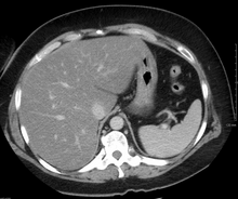
Liver disease, or hepatic disease, is any of many diseases of the liver. If long-lasting it is termed chronic liver disease. Although the diseases differ in detail, liver diseases often have features in common.

<span class="mw-page-title-main">Steatohepatitis</span> Medical condition

Steatohepatitis is a type of fatty liver disease, characterized by inflammation of the liver with concurrent fat accumulation in liver. Mere deposition of fat in the liver is termed steatosis, and together these constitute fatty liver changes.

<span class="mw-page-title-main">Metabolic dysfunction–associated steatotic liver disease</span> Excessive fat buildup in the liver with other metabolic disease

Metabolic dysfunction–associated steatotic liver disease (MASLD) is the name adopted in 2023 for the condition previously known as non-alcoholic fatty liver disease (NAFLD). This condition is diagnosed when there is excessive fat build-up in the liver, and at least one metabolic risk factor. When there is also moderate alcohol use, the term MetALD is used, and these are differentiated from alcoholic liver disease (ALD) when this is the sole cause of steatotic liver disease. The terms non-alcoholic fatty liver (NAFL) and non-alcoholic steatohepatitis have been used to describe different severities, the latter indicating the presence of further liver inflammation. NAFL is less dangerous than NASH and usually does not progress to it, but this progression may eventually lead to complications, such as cirrhosis, liver cancer, liver failure, and cardiovascular disease.

Acute fatty liver of pregnancy is a rare life-threatening complication of pregnancy that occurs in the third trimester or the immediate period after delivery. It is thought to be caused by a disordered metabolism of fatty acids by mitochondria in the fetus, caused by long-chain 3-hydroxyacyl-coenzyme A dehydrogenase deficiency. This leads to decreased metabolism of long chain fatty acids by the feto-placental unit, causing subsequent rise in hepatotoxic fatty acids in maternal plasma. The condition was previously thought to be universally fatal, but aggressive treatment by stabilizing the mother with intravenous fluids and blood products in anticipation of early delivery has improved prognosis.

<span class="mw-page-title-main">Phosphatidylethanolamine N-methyltransferase</span> Protein-coding gene in the species Homo sapiens

Phosphatidylethanolamine N-methyltransferase is a transferase enzyme which converts phosphatidylethanolamine (PE) to phosphatidylcholine (PC) in the liver. In humans it is encoded by the PEMT gene within the Smith–Magenis syndrome region on chromosome 17.

<span class="mw-page-title-main">Cirrhosis</span> Chronic disease of the liver, characterized by fibrosis

Cirrhosis, also known as liver cirrhosis or hepatic cirrhosis, and end-stage liver disease, is the impaired liver function caused by the formation of scar tissue known as fibrosis due to damage caused by liver disease. Damage to the liver leads to repair of liver tissue and subsequent formation of scar tissue. Over time, scar tissue can replace normal functioning tissue, leading to the impaired liver function of cirrhosis. The disease typically develops slowly over months or years. Early symptoms may include tiredness, weakness, loss of appetite, unexplained weight loss, nausea and vomiting, and discomfort in the right upper quadrant of the abdomen. As the disease worsens, symptoms may include itchiness, swelling in the lower legs, fluid build-up in the abdomen, jaundice, bruising easily, and the development of spider-like blood vessels in the skin. The fluid build-up in the abdomen may develop into spontaneous infections. More serious complications include hepatic encephalopathy, bleeding from dilated veins in the esophagus, stomach, or intestines, and liver cancer.Stages of cirrhosis include compensated cirrhosis and decompensated cirrhosis.

<span class="mw-page-title-main">Lipotoxicity</span> Metabolic syndrome

Lipotoxicity is a metabolic syndrome that results from the accumulation of lipid intermediates in non-adipose tissue, leading to cellular dysfunction and death. The tissues normally affected include the kidneys, liver, heart and skeletal muscle. Lipotoxicity is believed to have a role in heart failure, obesity, and diabetes, and is estimated to affect approximately 25% of the adult American population.

<span class="mw-page-title-main">Obeticholic acid</span> Chemical compound

Obeticholic acid (OCA), sold under the brand name Ocaliva, is a semi-synthetic bile acid analogue which has the chemical structure 6α-ethyl-chenodeoxycholic acid. It is used as a medication used to treat primary biliary cholangitis. Intercept Pharmaceuticals Inc. hold the worldwide rights to develop OCA outside Japan and China, where it is licensed to Dainippon Sumitomo Pharma.

<span class="mw-page-title-main">Aramchol</span> Chemical compound

Aramchol is an investigational drug being developed by Galmed Pharmaceuticals as a first-in-class, potentially disease modifying treatment for nonalcoholic steatohepatitis, or NASH, a more advanced condition of non-alcoholic fatty liver disease.

<span class="mw-page-title-main">Christos Socrates Mantzoros</span> Greek American physician and scientist

Christos Socrates Mantzoros is a Greek American physician-scientist, practicing internist-endocrinologist, teacher and researcher. He is a professor of medicine at Harvard Medical School and an adjunct professor at Boston University School of Medicine. He currently serves as the chief of endocrinology, diabetes and metabolism at the VA Boston Healthcare System, where he created de novo a leading academic division true to its tripartite mission and as the founding director of human nutrition at Beth Israel Deaconess Medical Center (BIDMC), Harvard Medical School. Finally, he holds the editor-in-chief position of the journal Metabolism: Clinical and Experimental.

<span class="mw-page-title-main">Melissa Palmer</span> American hepatologist

Melissa Palmer is an American hepatologist. She is recognized for her research and treatment of hepatitis and liver disease. Palmer is the Chief Medical Officer of Gannex Pharma, a wholly owned company of Ascletis Pharma.

<span class="mw-page-title-main">Claudio Tiribelli</span> Italian hepatologist (born 1946)

Claudio Tiribelli is an Italian hepatologist best known for his studies on bilirubin and Kernicterus, a bilirubin-induced neurological condition.

<span class="mw-page-title-main">Cilofexor</span> Drug in clinical trials

Cilofexor is a nonsteroidal farnesoid X receptor (FXR) agonist in clinical trials for the treatment of non-alcoholic fatty liver disease (NAFLD), non-alcoholic steatohepatitis (NASH), and primary sclerosing cholangitis (PSC). It is being investigated for use alone or in combination with firsocostat, selonsertib, or semaglutide. In rat models and human clinical trials of NASH it has been shown to reduce fibrosis and steatosis, and in human clinical trials of PSC it improved cholestasis and reduced markers of liver injury.

Hepatokines are proteins produced by liver cells (hepatocytes) that are secreted into the circulation and function as hormones across the organism. Research is mostly focused on hepatokines that play a role in the regulation of metabolic diseases such as diabetes and fatty liver and include: Adropin, ANGPTL4, Fetuin-A, Fetuin-B, FGF-21, Hepassocin, LECT2, RBP4,Selenoprotein P, Sex hormone-binding globulin.

AXA1125 is an experimental drug developed by Axcella Health that "increased β-oxidation and improved bioenergetics in preclinical models". It was studied as a treatment for non-alcoholic fatty liver disease and long COVID.

References

  1. 1 2 3 4 5 6 7 8 9 10 11 12 13 14 15 16 17 18 19 20 21 22 23 "Nonalcoholic Fatty Liver Disease & NASH". National Institute of Diabetes and Digestive and Kidney Diseases. November 2016. Retrieved 7 November 2018.
  2. 1 2 3 4 5 Singh S, Osna NA, Kharbanda KK (28 September 2017). "Treatment options for alcoholic and non-alcoholic fatty liver disease: A review". World Journal of Gastroenterology. 23 (36): 6549–6570. doi: 10.3748/wjg.v23.i36.6549 . PMC   5643281 . PMID   29085205.
  3. 1 2 3 4 5 6 7 8 Antunes C, Azadfard M, Hoilat GJ, Gupta M (2022). "Fatty Liver". StatPearls. StatPearls Publishing. PMID   28723021.
  4. 1 2 Basra S (2011). "Definition, epidemiology and magnitude of alcoholic hepatitis". World Journal of Hepatology. 3 (5): 108–113. doi: 10.4254/wjh.v3.i5.108 . PMC   3124876 . PMID   21731902.
  5. 1 2 Rinella ME, Lazarus JV, Ratziu V, Francque SM, Sanyal AJ, Kanwal F, Romero D, Abdelmalek MF, Anstee QM, Arab JP, Arrese M, Bataller R, Beuers U, Boursier J, Bugianesi E (2023). "A multi-society Delphi consensus statement on new fatty liver disease nomenclature". Hepatology. 78 (6): 1966–1986. doi: 10.1097/HEP.0000000000000520 . hdl: 10807/245116 . ISSN   0270-9139. PMC   10653297 . PMID   37363821. S2CID   259260747.
  6. 1 2 Iser D, Ryan M (July 2013). "Fatty liver disease—a practical guide for GPs". Australian Family Physician. 42 (7): 444–7. PMID   23826593.
  7. "A Liver Disease Gets a New Name, Diagnostic Criteria". Medscape. Retrieved 2023-09-04.
  8. 1 2 3 4 5 6 7 Chalasani N, Younossi Z, Lavine JE, Charlton M, Cusi K, Rinella M, Harrison SA, Brunt EM, Sanyal AJ (January 2018). "The diagnosis and management of nonalcoholic fatty liver disease: Practice guidance from the American Association for the Study of Liver Diseases". Hepatology. 67 (1): 328–357. doi: 10.1002/hep.29367 . hdl: 1805/14037 . PMID   28714183.
  9. Singh S, Allen AM, Wang Z, Prokop LJ, Murad MH, Loomba R (April 2015). "Fibrosis Progression in Nonalcoholic Fatty Liver vs Nonalcoholic Steatohepatitis: A Systematic Review and Meta-analysis of Paired-Biopsy Studies". Clinical Gastroenterology and Hepatology. 13 (4): 643–654.e9. doi:10.1016/j.cgh.2014.04.014. PMC   4208976 . PMID   24768810.
  10. 1 2 3 Younossi Z, Anstee QM, Marietti M, Hardy T, Henry L, Eslam M, George J, Bugianesi E (20 September 2017). "Global burden of NAFLD and NASH: trends, predictions, risk factors and prevention". Nature Reviews Gastroenterology & Hepatology. 15 (1): 11–20. doi:10.1038/nrgastro.2017.109. hdl: 2318/1659230 . PMID   28930295. S2CID   31345431.
  11. Qian Y, Fan JG (May 2005). "Obesity, fatty liver and liver cancer". Hepatobiliary & Pancreatic Diseases International. 4 (2): 173–7. PMID   15908310.
  12. Bellentani S (January 2017). "The epidemiology of non-alcoholic fatty liver disease". Liver International. 37: 81–84. doi: 10.1111/liv.13299 . PMID   28052624.
  13. 1 2 3 Reddy JK, Rao MS (May 2006). "Lipid metabolism and liver inflammation. II. Fatty liver disease and fatty acid oxidation". American Journal of Physiology. Gastrointestinal and Liver Physiology. 290 (5): G852-8. doi:10.1152/ajpgi.00521.2005. PMID   16603729.
  14. 1 2 Angulo P (18 April 2002). "Nonalcoholic Fatty Liver Disease". New England Journal of Medicine. 346 (16): 1221–1231. doi:10.1056/NEJMra011775. PMID   11961152.
  15. 1 2 3 Bayard M, Holt J, Boroughs E (June 2006). "Nonalcoholic fatty liver disease". American Family Physician. 73 (11): 1961–8. PMID   16770927.
  16. Osman KA, Osman MM, Ahmed MH (January 2007). "Tamoxifen-induced non-alcoholic steatohepatitis: where are we now and where are we going?". Expert Opinion on Drug Safety. 6 (1): 1–4. doi:10.1517/14740338.6.1.1. PMID   17181445. S2CID   33505288.
  17. Marciano F, Savoia M, Vajro P (February 2016). "Celiac disease-related hepatic injury: Insights into associated conditions and underlying pathomechanisms". Digestive and Liver Disease. 48 (2): 112–9. doi:10.1016/j.dld.2015.11.013. PMID   26711682.
  18. Valenti L, Dongiovanni P, Piperno A, Fracanzani AL, Maggioni M, Rametta R, Loria P, Casiraghi MA, Suigo E, Ceriani R, Remondini E, Trombini P, Fargion S (October 2006). "Alpha 1-antitrypsin mutations in NAFLD: high prevalence and association with altered iron metabolism but not with liver damage". Hepatology. 44 (4): 857–64. doi: 10.1002/hep.21329 . PMID   17006922. S2CID   26068505.
  19. Goldman L (2003). Cecil Textbook of Medicine – 2-Volume Set, Text with Continually Updated Online Reference. Philadelphia: W.B. Saunders Company. ISBN   978-0-7216-4563-6.[ page needed ]
  20. Adams LA, Lymp JF, St Sauver J, Sanderson SO, Lindor KD, Feldstein A, Angulo P (July 2005). "The natural history of nonalcoholic fatty liver disease: a population-based cohort study". Gastroenterology. 129 (1): 113–21. doi: 10.1053/j.gastro.2005.04.014 . PMID   16012941.
  21. Crabb DW, Galli A, Fischer M, You M (August 2004). "Molecular mechanisms of alcoholic fatty liver: role of peroxisome proliferator-activated receptor alpha". Alcohol. 34 (1): 35–8. doi:10.1016/j.alcohol.2004.07.005. PMID   15670663.
  22. Medina J, Fernández-Salazar LI, García-Buey L, Moreno-Otero R (August 2004). "Approach to the pathogenesis and treatment of nonalcoholic steatohepatitis". Diabetes Care. 27 (8): 2057–66. doi: 10.2337/diacare.27.8.2057 . PMID   15277442.
  23. Day CP, James OF (April 1998). "Steatohepatitis: a tale of two "hits"?". Gastroenterology. 114 (4): 842–5. doi: 10.1016/S0016-5085(98)70599-2 . PMID   9547102.
  24. Gramlich T, Kleiner DE, McCullough AJ, Matteoni CA, Boparai N, Younossi ZM (February 2004). "Pathologic features associated with fibrosis in nonalcoholic fatty liver disease". Human Pathology. 35 (2): 196–9. doi:10.1016/j.humpath.2003.09.018. PMID   14991537.
  25. Zafrani ES (January 2004). "Non-alcoholic fatty liver disease: an emerging pathological spectrum". Virchows Archiv. 444 (1): 3–12. doi:10.1007/s00428-003-0943-7. PMID   14685853. S2CID   7708476.
  26. Adams LA, Angulo P, Lindor KD (29 March 2005). "Nonalcoholic fatty liver disease". Canadian Medical Association Journal. 172 (7): 899–905. doi:10.1503/cmaj.045232. PMC   554876 . PMID   15795412.
  27. Reid AE (2006). "Chapter 82: Nonalcoholic Fatty Liver Disease". In Feldman M, Friedman LS, Brandt LJ (eds.). Sleisenger and Fordtran's Gastrointestinal and Liver Disease (8th ed.). Philadelphia: W.B. Saunders Company. ISBN   978-1-4160-0245-1 . Retrieved 4 July 2023 via Internet Archive.
  28. Peleg N, Issachar A, Sneh-Arbib O, Shlomai A (October 2017). "AST to Platelet Ratio Index and fibrosis 4 calculator scores for non-invasive assessment of hepatic fibrosis in patients with non-alcoholic fatty liver disease". Digestive and Liver Disease. 49 (10): 1133–1138. doi:10.1016/j.dld.2017.05.002. PMID   28572039.
  29. Peleg N, Sneh Arbib O, Issachar A, Cohen-Naftaly M, Braun M, Shlomai A (14 August 2018). "Noninvasive scoring systems predict hepatic and extra-hepatic cancers in patients with nonalcoholic fatty liver disease". PLOS ONE. 13 (8): e0202393. Bibcode:2018PLoSO..1302393P. doi: 10.1371/journal.pone.0202393 . PMC   6091950 . PMID   30106985.
  30. Miles DA, Levi CS, Uhanova J, Cuvelier S, Hawkins K, Minuk GY. Pocket-Sized Versus Conventional Ultrasound for Detecting Fatty Infiltration of the Liver. Dig Dis Sci. 2020 Jan;65(1):82-85. doi: 10.1007/s10620-019-05752-x. Epub 2019 Aug 2. PMID 31376083.
  31. Costantino A, Piagnani A, Caccia R, Sorge A, Maggioni M, Perbellini R, Donato F, D'Ambrosio R, Sed NPO, Valenti L, Prati D, Vecchi M, Lampertico P, Fraquelli M. Reproducibility and accuracy of a pocket-size ultrasound device in assessing liver steatosis. Dig Liver Dis. 2023 Nov 27:S1590-8658(23)01032-0. doi: 10.1016/j.dld.2023.11.014. Epub ahead of print. PMID 38016894.
  32. Singh S, Venkatesh SK, Loomba R, Wang Z, Sirlin C, Chen J, Yin M, Miller FH, Low RN, Hassanein T, Godfrey EM, Asbach P, Murad MH, Lomas DJ, Talwalkar JA, Ehman RL (28 August 2015). "Magnetic resonance elastography for staging liver fibrosis in non-alcoholic fatty liver disease: a diagnostic accuracy systematic review and individual participant data pooled analysis". European Radiology. 26 (5): 1431–1440. doi:10.1007/s00330-015-3949-z. PMC   5051267 . PMID   26314479.
  33. Benedict M, Zhang X (June 2017). "Non-alcoholic fatty liver disease: An expanded review". World Journal of Hepatology. 9 (16): 715–732. doi: 10.4254/wjh.v9.i16.715 . PMC   5468341 . PMID   28652891.
  34. Fatty Liver at eMedicine
  35. Buchman AL, Dubin MD, Moukarzel AA, Jenden DJ, Roch M, Rice KM, Gornbein J, Ament ME (November 1995). "Choline deficiency: a cause of hepatic steatosis during parenteral nutrition that can be reversed with intravenous choline supplementation". Hepatology. 22 (5): 1399–403. doi:10.1002/hep.1840220510. PMID   7590654. S2CID   20227016.
  36. Buchman AL, Dubin M, Jenden D, Moukarzel A, Roch MH, Rice K, Gornbein J, Ament ME, Eckhert CD (April 1992). "Lecithin increases plasma free choline and decreases hepatic steatosis in long-term total parenteral nutrition patients". Gastroenterology. 102 (4 Pt 1): 1363–70. doi:10.1016/0016-5085(92)70034-9. PMID   1551541.
  37. Buchman AL, Ament ME, Sohel M, Dubin M, Jenden DJ, Roch M, Pownall H, Farley W, Awal M, Ahn C (2016). "Choline deficiency causes reversible hepatic abnormalities in patients receiving parenteral nutrition: proof of a human choline requirement: a placebo-controlled trial". Journal of Parenteral and Enteral Nutrition. 25 (5): 260–8. doi:10.1177/0148607101025005260. PMID   11531217.
  38. Hollenbeck CB (August 2010). "The importance of being choline". Journal of the American Dietetic Association. 110 (8): 1162–5. doi:10.1016/j.jada.2010.05.012. PMID   20656090.
  39. 1 2 Liu J, Ayada I, Zhang X, Wang L, Li Y, Wen T, Ma Z, Bruno MJ, de Knegt RJ, Cao W, Peppelenbosch MP, Ghanbari M, Li Z, Pan Q (February 2021). "Estimating Global Prevalence of Metabolic Dysfunction-Associated Fatty Liver Disease in Overweight or Obese Adults". Clinical Gastroenterology and Hepatology. 20 (3): e573–e582. doi: 10.1016/j.cgh.2021.02.030 . PMID   33618024. S2CID   232018678.
  40. Hamaguchi M, Kojima T, Takeda N, Nakagawa T, Taniguchi H, Fujii K, Omatsu T, Nakajima T, Sarui H, Shimazaki M, Kato T, Okuda J, Ida K (15 November 2005). "The Metabolic Syndrome as a Predictor of Nonalcoholic Fatty Liver Disease". Annals of Internal Medicine. 143 (10): 722–728. doi:10.7326/0003-4819-143-10-200511150-00009. PMID   16287793. S2CID   22475943.
  41. Sarah Boseley (12 April 2019). "Experts warn of fatty liver disease 'epidemic' in young people". The Guardian. Retrieved 4 July 2023.
  42. SPINK HEALTH (11 April 2019). "Nonalcoholic fatty liver disease found in large numbers of teenagers and young adults". EurekAlert! (Press release). American Association for the Advancement of Science. Retrieved 4 July 2023.
  43. Cinque F, Cespiati A, Lombardi R, Costantino A, Maffi G, Alletto F, Colavolpe L, Francione P, Oberti G, Fatta E, Bertelli C, Sigon G, Dongiovanni P, Vecchi M, Fargion S, Fracanzani AL (January 2022). "Interaction between Lifestyle Changes and PNPLA3 Genotype in NAFLD Patients during the COVID-19 Lockdown". Nutrients. 14 (3): 556. doi: 10.3390/nu14030556 . ISSN   2072-6643. PMC   8838646 . PMID   35276911. CC-BY icon.svg Text was copied from this source, which is available under a Creative Commons Attribution 4.0 International License.
  44. Liu, Y.L.; Patman, G.L.; Leathart, J.B.; Piguet, A.C.; Burt, A.D.; Dufour, J.F.; Day, C.P.; Daly, A.K.; Reeves, H.L.; Anstee, Q.M. Carriage of the PNPLA3 rs738409 C >G polymorphism confers an increased risk of non-alcoholic fatty liver disease associated hepatocellular carcinoma. J. Hepatol. 2014, 61, 75–81.
  45. Eslam, M.; Valenti, L.; Romeo, S. Genetics and epigenetics of NAFLD and NASH: Clinical impact. J. Hepatol. 2018, 68, 268–279.
  46. Lock B (8 August 2017). "Hepatic Lipidosis (Fatty Liver Disease) in Reptiles". Vin.com. Retrieved 29 December 2020.
  47. "Fatty Liver Disease in Birds". Animal House of Chicago. Retrieved 29 December 2020.
  48. "Fatty Liver Disease in Cats". PetMD. Retrieved 29 December 2020.
  49. Kalyesubula M, Mopuri R, Rosov A, Alon T, Edery N, Moallem U, Dvir H (December 2020). "Hyperglycemia-stimulating diet induces liver steatosis in sheep". Scientific Reports. 10 (1): 12189. Bibcode:2020NatSR..1012189K. doi:10.1038/s41598-020-68909-z. PMC   7376193 . PMID   32699301.
  50. Kalyesubula M, Mopuri R, Asiku J, Rosov A, Yosefi S, Edery N, Bocobza S, Moallem U, Dvir H (1 March 2021). "High-dose vitamin B1 therapy prevents the development of experimental fatty liver driven by overnutrition". Disease Models & Mechanisms. 14 (3): dmm048355. doi:10.1242/dmm.048355. PMC   7988776 . PMID   33608323.